The neuropsychological assessment of emotion is typically overshadowed by assessment of language, executive processes, learning, and so on, as affect is difficult to assess using traditional approaches. Despite the challenges, emotion is an aspect of brain function that is a critical determinant of human behavior and so fundamental to neuropsychology. The past 3 decades has seen growing momentum in understanding its neural underpinnings and clinical implications.
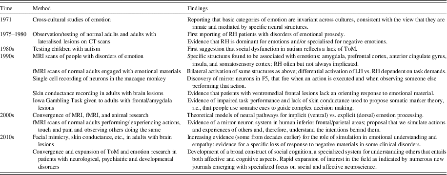
In 1872, Darwin described emotions as behavioral patterns that are important for the species’ survival (Darwin, Reference Darwin1872). Supporting this, seminal cross-cultural research (Ekman and Friesen, Reference Ekman and Friesen1971; Izard, Reference Izard1971) revealed that people of different cultures are highly consistent in their expression of anger, fear, happiness, surprise, disgust, and sadness, suggesting these are innate and integral to communication. This set the stage for a generation of research that almost exclusively focused on the perception, expression, and experience of these six basic emotions. Increasingly, the social nature of emotions is coming into focus and other emotions (trust, envy, flirtation, etc.) are being examined (Adolphs, Tranel, & Damasio, Reference Adolphs, Tranel and Damasio1998; Reynders, Broks, Dickson, Lee, & Turpin, Reference Reynders, Broks, Dickson, Lee and Turpin2005; Rosenberg, McDonald, Rosenberg, & Westbrook, Reference Rosenberg, McDonald, Rosenberg and Westbrook2016). These are more subject to learning and cultural variation but, none-the-less, critical when considering how emotion disorders disrupt social competence. The following overview (see Table 1 for summary) considers major milestones in research of the basic emotions in the past 30 years
Major milestones in emotion research over the past 30 years

Note. “Patients” refer to adults with brain lesions.
RH=right hemisphere; LH=left hemisphere; CT=computed tomography; MRI=magnetic resonance imaging; fMRI=functional MRI; ToM=Theory of Mind.
THE 1980s: THE RIGHT HEMISPHERE IN EMOTION PROCESSING
In 1975, Heilman and colleagues reported several patients with right hemisphere lesions who could not comprehend emotional prosody of speech (Heilman, Scholes, & Watson, Reference Heilman, Scholes and Watson1975). In 1979, Ross and Mesulam described two cases with right hemisphere lesions who lost their capacity to impart affect in speech. Thus, the right hemisphere was attributed a role in affective speech processing, potentially mirroring the role of the left hemisphere in propositional speech (Ross, Reference Ross1981). The 1980s onward saw sustained research extending the role of the right hemisphere in emotion to include expressivity and perception of face Borod, Koff, Lorch, & Nicholas, Reference Borod, Koff, Lorch and Nicholas1985, Reference Borod, Koff, Lorch and Nicholas1986) and gestures (e.g., crying and laughing) (Ross & Mesulam, Reference Ross and Mesulam1979). During the 1980s, for the first time, it was demonstrated that people with right hemisphere damage had broader problems extracting emotional content from pictures (Cicone, Wapner, & Gardner, Reference Cicone, Wapner and Gardner1980) and words (Brownell, Michel, Powelson, & Gardner, 1983; Wapner, Hamby, & Gardner, Reference Wapner, Hamby and Gardner1981), leading to the view that the right hemisphere is dominant for emotional processing generally (Heilman, Bowers, & Valenstien, Reference Heilman, Bowers and Valenstein1985). Research in healthy adults supported this, finding a right hemisphere advantage for emotion perception via tachistoscopic viewing (Heller & Levy, Reference Heller and Levy1981) and dichotic listening (Bryden, Ley, & Sugarman, Reference Bryden, Ley and Sugarman1982) and for facial expressivity (Borod & Caron, Reference Borod and Caron1980).
A contemporaneous, alternative view was the “valence hypothesis,” that is, that the left hemisphere is specialized for positive and the right hemisphere specialized for negative emotions (Silberman & Weingartner, Reference Silberman and Weingartner1986) or the related “approach (left) - withdrawal (right) hypothesis” (Davidson, Reference Davidson1984). Rapid presentation of facial expressions to one hemisphere in normal adults (Ley & Bryden, Reference Ley and Bryden1979; Reuter-Lorenz & Davidson, Reference Reuter-Lorenz and Davidson1981) demonstrated a hemispheric bias for processing positive (left) and negative (right) emotions (Silberman & Weingartner, Reference Silberman and Weingartner1986). Patients with right hemisphere damage were reportedly indifferent or euphoric while those with left hemisphere damage commonly experienced depression (Gainotti, Reference Gainotti1972; Sackeim et al., Reference Sackeim, Greenberg, Weiman, Gur, Hungerbuhler and Geschwind1982). These responses were thought to reflect the disablement of normal positive, approach related responses following left hemisphere lesions (thus a swing to depression) and conversely the disablement of negative, withdrawal responses following right hemisphere lesions (thus the swing to euphoria) (Davidson, Pizzagalli, Nitschke, & Kalin, Reference Davidson, Pizzagalli, Nitschke and Kalin2003). Research into emotion perception has also suggested a valence effect with the right hemisphere dominant for negative emotions while both hemispheres played a role in positive (Adolphs, Jansari, & Tranel, Reference Adolphs, Jansari and Tranel2001).
THE 1990s: MRI IDENTIFIES SPECIFIC STRUCTURES UNDERPINNING EMOTION
Lesion-based research in the 1980s was hampered by two issues. First, most work focused on patients with middle cerebral artery stroke who were heterogeneous with respect to intra-hemispheric pathology. At the same time, computed tomography (CT) produced low-resolution images. In the 1990s, magnetic resonance imaging (MRI) provided dramatic improvements in the visualization of brain lesions and identification of specific structures associated with emotion processing.
The amygdala, interconnected with the hypothalamus, thalamus, basal forebrain, and brainstem, as well as neocortex, reciprocally interacts with the autonomic nervous system (Emery & Amaral, Reference Emery and Amaral2000) making it integral to emotional behavior. Using MRI, rare patients with focal amygdala damage were identified who had impairments in recognizing fear and other negative emotions in faces (Adolphs, Tranel, Damasio, & Damasio, Reference Adolphs, Tranel, Damasio and Damasio1994) and voices (Scott et al., Reference Scott, Young, Calder, Hellawell, Aggleton and Johnson1997) and loss of the autonomic fear response to aversive stimuli (Bechara et al., Reference Bechara, Tranel, Damasio, Adolphs, Rockland and Damasio1995). Lesions were usually bilateral, although in some studies right amygdala lesions produced poorer performance than left (Adolphs & Tranel, Reference Adolphs and Tranel2004; Adolphs, Tranel, & Damasio, Reference Adolphs, Tranel and Damasio2001).
The prefrontal lobes also represented an important candidate for mediating emotions. It was long known that frontal lobe injury led to changes to socio-emotional behavior (Benton, Reference Benton1968; Stuss & Benson, Reference Stuss and Benson1986; Weinstein & Kahn, Reference Weinstein and Kahn1955). In the 1990s, circumscribed lesions in the orbital/medial prefrontal cortex and ventral, anterior cingulate cortex were identified as specific to emotional behavior, associated with poor emotion perception, lowered self-reported emotional experience, especially for negative emotions, and apathy, disinhibition, lability, aggression, and personality change (Barrash, Tranel, & Anderson, Reference Barrash, Tranel and Anderson2000; Grafman et al., Reference Grafman, Schwab, Warden, Pridgen, Brown and Salazar1996; Hornak, Rolls, & Wade, Reference Hornak, Rolls and Wade1996). This pattern led to views that the medial prefrontal system (especially the right) has a role in regulating “high autonomic arousal” emotions such as anger and fear (Adolphs, Reference Adolphs2002a). Lesions to the (left) anterior insula cortex along with the basal ganglia were also implicated, specifically perception of disgust in patients with focal brain lesions (Calder, Keane, Manes, Antoun, & Young, Reference Calder, Keane, Manes, Antoun and Young2000) and in Huntington’s disease (Sprengelmeyer et al., Reference Sprengelmeyer, Young, Calder, Karnat, Lange, Homber and Rowland1996). Selective impairment in fear (amygdala) and disgust (insula) were regarded as evidence for a double dissociation, demonstrating that specific emotions were associated with specific structures (Adolphs, 2002b).
A major stepping stone in current emotion theories came from evidence that emotion perception could be disrupted by lesions in somatosensory and motor cortex (especially in the right hemisphere: Adolphs, Damasio, & Tranel, Reference Adolphs, Damasio and Tranel2002; Adolphs, Damsio, Tranel, & Damsio, Reference Adolphs, Damasio, Tranel and Damasio1996). These studies suggested that processing visual or auditory affective stimuli, “as if” it were one’s own, might play a role in its identification. Dissociations were also reported suggesting that prosody and facial affect engaged overlapping but discrete cerebral systems (Adolphs et al., Reference Adolphs, Damasio and Tranel2002) as did moving versus static emotional stimuli (Adolphs, Tranel, & Damasio, Reference Adolphs, Tranel and Damasio2003).
THE 1990s: FUNCTIONAL IMAGING REVEALS NEURAL ACTIVATION
In parallel to MRI lesion research, functional imaging rapidly emerged as a powerful adjunct, providing opportunities to observe localized brain activity in normal adults when viewing emotional materials. Using positron emission tomography (PET) normal adults showed left amygdala activation to angry and fearful faces even when their attention was diverted from the expression (Morris et al., Reference Morris, Frith, Perrett, Rowland, Young, Calder and Dolan1996). This was even found in a patient with unilateral brain damage and no conscious awareness of an image transmitted to his blinded right hemi-field (Morris, DeGelder, Weiskrantz, & Dolan, Reference Morris, DeGelder, Weiskrantz and Dolan2001). Functional magnetic resonance imaging (fMRI; Belliveau et al., Reference Belliveau, Kennedy, McKinstry, Buchbinder, Weisskoff, Cohen and Rosen1991), a cheaper, less-invasive technique than PET, led to an explosion of interest in emotion. There have been literally hundreds of fMRI studies of normal adults exposed to emotional stimuli.
Meta-analyses of these generally suggest bilateral activation of specific structures in response to emotional stimuli. Amygdala activation was found when participants passively observe facial expressions. Despite earlier reports, there was little evidence of greater activation to negative expressions (Baas, Aleman, & Kahn, Reference Baas, Aleman and Kahn2004; Costafreda, Brammer, David, & Fu, Reference Costafreda, Brammer, David and Fu2008; Sergerie, Chocol, & Armony, Reference Sergerie, Chochol and Armony2008). Left and right amygdala activation varied due to task. Language-based tasks activate the left amygdala, whereas masked emotions are more likely to activate the right amygdala briefly with rapid habituation (Costafreda et al., Reference Costafreda, Brammer, David and Fu2008; Sergerie et al., Reference Sergerie, Chochol and Armony2008).
Using fMRI meta-analysis, Lindquist, Wager, Kober, Bliss-Moreau, and Barrett (Reference Lindquist, Wager, Kober, Bliss-Moreau and Barrett2012) also challenged traditional views that certain types of emotion (such as fear) were mediated by specific structures (such as the amygdala). Their results suggested key structures are not activated by specific emotions but, rather, play a role that is important for specific emotions. Thus, the amygdalae are engaged in orienting responses to novel and arousing events rather than fear specifically, the insula is engaged in self-awareness of body sensations rather than disgust in particular, and the ventromedial frontal lobes appears to integrate external and internal high arousal sensations rather than simply mediating anger. It has also been shown that early sensory cortices are activated by affective (vs. neutral) sensory input, suggesting a role in emotional experience beyond simply processing sensory inputs (Satpute et al., Reference Satpute, Kang, Bickart, Yardley, Wager and Barrett2015).
2000s: TWO NEURAL MODELS OF EMOTION PROCESSES
The explosion of MRI, fMRI, animal, and experimental research during the 1990s provided the impetus to develop two complementary models of neural systems of emotion that emerged in the 2000s.
Explicit Versus Implicit (Autonomic) Processing
Animal research by LeDoux and colleagues suggested that the “flight or fight” response is mediated by very rapid, coarse evaluation of potential sources of danger in the environment processed mainly by subcortical routes (LeDoux, Reference LeDoux1995). In humans, a ventral system was identified that entails the amygdalae, insula, ventral striatum, and anterior cingulate and prefrontal cortex. This system identifies emotionally significant stimuli; produces affective, autonomic responses; and provides automatic regulation (Lieberman, Reference Lieberman2007; Phillips, Drevets, Rauch, & Lane, Reference Phillips, Drevets, Rauch and Lane2003).
In contrast, complex emotional judgements occur more slowly, engaging the dorsal lateral and medial prefrontal cortex and anterior cingulate gyrus as well as hippocampus and temporo-parietal regions (Lieberman, Reference Lieberman2007; Phillips et al., Reference Phillips, Drevets, Rauch and Lane2003). This system mediates effortful processing of emotional stimuli and engagement of relevant cognitive processes (e.g., language). The two systems are intimately and reciprocally associated. Emotion tasks that require verbal mediation, for example, labeling, increase activation of dorsolateral prefrontal cortex (Hariri, Bookheimer, & Mazziotta, Reference Hariri, Bookheimer and Mazziotta2000; Keightly et al., Reference Keightly, Winocur, Graham, Matyberg, Hevenor and Grady2003) and reduce activation in both amygdalae (Hariri et al., Reference Hariri, Bookheimer and Mazziotta2000).
Shared Neural Representations for Self Versus Other
In 1992, a major breakthrough occurred with the discovery of mirror neurons in the F5 area of the macaque’s frontal cortex (approximating the human Broca’s area), which activated when the subject both undertook a meaningful action and observed that same action in others (di Pellegrino, Fadiga, Fogassi, Gallese, & Rizzolatti, Reference di Pellegrino, Fadiga, Fogassi, Gallese and Rizzolatti1992). It has since been revealed that, in humans also, overlapping areas in the inferior frontal lobe, including the premotor area, and inferior and superior parietal lobes activate when both observing and performing actions (Grèzes & Decety, Reference Grèzes and Decety2001; Molenberghs, Cunnington, & Mattingley, Reference Molenberghs, Cunnington and Mattingley2012) or even hearing sounds associated with actions (Gazzola, Aziz-Zadeh, & Keysers, Reference Gazzola, Aziz-Zadeh and Keysers2006).
Such “mirroring” only occurs to meaningful actions and is thought to provide a mental representation of an action and its goal, allowing the observer to anticipate and understand the intention behind it (Rizzolatti & Sinigaglia, Reference Rizzolatti and Sinigaglia2010). Emotional stimuli lead to similar patterns of activation extending to the insula, amygdalae, and the superior temporal sulcus (Carr, Iacoboni, Dubeau, Maxzziotta, & Lenzi, Reference Carr, Iacoboni, Dubeau, Maxzziotta and Lenzi2003; Pfeifer, Iacoboni, Mazziotta, & Dapretto, Reference Pfeifer, Iacoboni, Mazziotta and Dapretto2008), suggesting a role for action simulation in emotion understanding (Iacoboni, Reference Iacoboni2009).
Whether this co-activation reflects mirror neurons in humans remains controversial. Unlike single cell recordings in monkeys (Rizzolatti, Fadiga, Gallese, & Fogassi, Reference Rizzolatti, Fadiga, Gallese and Fogassi1996), most evidence in humans is indirect arising from voxel-based patterns of activations, although single-cell recordings in humans support their existence (Mukamel, Ekstrom, Kaplan, Iacoboni, & Fried, Reference Mukamel, Ekstrom, Kaplan, Iacoboni and Fried2010). Even so, the notion that overlapping areas mediate both direct emotional experience and observed experience has gained momentum. Furthermore, simulation is not limited to observing actions. Both simple touch to the hand and observation of touch activate the somatosensory cortex (Kuehn, Mueller, Turner, & Schutz-Bosbach, Reference Kuehn, Mueller, Turner and Schutz-Bosbach2014; Schaefer, Heinze, & Rotte, Reference Schaefer, Heinze and Rotte2012). The anterior cingulate gyrus, thalamus, somatosensory cortices, and insula are activated when experiencing painful stimuli in oneself with overlapping but not identical structures involved when observing others in pain (Jackson, Rainville, & Decety, Reference Jackson, Rainville and Decety2006).
PHYSIOLOGICAL EVIDENCE FOR MODELS OF EMOTION PROCESSES
fMRI and other techniques, such as electroencephalography (Fox et al., Reference Fox, Bakermans-Kranenburg, Yoo, Bowman, Cannon, Vanderwert and van Ijzendoorn2016), provide unprecedented opportunity to observe brain activation. However, as they are correlational in nature, they give only indirect evidence of the functional significance and, on occasion, this may be erroneous. For example, Feinstein et al. (Reference Feinstein, Khalsa, Salomons, Prkachin, Frey-Law, Lee and Rudrauf2016) reported a rare patient with bilateral lesions of the “pain matrix,” that is, the insula, anterior cingulate, and amygdala, who still experienced pain, sometimes excessively. They argued that the common interpretation that the pain matrix mediates the emotional significance of pain may be misdirected, and it may have another role, for example, regulation.
Psychophysiological measurements provide a complementary approach to understanding emotional processing. Although such measures had a long tradition in Russian neuropsychology from the 1960s, much of this literature was never published in English (e.g., see Luria, Reference Luria1973). Western psychophysiological research, which has accelerated since the 2000s, has converged with theorizing from structural and functional imaging to refine understanding of emotion processes.
EVIDENCE FOR AUTONOMIC RESPONSES TO EMOTIONAL STIMULI
In normal adults, skin conductance responses (SCR, indicating the orientation reflex) occur in response to fearful stimuli even when participants are not overtly aware of them (Esteves, Dimberg, & Ohman, Reference Esteves, Dimberg and Ohman1994; Ohman & Soares, Reference Ohman and Soares1994). Damasio, Tranel, and Damasio (Reference Damasio, Tranel and Damasio1990) reported that five patients with ventromedial frontal lesions lacked this orienting response to emotionally charged pictures. Furthermore, people with traumatic brain injuries (TBIs), many of whom experience ventromedial frontal pathology, have abnormally low arousal (skin conductance level) at baseline (Fisher, Rushby, McDonald, Parks, & Piguet, Reference Fisher, Rushby, McDonald, Parks and Piguet2015; McDonald, Rushby, et al., Reference McDonald, Flanagan and Rollins2011) and habituate unusually rapidly to angry faces (McDonald, Rushby, et al., Reference McDonald, Flanagan and Rollins2011). Their startle response is also abnormally unresponsive to negative images (Saunders, McDonald, & Richardson, Reference Saunders, McDonald and Richardson2006; Williams & Wood, Reference Williams and Wood2012). A similar lack of startle response was reported in people with amygdala lesions (Angrilli et al., Reference Angrilli, Mauri, Palomba, Flor, Birbaumer, Sartori and di Paola1996; Buchanan, Tranel, & Adolphs, Reference Buchanan, Tranel and Adolphs2004; Funayama, Grillon, Davis, & Phelps, Reference Funayama, Grillon, Davis and Phelps2001) supporting the notion that the amygdalae, in concert with the ventromedial frontal lobes, mediate autonomic responses to emotional events with or without conscious awareness.
Evidence for Simulation
Psychophysiological techniques have also shed light on the role of simulation in emotion. Studies of normal adults in the 1990s demonstrated that observers experience activation of their own cheek (zygomaticus major) and brow (corrugator supercilii) muscles when observing another person expressing happy or angry emotions, respectively. Mimicry occurs spontaneously and rapidly, even without conscious awareness (Dimberg & Thunberg, Reference Dimberg and Thunberg1998; Dimberg, Thunberg, & Elmehed, Reference Dimberg, Thunberg and Elmehed2000), suggesting an automatic process. This observation re-invigorated a view initially proposed by Lipps (1907, cited in Hoffman, Reference Hoffman1984) that facial feedback engenders a shared emotional experience (emotional contagion) that helps identify the emotion being observed. This view clearly dovetails into implications of the mirror neuron system. Furthermore, deficits in facial mimicry have been reported in people with low empathic ability (Sonnby-Borgström, Jönsson, & Svensson, Reference Sonnby-Borgström, Jönsson and Svensson2003) as well as clinical conditions associated with low empathy, such as schizophrenia (Park, Mathews, & Gibson, Reference Park, Matthews and Gibson2008).
Clinical evidence based on people with lesions to ventromedial frontal cortex or as a result of TBI suggests that loss of emotion simulation extends beyond facial mimicry. While there can be loss of facial expressivity (Dethier, Blairy, Rosenberg, & McDonald, Reference Dethier, Blairy, Rosenberg and McDonald2012) and spontaneous facial mimicry (Angrilli, Palomba, Cantagallo, Maietti, & Stegagno, Reference Angrilli, Palomba, Cantagallo, Maietti and Stegagno1999; McDonald, Li, et al., Reference McDonald, Flanagan and Rollins2011), there is also lowered autonomic arousal to facial expressions (Blair & Cipolotti, Reference Blair and Cipolotti2000; de Sousa et al., Reference de Sousa, McDonald, Rushby, Li, Dimoska and James2011; Hopkins, Dywan, & Segalowitz, Reference Hopkins, Dywan and Segalowitz2002) and reduced subjective appraisal of emotional material (Hornak et al., Reference Hornak, Rolls and Wade1996; Saunders et al., Reference Saunders, McDonald and Richardson2006) and the effects of postural feedback (Dethier, Blairy, Rosenberg, & McDonald, Reference Dethier, Blairy, Rosenberg and McDonald2013).
Thus, disruption of emotional contagion can occur at numerous points in a complex system from motor mimicry through to arousal and the subjective evaluation of the emotional experience. Of interest, when emotional contagion is impaired, this is usually limited to negative emotions (Blair & Cipolotti, Reference Blair and Cipolotti2000; McDonald, Rushby, et al., Reference McDonald, Rushby, Li, de Sousa, Dimoska, James and Togher2011). This valence specific effect suggests that disruption occurs at a different stage to mirroring which, otherwise, should affect all emotions equally. It does, however, fit with the notion that autonomic responses are mediated by the ventral system (Phillips et al., Reference Phillips, Drevets, Rauch and Lane2003) that has evolved to detect potential danger and elicit an immediate arousal reponse.
While both emotion perception and emotional contagion can be disrupted following brain lesions, evidence for a causal link is far from established. On the one hand, accumulating behavioral evidence suggests that loss of mimicry impedes emotion recognition. Disrupting the (left) premotor cortex using transient magnetic stimulation decreases emotion recognition accuracy and increases reaction time (Balconi & Bortolotti, Reference Balconi and Bortolotti2013). Botulin toxin that impairs facial movement also reduces emotional experience and emotion recognition (Davis, Senghas, Brandt, & Ochsner, Reference Davis, Senghas, Brandt and Ochsner2010). People with locked in syndrome and facial paralysis are less efficient than normal when recognizing (negative) emotions (Pistoia et al., Reference Pistoia, Conson, Trojano, Grossi, Ponari, Colonnese and Sara2010) as are normal adults prevented from mimicry (Niedenthal, Brauer, Halberstadt, & Innes-Ker, Reference Niedenthal, Brauer, Halberstadt and Innes-Ker2001).
However, there is also evidence to the contrary. While loss of contagion and poor emotion recognition do co-occur in individual patients (Blair & Cipolotti, Reference Blair and Cipolotti2000), they are not correlated in people with brain injury (McDonald, Li, et al., Reference McDonald, Li, De Sousa, Rushby, Dimoska, James and Tate2011; McDonald, Rushby, et al., Reference McDonald, Rushby, Li, de Sousa, Dimoska, James and Togher2011) nor in adults with a complete disorder of peripheral autonomic function (Heims, Critchley, Dolan, Mathias, & Cipolotti, Reference Heims, Critchley, Dolan, Mathias and Cipolotti2004). A similar lack of relation is seen in normal adults (Blairy, Herrera, & Hess, Reference Blairy, Herrera and Hess1999; Hess & Blairy, Reference Hess and Blairy2001). Thus, while mimicry may facilitate emotion perception, there is insufficient evidence that it is essential. It may have other functions, for example, to communicate empathic understanding (Bavelas, Black, Chovil, Lemery, & Mullett, Reference Bavelas, Black, Chovil, Lemery and Mullett1988).
ATTRIBUTING EMOTIONS WITH A ROLE IN COGNITION
A major development in contemporary neuropsychology has been the recognition that emotion has a role in cognition. Two important theoretical developments have been the somatic marker theory (1990s) and the construct of social cognition (2000s).
Somatic Marker Theory
The influential “somatic marker” hypothesis arose in the 1990s (Damasio, Tranel, & Damsio, Reference Damasio, Tranel and Damasio1991), suggesting that emotional responses can guide understanding of complex response contingencies even before conscious awareness (Bechara, Damasio, Tranel, & Damasio, Reference Bechara, Damasio, Tranel and Damasio1997). Most work used the Iowa Gambling Task (IGT) in which people learn reward contingencies given feedback. Both normal adults and those with ventromedial frontal lobe lesions have a SCR following feedback about their choices. Normal adults reportedly demonstrate these changes in anticipation of their choice, even before they can verbalize the rules. People with ventromedial damage do not (Bechara et al., Reference Bechara, Damasio, Tranel and Damasio1997; Bechara, Tranel, Damasio, & Damasio, Reference Bechara, Tranel, Damasio and Damasio1996; Naccache et al., Reference Naccache, Dehaene, Cohen, Habert, Guichart-Gomez, Galanaud and Willer2005). Similar results were found with people with specific amygdala damage, except, in these cases, there was no SCR at all, even after feedback (Bechara, Damasio, Damsio, & Lee, Reference Bechara, Damasio, Damasio and Lee1999).
As a result of this work, it was argued that somatic responses assist with decision making, especially in personal and social situations, where precise calculations of the outcome of a course of action is not possible (Bechara, Reference Bechara2004). Although interest in the somatic marker theory has been high, poor performance on the IGT is not limited to those with frontal lesions (Levine et al., Reference Levine, Black, Cheung, Campbell, O’Toole and Schwartz2005). Other cognitive abilities, such as working memory (Hinson, Jameson, & Whitney, Reference Hinson, Jameson and Whitney2002), intelligence (Toplak, Sorge, Benoit, West, & Stanovich, Reference Toplak, Sorge, Benoit, West and Stanovich2010), and reversal learning (Fellows & Farah, Reference Fellows and Farah2005) influence IGT performance.
Whether physiological responses occur before conscious awareness also remains controversial. Densely amnesic patients who cannot learn the IGT do not display anticipatory SCRs (despite having intact SCRs to punishment), and healthy adults have demonstrated differential anticipatory SCRs only after explicit knowledge of IGT contingencies was attained (Fernie & Tunney, Reference Fernie and Tunney2013; Gutbrod et al., Reference Gutbrod, Krouzel, Hofer, Muri, Perrig and Ptak2006). In all, it appears that somatic markers are involved in decision making on the IGT, as is conscious processing (Guillaume et al., Reference Guillaume, Jollant, Jaussent, Lawrence, Malafosse and Courtet2009), but are not necessarily critical to successful performance.
Social Cognition
Since the 2000s, there has been a groundswell of interest in the role of emotional processes in social performance. This was heralded by the 1980s research describing how emotional and humorous materials were poorly processed following right hemisphere lesions. In 1985, Baron Cohen, Leslie, and Frith proposed that children with autism lack the ability to impute mental states, that is, lack Theory of Mind (ToM; Baron Cohen, Leslie, and Frith, Reference Baron-Cohen, Leslie and Frith1985). More recently, these attributes have been subsumed under the construct of social cognition, that is, the ability to understand the minds of other people in relation to ourselves (Amodio & Frith, Reference Amodio and Frith2006), including judgments about what they are thinking, feeling, and intending by their behavior.
Social cognition also includes empathy, social connectedness, and making broader social judgments, such as attitudes about social groups and morality. Not all social cognition is related to emotion. Cognitive ToM tasks simply require understanding what another person might think and believe. On the other hand, affective ToM refers to understanding what another person feels (Mitchell & Phillips, Reference Mitchell and Phillips2015). fMRI research has demonstrated activation of the anterior dorsal medial prefrontal cortex and bilateral temporo-parietal junctions when performing any mental judgment but with additional activation of the orbitofrontal cortex, temporal poles, and bilateral premotor cortex for affective judgements (Molenberghs, Johnson, Henry, & Mattingley, Reference Molenberghs, Johnson, Henry and Mattingley2016). Lesion studies suggest that ventromedial (especially right) lesions are associated with impaired affective ToM judgments, such as understanding irony and faux pas (Shamay-Tsoory, Tomer, Berger, Goldsher, & Aharon-Peretz, Reference Shamay-Tsoory, Tomer, Berger, Goldsher and Aharon-Peretz2005).
IMPLICATIONS FOR CONTEMPORARY CLINICAL PRACTICE
Since the late 1990s, it has been recognized that impairments in emotion processing are pivotal to understanding neuropsychiatric disorders (Borod et al., Reference Borod, Welkowitz, Alpert, Brozgold, Martin, Peselow and Diller1990), including schizophrenia (Kohler, Walker, Martin, Healey, & Moberg, Reference Kohler, Walker, Martin, Healey and Moberg2010; Phillips, Reference Phillips2003), bipolar and other mood disorders (Cusi, Nazarov, Holshausen, MacQueen, & McKinnon, Reference Cusi, Nazarov, Holshausen, MacQueen and McKinnon2012; Green, Cahill, & Mahli, Reference Green, Cahill and Malhi2007), and intellectual and genetic developmental disabilities (Roelofs, Wingbermühle, Egger, & Kessels, Reference Roelofs, Wingbermühle, Egger and Kessels2017), as well as people with acquired brain injury. Characterization or socio-emotional impairments has greatly aided diagnosis and understanding of these disorders. For example, psychosocial changes following severe TBI are common (Brooks, Campsie, Symington, Beattie, & McKinlay, Reference Brooks, Campsie, Symington, Beattie and McKinlay1986) but were poorly understood before systematic research into emotional impairments.
Recently it has been estimated that up to 39% of adults with moderate-severe TBI suffer problems of emotion perception (Babbage et al., Reference Babbage, Yim, Zupan, Neumann, Tomita and Willer2011). They also self-report disproportionately low levels of empathy (Williams & Wood, Reference Williams and Wood2010; Wood & Williams, Reference Wood and Williams2008), blunted emotional experience (Croker & McDonald, Reference Croker and McDonald2005), and low self-awareness of their own emotions (i.e., alexithymia; Williams & Wood, Reference Williams and Wood2010). Up to 70% of adults with TBI demonstrate apathy (Kant, Duffy, & Pivovarnik, Reference Kant, Duffy and Pivovarnik1998), and irritability is one of the most common behavioral complaints (Demark & Gemeinhardt, Reference Demark and Gemeinhardt2002) along with impulsivity and aggression (Kinsella, Packer, & Olver, Reference Kinsella, Packer and Olver1991). Emotion disorders are not necessarily correlated to standard neuropsychological tests (Spikman, Timmerman, Milders, Veenstra, & van der Naalt, Reference Spikman, Timmerman, Milders, Veenstra and van der Naalt2012), but do predict social behavioral problems in adults (McDonald, Flanagan, Martin, & Saunders, Reference McDonald, Flanagan, Martin and Saunders2004; Spikman et al., Reference Spikman, Milders, Visser-Keizer, Westerhof-Evers, Herben-Dekker and van der Naalt2013) and children (Yeates et al., Reference Yeates, Swift, Taylor, Wade, Drotar, Stancin and Minich2004).
In frontotemporal dementia, different constellations of emotion impairment characterize sub-types. Semantic dementia, arising from initial asymmetric deterioration of the temporal lobes (primarily left), causes problems, not only with language and semantic comprehension, but restricted food interests; changed eating behavior; increased apathy; impaired emotion perception, insight, and empathy; poor mental state judgements; and poor emotional memories (Landin-Romero, Tan, Hodges, & Kumfor, Reference Landin-Romero, Tan, Hodges and Kumfor2016). Patients can also present with asymmetric deterioration of the right temporal lobe, resulting in even more severe emotion perception and behavioral problems, which suggests that it is the right temporal lobe that mediates these deficits in both groups (Kumfor et al., Reference Kumfor, Landin-Romero, Devenney, Hutchings, Grasso, Hodges and Piguet2016).
Behavioral variant frontotemporal dementia is similarly characterized by early behavior disturbance, disinhibition, poor emotion perception, and low empathy; although, in this case, these difficulties appear to arise from bilateral orbitofrontal and left temporal deterioration (Kamminga et al., Reference Kamminga, Kumfor, Burrell, Piguet, Hodges and Irish2015). Impaired empathy and emotional perception predict carer distress in the frontotemporal dementias (Hazelton, Irish, Hodges, Piguet, & Kumfor, Reference Hazelton, Irish, Hodges, Piguet and Kumfor2016).
Schizophrenia is defined by changes in socioemotional processes (Penn, Corrigan, Bentall, Racenstein, & Newman, Reference Penn, Corrigan, Bentall, Racenstein and Newman1997), including emotional flattening, anhedonia and persecutory delusions (Phillips, Reference Phillips2003), disorders of social cognition broadly (Savla, Vella, Armstrong, Penn, & Twamley, Reference Savla, Vella, Armstrong, Penn and Twamley2013), and emotion process specifically (Irani, Seligman, Kamath, Kohler, & Gur, Reference Irani, Seligman, Kamath, Kohler and Gur2012). Poor social cognitive abilities in schizophrenia more accurately predict community function than traditional cognitive measures (Fett et al., Reference Fett, Viechtbauer, Dominguez, Penn, van Os and Krabbendam2011).
Assessment
Given the relevance of emotional processes to interpersonal function and outcome, they should be assessed routinely. However, while some clinical measures have been available for decades (Borod, Tabert, Santschi, & Strauss, Reference Borod, Tabert, Santschi and Strauss2000), this is one area where change has been slow. Despite 84% of clinicians working in brain injury reporting that at least half of their clients had problems in social cognition (including multiple facets of emotion), less than 22% reported using any kind of formal assessment (Kelly, McDonald, & Frith, in press). Furthermore, professions differ in terms of who they believe to be responsible, with psychologists, neuropsychologists, occupational therapists, and speech pathologists each of the view that the responsibility lies with the other (Kelly, McDonald, & Frith, Reference Kelly, McDonald and Frith2017).
Current, consensus-based recommendations about outcome measures in brain injury include self- and proxy questionnaires to assess emotional behaviors (such as aggression and irritability), psychological status (such as negative and positive mood states), and arousal (apathy, disinhibition) (Wilde et al., Reference Wilde, Whiteneck, Bogner, Bushnik, Cifu, Dikmen and von Steinbuechel2010), and only recently has emotion perception and social cognition been added (Honan et al., in press). In contrast, in the field of schizophrenia, emotion perception has been recognized as a core cognitive ability and included in the National Institute of Mental Health (USA) MATRICS cognition battery (Kern et al., Reference Kern, Nuechterlein, Green, Baade, Fenton, Gold and Marder2008; Nuechterlein et al., Reference Nuechterlein, Green, Kern, Baade, Barch, Cohen and Marder2008).
Although clinicians do not typically assess emotion, there are tests available with normative data. The Florida Affect Battery (Bowers, Blonder, & Heilman, Reference Bowers, Blonder and Heilman1991) and the Diagnostic Analysis of Non-Verbal Accuracy (Nowicki, Reference Nowicki2010) assess emotion perception as does the social perception subtests in the Advanced Clinical Solutions battery (Wechsler, Reference Wechsler2009). The Awareness of Social Inference Test (TASIT) (McDonald, Flanagan, & Rollins, Reference McDonald, Flanagan and Rollins2011; McDonald, Flanagan, Rollins, & Kinch, Reference McDonald, Flanagan, Rollins and Kinch2003) uses audiovisual vignettes of actors to assess emotion, mental judgments, and conversational inference (McDonald, Reference McDonald2012) in adults and also adolescents (McDonald et al., Reference McDonald, Fisher, Togher, Tate, Rushby, English and Francis2015), and a brief version has recently been developed (Honan, McDonald, Sufani, Hine, & Kumfor, Reference Honan, McDonald, Sufani, Hine and Kumfor2016). The NEPSY-II (Korkman, Kirk, & Kemp, Reference Korkman, Kirk and Kemp2007) assesses both emotion and ToM in children from 3 to 16 years while the Social Language Development Test (Bowers, Huisingh, & LoGiudice, Reference Bowers, Huisingh and LoGiudice2010) and the Clinical Evaluation of Language Fundamentals (Wiig & Secord, Reference Wiig and Secord2014) assess conversational and pragmatic inference from child to early adulthood.
Rehabilitation For Emotional Disorders
Research into remediation for emotional and social perception deficits started to emerge in the 2000s for people with autism spectrum disorders (Bölte, Feineis-Matthews, & Poustka, Reference Bölte, Feineis-Matthews and Poustka2008), schizophrenia (Combs et al., Reference Combs, Adams, Penn, Roberts, Tiegreen and Stem2007; Horan, Kern, Green, & Penn, Reference Horan, Kern, Green and Penn2008), and acquired brain injury (Bornhofen & McDonald, Reference Bornhofen and McDonald2008a, Reference Bornhofen and McDonald2008b). Accruing evidence, especially in schizophrenia, suggests that training of emotion perception, via direct instruction, repetitive practice, and role plays, can be effective (Cassel, McDonald, Kelly, & Togher, Reference Cassel, McDonald, Kelly and Togher2016; Kurtz & Richardson, Reference Kurtz and Richardson2012; Roelofs et al., Reference Roelofs, Wingbermühle, Egger and Kessels2017).
Training of emotion regulation such as anger management has traditionally followed cognitive-behavioral principles (Demark & Gemeinhardt, Reference Demark and Gemeinhardt2002; Golden & Consorte, Reference Golden and Consorte1982). The use of autonomic measures to monitor deficits in arousal and regulation opens the horizons to investigate other techniques, such as biofeedback, to improve heart rate variability and consequently emotional balance (Francis, Fisher, Rushby, & McDonald, Reference Francis, Fisher, Rushby and McDonald2016). There is also potential in using direct cortical stimulation to improve depression, psychomotor speed (Boggio et al., Reference Boggio, Rigonatti, Ribeiro, Myczkowski, Nitsche, Pascual-Leone and Fregni2008; Loo et al., Reference Loo, Alonzo, Martin, Mitchell, Galvez and Sachdev2012), and inhibitory control (Jacobson, Javitt, & Lavidor, Reference Jacobson, Javitt and Lavidor2011).
CONCLUSION
The past 30 years has witnessed a seismic shift in understanding the neural and functional nature of human emotion. From broad sweep notions of right hemisphere dominance, new methodologies have specified brain structures and networks. Breakthroughs have arisen from a convergence of methodologies to identify damaged structures, observe normal neural activation, and measure psychophysiological responses at different stages in emotional processing. Emotion research has expanded neuropsychology to consider the role of emotions in cognitive processes such as decision making (Bechara, Tranel, & Damasio, Reference Bechara, Tranel and Damasio2000) and memory and learning (Rolls, Reference Rolls2000). The construct of social cognition has emerged that addresses how emotional processes affect social judgements more broadly.
Despite this, many questions remain. One conundrum is the contrast between apparent bilateral activation for emotion perception and social cognition as revealed by fMRI and yet specific right hemisphere involvement suggested by lesion research. This parallels language research where fMRI activation consistently implicates bilateral activation in language tasks in contrast to lesion work that consistently implicates the left hemisphere (see Price, Reference Price2012). So what exactly is the contribution of right hemisphere processes? Possibly the right hemisphere plays the larger role in emotion processing simply because the left hemisphere has re-organized to accommodate other functions such as language (De Winter et al., Reference De Winter, Zhu, Van den Stock, Nelissen, Peeters, de Gelder and Vandenbulcke2015). Alternatively, the right hemisphere may mediate arousal and attention to emotional significant events (Langner & Eickhoff, Reference Langner and Eickhoff2013).
While simulation has captured the research community’s imagination, its neural and functional nature is yet to be articulated. Observation of another’s emotional state causes resonance throughout the brain, from early sensory cortices, through to association and motor cortex, frontal, amygdala, and subcortical processing. This suggests that emotional experience develops continuously, reflecting the fluctuating and dynamic interaction of multiple sources (Raz et al., Reference Raz, Jacob, Gonen, Winetraub, Flash, Soreq and Hendler2013). However, exactly how feed-forward and feedback interactions occur and their functional significance remains underspecified. Ongoing research focusing on the temporal dynamics of emotion systems should provide new insights. In the meantime, emotion appears to influences almost every perceptual, mnemonic, cognitive, and executive process in the brain.
The past 15 years has seen a groundswell of interest in the emotional sequelae of various clinical disorders, useful not only diagnostically but because they add to the capacity of neuropsychological testing to predict everyday difficulties (Cattran, Oddy, Wood, & Moir, Reference Cattran, Oddy, Wood and Moir2011; Fett et al., Reference Fett, Viechtbauer, Dominguez, Penn, van Os and Krabbendam2011; Shimokawa et al., Reference Shimokawa, Yatomi, Anamizu, Torii, Isono, Sugai and Kohno2001). Emotion is also the target for a new era in remediation research. Despite this, neuropsychological practice has changed little. Assessment of emotion perception, emotional experience (other than mood disorders), and/or emotion regulation using available tests/questionnaires is not the norm. Hopefully, this will change as clinical training embraces the latest evidence of the role of emotions and a new generation of clinicians step forward to take up the mantle of assessors and remediators in the complex and vital field of emotion.
Acknowledgments
The Awareness of Social Inference Test (TASIT), mentioned in this manuscript, is sold commercially by Pearson Assessment, and the author receives royalties for this.