Introduction
Recently, three-dimensional fluid-attenuated inversion recovery (3D-FLAIR) magnetic resonance imaging (MRI) has been used to detect alterations in the composition of inner-ear fluid that are difficult to detect using T1- or T2-weighted MRI.Reference Tanigawa, Tanaka, Sato, Nakao, Katahira and Tsuchiya1–Reference Lee, Jung, Park, Yeo, Lee and Lee6 Yoshida et al. have reported that 3D-FLAIR MRI showed high-intensity signals in the affected ears in 31 of 48 patients (65 per cent) with sudden-onset severe sensorineural hearing loss (SNHL).Reference Yoshida, Sugiura, Naganawa, Teranishi, Nakata and Nakashima2 If high-intensity signals are observed near the round window, such as in the vestibule or cochlear basal turn, transtympanic steroid administration for the treatment of sudden-onset SNHL seems justified.Reference Berrettini, Seccia, Fortunato, Forli, Bruschini and Piaggi5 However, there are only a few studies available to confirm the potential value of 3D-FLAIR MRI in such patients.
We know that high-intensity signals in the affected ears have not been shown in patients with sudden-onset mild-to-moderate SNHL (personal communication). In our practice we have also observed that 3D-FLAIR MRI does not show high-intensity signals in patients with sudden-onset mild-to-moderate SNHL. However, so far, no study evaluating these patients has been published. This study aimed to investigate the association between hearing levels and the signal intensity of pre- and post-contrast 3D-FLAIR MRI in patients with SNHL, and to assess the clinical relevance of the findings.
Materials and methods
Patients
We evaluated 18 patients who presented at the Aichi Medical University Hospital or affiliated hospitals with complaints of unilateral sudden-onset SNHL. Patients who could describe the day of onset of SNHL but did not know its cause were included in the study. Before the onset of SNHL, no hearing loss had been noted. We excluded patients with acute low-tone SNHL, fluctuating hearing loss or progressive hearing loss.Reference Tanigawa, Tanaka, Sato, Nakao, Katahira and Tsuchiya1, Reference Chen, Zhang, Gu, Fang and Zhang7 The study was conducted in accordance with the Declaration of Helsinki and approved by the local ethical committee.
Audiological assessment
Hearing levels were evaluated using an audiometer (models AA-71, AA-73 or AA-75; Rion, Tokyo, Japan) in a sound-insulated chamber. If the patient did not respond to the maximum sound level produced by the audiometer, we defined the threshold as 5 dB added to the maximum level. The average hearing level was calculated as the mean of the hearing levels measured at 250, 500, 1000, 2000 and 4000 Hz.
Patients were graded according to hearing level: grade 1, hearing level of 40 dB or less; grade 2, hearing level of 41–60 dB; grade 3, hearing level of 61–90 dB; and grade 4, hearing level of 91 dB or more.Reference Sato, Matsunaga, Kanzaki, Ogawa, Inoue and Houya8 Patients were then further classified according to two categories of SNHL: mild-to-moderate (grades 1 and 2) and severe-to-profound (grades 3 and 4).
Magnetic resonance imaging
All scans were performed using a 3-T MRI scanner (Trio; Siemens, Erlangen, Germany) using a receive-only, eight-channel, phased-array coil. Three-dimensional fluid-attenuated inversion recovery (3D-FLAIR) MRI was performed before and after the intravenous administration of a single dose of a gadolinium-based contrast agent (Omniscan, 0.1 mmol/kg; Daiichi Sankyo Pharmaceutical, Tokyo, Japan). Heavily T2-weighted three-dimensional and constructive interference in steady-state sequences were performed before the contrast agent was administered, in order to delineate the anatomy of the cerebrospinal fluid space. All patients were also examined using T1- and T2-weighted MRI. These methods have been described in detail previously.Reference Tanigawa, Tanaka, Sato, Nakao, Katahira and Tsuchiya1–Reference Sugiura, Naganawa, Teranishi and Nakashima3
The scan parameters for 3D-FLAIR were as follows: repetition time of 9000 ms; effective echo time of 638 ms; variable flip angle echo train with an average flip angle of 151° for turbo spin echo refocusing echo train; echo train length of 171; matrix size of 384 × 384; 48 axial 0.8 mm thick slices to cover the labyrinth, with a 25.6 cm2 field of view; and acceleration factor of 2 using the parallel imaging technique (generalised auto-calibrating partially parallel acquisitions). The voxel size was 0.7 mm × 0.7 mm × 0.8 mm. The readout bandwidth was 592 Hz/pixel, and the echo spacing was 3.64 ms. Non-selective inversion pulse and slab-selective excitation pulse were used.
The MRI findings for the inner ear were evaluated by two independent investigators who were blinded to patients' characteristics. The signal was considered as positive when the investigators agreed that the signal intensity of the inner ear was equal to or higher than that of the cerebellum.
Statistics
The mid-p test (e.g. AgrestiReference Agresti9 and Karim et al.Reference Karim, Shu-Jane and Robert10) was used to compare the occurrence of high-intensity signals between the mild-to-moderate SNHL and severe-to-profound SNHL groups. The results were considered statistically significant at a p value of less than 0.05. All statistical analyses were performed with SAS 9.3 software (SAS Institute, Cary, North Carolina, USA).
Results
Patient data
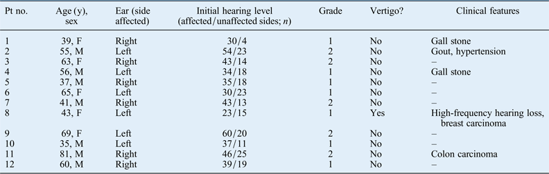
The clinical findings (e.g. patient age, sex, the side affected, average baseline hearing level, severity of hearing loss, and the presence or absence of vertigo) of 18 patients with sudden-onset SNHL are presented in Tables I and II.
Characteristics of sudden snhl patients with mild-to-moderate impairment

SNHL = sensorineural hearing loss; pt no. = patient number; y = years; F = female; M = male
Characteristics of sudden snhl patients with severe-to-profound impairment
SNHL = sensorineural hearing loss; pt no. = patient number; y = years; F = female; M = male
In the group with mild-to-moderate SNHL (7 men and 5 women; mean age, 54 years), 7 patients were classified as having grade 1 SNHL and 5 patients as having grade 2 SNHL (Table I). In the group with severe-to-profound SNHL (5 men and 1 woman; mean age, 60 years), 3 patients were classified as having grade 3 SNHL and 3 patients as having grade 4 SNHL (Table II). Four patients (patients 8, 13, 17 and 18) experienced sudden-onset SNHL with vertigo. Nine patients had a history of hypertension, renal failure and other disorders.
Magnetic resonance imaging findings
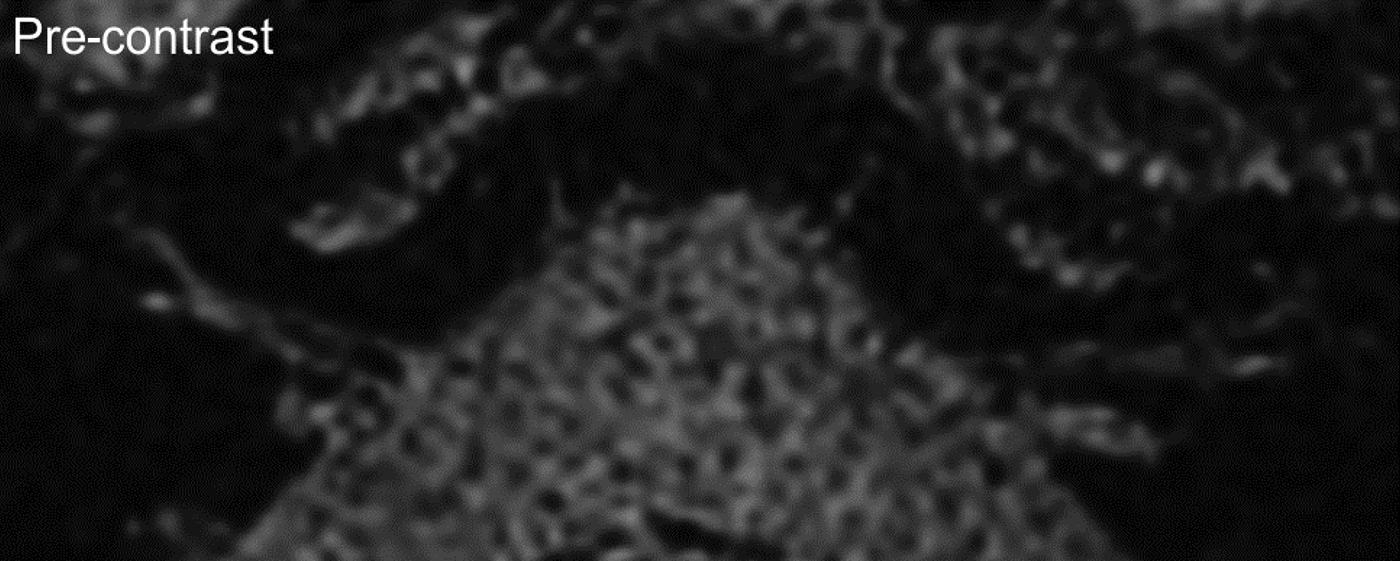
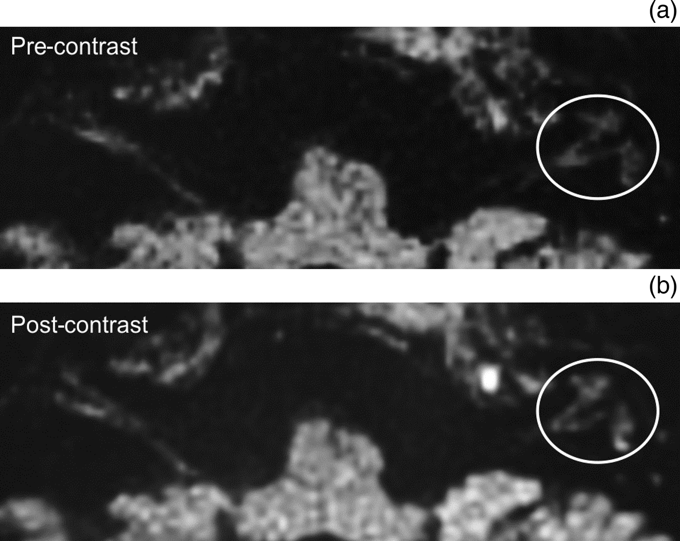
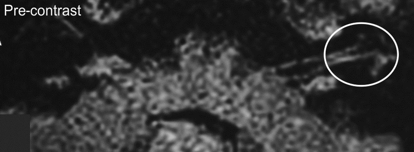
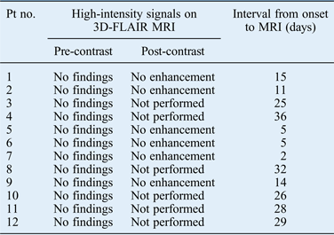
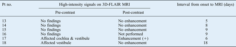
High-intensity signals were not detected in the inner ear on pre- and post-contrast three-dimensional fluid-attenuated inversion recovery (3D-FLAIR) MRI in any of the patients with mild-to-moderate SNHL (Table III, Figure 1). In contrast, two of the six patients (33 per cent) with severe-to-profound SNHL showed high-intensity signals in the affected inner ear on pre-contrast 3D-FLAIR MRI (Table IV, Figures 2 and 3). The occurrence of high-intensity signals was significantly lower in the mild-to-moderate SNHL group compared with the severe-to-profound SNHL group (p = 0.049, mid-p test). Of the two patients with high-intensity signals in the inner ear, one showed high-intensity signals both in the cochlea and vestibule (patient 17, Figure 2a), and the other only in the vestibule (patient 18, Figure 3). Furthermore, on post-contrast 3D-FLAIR MRI, patient 17 showed gadolinium enhancement in the affected inner ear (Figure 2b), but his hearing level showed remarkable improvement.
Patient 2, who had sudden-onset moderate sensorineural hearing loss in the left ear (Table I). No signals were shown either in the cochlea, vestibule or semicircular canals on axial, pre-contrast, three-dimensional fluid-attenuated inversion recovery magnetic resonance imaging (MRI). Contrast-enhanced MRI showed no increase in signal intensity in the inner-ear area.
Patient 17, who had sudden-onset profound sensorineural hearing loss with vertigo in the left ear (Table II). High-intensity signals (encircled) were shown in the left cochlea and vestibule on axial, pre-contrast, three-dimensional fluid-attenuated inversion recovery (3D-FLAIR) magnetic resonance imaging (MRI) (a). Contrast-enhanced MRI showed an increase in signal intensity (encircled) in the areas that had high-intensity signal on pre-contrast 3D-FLAIR MRI (b).
Patient 18, who had sudden-onset severe sensorineural hearing loss with vertigo in the left ear (Table II). High-intensity signals (encircled) were shown in the left vestibule, but no signals were shown in the left cochlea on axial, pre-contrast, three-dimensional fluid-attenuated inversion recovery (3D-FLAIR) magnetic resonance imaging (MRI). Contrast-enhanced MRI showed no increase in signal intensity in the areas that had high-intensity signal on pre-contrast 3D-FLAIR MRI.
3D-Flair mri inner-ear findings for sudden snhl patients with mild-to-moderate impairment
3D-FLAIR = three-dimensional fluid-attenuated inversion recovery; MRI = magnetic resonance imaging; SNHL = sensorineural hearing loss; pt no. = patient number
3D-flair mri inner-ear findings for sudden snhl patients with severe-to-profound impairment
3D-FLAIR = three-dimensional fluid-attenuated inversion recovery; MRI = magnetic resonance imaging; SNHL = sensorineural hearing loss; pt no. = patient number
• This study investigated sudden-onset sensorineural hearing loss (SNHL) using three-dimensional fluid-attenuated inversion recovery (3D-FLAIR) magnetic resonance imaging (MRI)
• It focused on the association between hearing level and the signal intensity of pre- and post-contrast 3D-FLAIR MRI
• High-intensity signals in the inner ear were shown only in severe-to-profound SNHL patients
• Hence, 3D-FLAIR MRI is not a useful tool for detecting inner-ear abnormalities in mild SNHL patients
The 3D-FLAIR images can be used to detect subtle changes, even if no hearing loss is shown by pure tone audiometry.Reference Tanigawa, Tanaka, Sato, Nakao, Katahira and Tsuchiya1 In our study, 3D-FLAIR MRI showed negative findings for all contralateral ears, even in patients with high-intensity signals in the affected ear. High-intensity signal areas observed on 3D-FLAIR MRI were not detected by T1- or T2-weighted MRI in any of those patients.
The interval between the onset of SNHL and MRI testing ranged from 2 to 36 days (average, 19 days) in the mild-to-moderate group, and from 5 to 18 days (average, 9.2 days) in the severe-to-profound group (Tables III and IV). In the two patients with high-intensity signals, these signals were observed in MRI scans performed 6 or 18 days (patients 17 and 18, respectively) after SNHL onset.
Discussion
In the present study, three-dimensional fluid-attenuated inversion recovery (3D-FLAIR) MRI revealed no high-intensity signals in the cochlea in any of the patients with sudden-onset mild-to-moderate SNHL (grades 1 and 2), but such signals were revealed in two patients with sudden-onset severe-to-profound SNHL (grades 3 and 4). Our results indicate that 3D-FLAIR MRI is not a useful tool for detecting alterations in the composition of the labyrinthine lymph fluid in patients with sudden-onset mild SNHL.
The reasons why patients with mild-to-moderate SNHL did not show high-intensity signals in the inner ear remain unclear. Several authors have speculated that high-intensity signals on 3D-FLAIR MRI may reflect an increased concentration of protein that originates from disrupted cells, or haemorrhages in the inner ear.Reference Tanigawa, Tanaka, Sato, Nakao, Katahira and Tsuchiya1–Reference Sugiura, Naganawa, Teranishi and Nakashima3 One possible explanation is that such damage to the inner ear was less severe in patients with SNHL grades 1 and 2 compared with those with SNHL grades 3 and 4. Berrettini et al. reported that the baseline hearing level was significantly worse in the group with positive 3D-FLAIR findings compared with those with negative findings.Reference Berrettini, Seccia, Fortunato, Forli, Bruschini and Piaggi5 The presence of a high-intensity signal is also associated with poorer hearing recoveryReference Yoshida, Sugiura, Naganawa, Teranishi, Nakata and Nakashima2–Reference Ryu, Yoon, Ahn, Kang, Choi and Lee4 and vestibular abnormalities.Reference Ryu, Yoon, Ahn, Kang, Choi and Lee4, Reference Lee, Jung, Park, Yeo, Lee and Lee6
Our study provides important data on the usefulness of 3D-FLAIR MRI for diagnosing inner-ear abnormalities in SNHL. However, the study has some limitations. We did not include an age-matched control group,Reference Berrettini, Seccia, Fortunato, Forli, Bruschini and Piaggi5 although all contralateral ears showed negative 3D-FLAIR findings. Moreover, our study comprised a relatively small number of patients. The results are preliminary; further studies are needed to determine the optimal interval period from the onset of SNHL to the performance of MRI.
In conclusion, 3D-FLAIR MRI may not be a useful tool for detecting inner-ear abnormalities in patients with sudden-onset mild SNHL. To our knowledge, this is the first study to have reported that 3D-FLAIR MRI does not detect inner-ear abnormalities in this patient group.