Rotaviruses are the primary aetiological agents of severe gastroenteritis in children less than 5 years of age. These viruses are estimated to be responsible for nearly 50% of hospitalisations due to acute gastrointestinal disease in young children throughout the world and approximately 600 000 deaths each year, most of which occur in developing nations (Ref. Reference Parashar1). Treatment of rotavirus illness is limited to supportive measures, such as oral or intravenous rehydration, and the only specific method to combat rotavirus disease is vaccination. Fortunately, between 2004 and 2006 two new rotavirus vaccine candidates became available, and licensing is proceeding across the world (Refs Reference Ruiz-Palacios2, Reference Vesikari3). Although both vaccine candidates have proven to be effective in developed as well as in less-well-developed nations, efficacy trials with either candidate have not been completed in a third-world nation where previous rotavirus vaccines have failed. Since the mechanisms of immunity to rotavirus are only partially understood, it has not been possible either to understand why these previous candidates failed or to predict whether the present candidates will be more successful. Even so, much has been learned about rotavirus immunity, both from human and animal-model studies. The purpose of this review is to present the most pertinent findings on the mechanisms of rotavirus immunity utilised by the present live rotavirus vaccine candidates being licensed worldwide, as well as second-generation non-living candidate vaccines that are under development.
The rotavirus particle
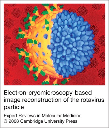
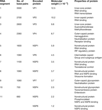
Before attempting to evaluate studies on the mechanism of immunity to rotavirus, several features of this virus need to be understood. The first is the structure of the rotavirus particle itself. Electron-cryomicroscopy-based image reconstructions (Fig. 1) show it is ~100 nm in diameter and has a capsid composed of three concentric protein layers (Refs Reference Shaw4, Reference Prasad and Chiu5). The outer layer contains 780 molecules of the viral protein 7 (VP7) glycoprotein and 60 dimers or trimers of the VP4 protein (Ref. Reference Yoder and Dormitzer6), which forms spike-like projections that extend through and 11–12 nm beyond the VP7 layer (Refs Reference Shaw4, Reference Prasad and Chiu5, Reference Yeager7). The VP4 protein is anchored to the intermediate layer of the particle, which is composed of 780 molecules of the VP6 protein. The innermost layer contains 120 molecules of the VP2 protein that interact with 12 molecules each of the viral transcriptase (VP1) and guanylyltransferase (VP3) along with the 11 segments of double-stranded RNA genome. These segments, which range in size from ~660 to 3300 base pairs, encode the six structural proteins of the virus as well as six nonstructural proteins, designated NSP1–NSP6 (Table 1). The smallest segment encodes both NSP5 and NSP6.
Electron-cryomicroscopy-based image reconstruction of the rotavirus particle. The cut-away diagram shows the outer capsid, which is composed of viral protein 4 (VP4) spikes (green), a VP7 layer (yellow) and an intermediate VP6 layer (blue) that surround the inner VP2 layer containing the 11 double-stranded RNA segments and VP1 and VP3 proteins. The background shows rotavirus particles as detected by electron microscopy (Courtesy of Dr B.V.V. Prasad, Baylor College of Medicine, Houston, TX, USA).

Size of rotavirus gene segments and properties of encoded proteins

Abbreviations: IRF, interferon regulatory factor; NSP, nonstructural protein; VP, viral protein.
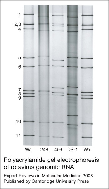
The genome segments of rotavirus can be extracted from viral particles and separated by polyacrylamide gel electrophoresis into 11 bands visualised by ethidium bromide or silver staining (Fig. 2). Each rotavirus strain has a characteristic RNA profile or electropherotype – a property that has been used extensively in epidemiological studies of these viruses. The characteristic RNA-electrophoretic pattern of group A rotaviruses, which cause the vast majority of rotavirus disease identified in humans, consists of four size classes (Fig. 2). The 11 RNA segments of strains belonging to less-well-characterised rotavirus groups (i.e. groups B to G) can also be separated into four size classes, but the distribution of segments within these classes differs from group to group.
Polyacrylamide gel electrophoresis of rotavirus genomic RNA. RNA from group A human rotaviruses was resolved by electrophoresis and visualised by silver staining. The patterns demonstrate the characteristic four size classes of RNA which, for group A rotaviruses, separate into groups of four (bands 1–4), two (bands 5 and 6), three (bands 7–9) and two (bands 10 and 11) segments each. Each rotavirus strain has its own characteristic PAGE pattern or electropherotype that can be used for identification in epidemiological studies. Human rotavirus strains (from left to right): lane 1, Wa; lane 2, 248 strain; lane 3, 456 strain; lane 4, DS-1; lane 5, Wa. Reproduced, with permission, from Ref. Reference Bernstein, Ward, Feigin and Cherry81 (© Elsevier).

Serotypes of rotavirus
Both the VP4 and VP7 outer-capsid proteins of rotavirus contain neutralisation epitopes (i.e. peptide regions within these proteins that stimulate neutralising antibody production) and both are involved in serotype determination, an important consideration in understanding the mechanisms of immunity. Originally, serotyping was based solely on differences in the VP7 protein because hyperimmunised animals developed almost all neutralising antibody to this protein (Refs Reference Wyatt8, Reference Hoshino9). When it was later found that VP4 could be the dominant neutralisation protein (Refs Reference Ward10, Reference Ward11), a dual-serotyping scheme was required. Although VP7 serotypes could be determined by crossneutralisation studies, this was more difficult for VP4. Therefore, two numeric systems were devised to classify the VP4 protein. One is based on sequence analyses (genotypes), which are designated in brackets; the second is based on neutralisation (serotypes). Rotavirus groupings based on VP4 and VP7 are classified as P and G types, referring to the protease sensitivity and glycosylated structure of these two proteins, respectively. Today, 15 G types and 26 P types had been reported, but only five G serotypes and three P genotypes are commonly associated with human rotavirus illnesses. Other G or P types, however, can be dominant in certain regions and may be considered as emerging pathogens.
Possible causes of rotavirus disease
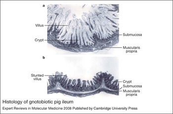
The symptoms associated with rotavirus disease typically are diarrhoea and vomiting accompanied by fever, nausea, anorexia, cramping and malaise. The disease can be mild and of short duration or produce severe dehydration, which is associated with destruction of the intestinal villi (Fig. 3). Although the events responsible for severe dehydrating disease are only partially understood and may be multifaceted, their identification could suggest targets for the development of immunity. An early study in piglets indicated that net sodium and calcium fluxes were not different between control and infected animals, but glucose-mediated sodium absorption was diminished by rotavirus infection (Ref. Reference Davidson12). On the basis of this and other physiological changes, the authors concluded that retarded differentiation of uninfected enterocytes that migrated at an accelerated rate from the crypts after the virus had invaded villus cells was responsible for absorptive abnormalities. Another study with piglets led to the conclusion that destruction of the villus-tip cells causes carbohydrate malabsorption and osmotic diarrhoea (Ref. Reference Graham, Sackman and Estes13). Studies in animals and humans concerning changes in the absorption of macromolecules across the intestinal surface after rotavirus infection have, however, revealed no general pattern.
Histology of gnotobiotic pig ileum. (a) Normal histological appearance of the ileum from an 8-day-old gnotobiotic pig. Normal mature vacuolate absorptive cells cover the villi. Haematoxylin and eosin (H & E) stain. (b) Ileum from an 8-day-old gnotobiotic pig after oral inoculation with virulent human rotavirus (Wa strain). Severe villous atrophy and early crypt hyperplasia are evident. H & E stain. (Courtesy of Dr L.A. Ward, Ohio Agricultural Research and Development Center, The Ohio State University, Wooster, OH, USA). Adapted, with permission, from Ref. 81 (© Elsevier).

Diarrhoea has been induced in infant mice and rats by intraperitoneal inoculation with the rotavirus NSP4 protein as well as with a 22 amino acid peptide derived from this protein (Refs Reference Ball14, Reference Morris15). Investigators observed that NSP4 and its peptide increased the levels of intracellular calcium by activating a calcium-dependent signal transduction pathway that mobilises transport of this ion from the endoplasmic reticulum (Refs Reference Dong16, Reference Tian17). Thus, binding of NSP4 to intestinal epithelium after its release from infected cells may contribute to altered ion transport and diarrhoea. Another possible target for secreted NSP4 is the enteric nervous system, which lies under the villus epithelium. It has been reported that rotavirus infection can activate this system in mice, and drugs that block nerve activity attenuate rotavirus-induced fluid secretion in vitro and attenuate diarrhoea in vivo (Ref. Reference Lundgren18). Whether this is a major cause of rotavirus diarrhoea and is therefore a logical target for rotavirus immunity or therapeutics remains to be determined.
Protection following natural rotavirus infection
All rotavirus vaccines evaluated in human trials have been live rotavirus strains that are delivered orally to mimic natural infection. When the first rotavirus vaccine candidates were developed, little was known about the ability of natural rotavirus infection to prevent subsequent rotavirus infections or disease. Although much more is known today, there are still major gaps in our knowledge, and what is learned in one setting may not be directly applicable to other settings. The overall general conclusions are that immunity after natural rotavirus infection depends on the time between exposures, the properties of the rotaviruses involved in those exposures and the immune status of the human host at the time of each exposure. The best protection seems to be in developed countries, especially when re-exposure is to rotaviruses serotypically related to those that caused the initial infection. For example, of 57 subjects enrolled in a study in Cincinnati, OH, who were initially infected with rotavirus in year 1, none experienced a symptomatic rotavirus infection in year 2 and only two had asymptomatic infections (Ref. Reference Bernstein19). By contrast, 11 of the 85 subjects who were not infected in the first year had symptomatic rotavirus infections in year 2, and 22 out of 85 were asymptomatically infected. The circulating rotavirus strains in both years belonged to serotype G1.
The picture was different in a less-well-developed country where several G types co-circulated. In this setting, it was observed that many children experienced two or more rotavirus infections within their first year of life (Ref. Reference Velazquez20). Even so, both the susceptibility to reinfection and the severity of illness decreased with each infection. In fact, a single symptomatic or asymptomatic infection provided 87% protection against subsequent severe rotavirus disease, and two infections provided 100% protection. A child's age, and therefore maturity of their immune system, at the time of first infection is also expected to play a role in the outcome following a second exposure. Studies conducted in both developed and developing nations have shown that neonatal infections can sometimes (Refs Reference Bishop21, Reference Bhan22, Reference Vethanayagam23), but not always (Ref. Reference Banerjee24), provide protection against heterotypic (i.e. serotypically unrelated) rotaviruses, but this protection again appears to be primarily against severe rotavirus illnesses.
Time between rotavirus infections is also expected to affect the severity of rotavirus illnesses. Although almost every child is expected to experience at least one rotavirus infection by 3 years of age, and severe disease is typically associated only with first infections, there are reports of outbreaks of rotavirus disease with high frequencies of hospitalisations in older children, adults and the elderly. These outbreaks are caused primarily by G2 rotaviruses that are heterotypic to rotaviruses responsible for more than 90% of illnesses worldwide, thus suggesting that heterotypic immunity may be the most susceptible to time-dependent attrition (Ref. Reference Griffin25).
In light of the imperfect protection provided by natural rotavirus infections, a realistic expectation for orally deliverable, live rotavirus vaccines is that they protect against severe disease during the first years of life, a time when reinfections that broaden and boost immunity are more common.
What is responsible for immunity after rotavirus infection?
Effectors of protection after natural human rotavirus infection
Rotavirus antibody has been examined as a possible correlate of immunity following natural infection in many human studies and numerous associations have been reported. However, in almost all studies, rotavirus antibody was the sole immunological component examined, so it could merely have been a surrogate for the true effector of protection. Also, the association between rotavirus antibody and protection levels was never perfect, thus suggesting that immune components other than or in addition to the antibodies examined have important roles. Levels of serum rotavirus IgG or IgA detected after natural infection have been correlated with protection in studies conducted in developed, developing and third-world nations (Refs Reference Velazquez26, Reference Ward27, Reference Clemens28, Reference O'Ryan29). Intestinal antibody is expected to have a larger role in protection against rotavirus illness than circulating antibody and, accordingly, levels of faecal (copro) rotavirus IgA have also been associated with protection (Refs Reference Matson30, Reference Coulson31).
The properties of these rotavirus antibodies, particularly whether they are able to neutralise the viruses, remain uncertain. In an early small study, serotype-specific serum-neutralising antibody titres of ∼1:128 were associated with protection following natural rotavirus infections (Ref. Reference Chiba32). In a much larger study conducted in Bangladesh, protection was associated with a previous rotavirus infection, determined by the presence of serum rotavirus IgG (Ref. Reference Clemens28); however, the only neutralising antibody that was independently associated with protection against subsequent severe rotavirus disease was heterotypic (Ref. Reference Ward33). In a comprehensive cohort study conducted in Mexico, protection against rotavirus illness after natural infection correlated with serum IgA but not IgG levels (Ref. Reference Velazquez26) and sequential illnesses were more likely to be caused by different rather than the same G serotypes of virus (Ref. Reference Velazquez20), thus suggesting better homotypic than heterotypic protection. However, since protection against severe rotavirus disease was 87% after one infection when all four major G serotypes of rotavirus were co-circulating, at least some of the observed protection against severe disease appeared to be due to effectors other than neutralising antibodies. Conflicting data concerning the requirement for neutralising antibody in protection has been generated in other studies, thus leaving no firm conclusions.
Immunological effectors associated with protection after oral immunisation with live rotaviruses
The controversy regarding the importance of neutralising antibody in protection after natural rotavirus infection has resulted in two very different approaches in the development of live rotavirus vaccines: one based on vaccines composed of single rotavirus strains and the other based on vaccine candidates composed of several strains belonging to different rotavirus serotypes. Just 10 years after the identification of rotavirus as the primary agent of severe diarrhoea in young children, the first vaccine trials were performed using RIT 4237, a G6P[Reference Parashar1] bovine rotavirus (Ref. Reference Vesikari34). This live virus vaccine was safe and effective when tested in Finland, providing protective efficacy of >80% against severe rotavirus disease due to heterotypic human rotaviruses (Ref. Reference Vesikari35). However, results obtained in later studies conducted in developing countries were disappointing and evaluation of this vaccine was discontinued.
A simian rotavirus called rhesus rotavirus (RRV) was developed as one of the first vaccine candidates, but the protection elicited was likewise inconsistent. The G3 serotype of RRV is shared with one of the dominant circulating serotypes of human rotaviruses, and protection elicited by RRV appeared to be more effective when this was the case (Ref. Reference Flores36). As a result, RRV was reassorted with human strains to incorporate VP7 genes of the other three dominant human G serotypes (G1, G2 and G4), thus creating the tetravalent Rotashield™ vaccine. In large, placebo-controlled trials with this vaccine in the USA, only about 20% of vaccinated individuals developed significant serum-neutralising-antibody responses to G1–G4 human rotaviruses, even though protection against severe rotavirus disease was greater than 70% (Ref. Reference Ward37). Furthermore, this protection did not correlate with serotype-specific neutralising-antibody levels. The Rotashield vaccine was licensed and incorporated into the USA infant immunisation program in 1998 but was withdrawn by its manufacturer in 1999 after post-licensure surveillance revealed a small but significant increase in intussusception rates in the week after vaccination (Ref. Reference Murphy38).
The development of a second bovine rotavirus vaccine candidate called WC3 followed a comparable pattern to that of RRV. An initial trial with this G6P[Reference Prasad and Chiu5] strain in Philadelphia provided protection against heterotypic rotaviruses (Ref. Reference Clark39), but subsequent trials did not show significant protection. This candidate vaccine was therefore also reassorted with human rotaviruses to select strains with human rotavirus VP4 or VP7 genes on a WC3 background. This resulted in the development of the pentavalent RotaTeq™ vaccine, which was licensed in the USA and Europe in 2006 after it was reported to be safe and to provide consistent protection. However, as was found with the RRV reassortant vaccine, no clear correlation between protection and serotype-specific neutralising-antibody levels has been reported with the WC3 reassortant vaccine.
The best correlation between rotavirus antibody responses following vaccination and subsequent protection has been with the monovalent G1P[Reference Wyatt8] Rotarix™ vaccine. Vaccinated individuals who did not develop serum rotavirus IgA responses after two doses of this vaccine were significantly more likely to develop rotavirus disease than subjects who did develop these responses (Ref. Reference De Vos40). Even so, the percentage of Rotarix vaccinees protected against severe rotavirus illnesses has been found to exceed the percentage with measurable serum rotavirus IgA responses after vaccination (Ref. Reference Salinas41). This suggests that serum rotavirus IgA levels may only partially correlate with the degree of protection stimulated by Rotarix immunisation. Explanations for this observation include, among others, the fact that serum and intestinal rotavirus IgA levels do not correlate directly, that levels of rotavirus IgA below the limit of detection can be protective or that immune effectors other than rotavirus IgA are involved in protection. It was also reported that Rotarix immunisation can elicit a high level of protection against fully heterotypic G2P[Reference Shaw4] strains (Ref. Reference Vesikari42), thus supporting suggestions that protection after live rotavirus infection might be elicited by immune components other than neutralising antibody.
If neutralising antibody is not required for protection after live rotavirus vaccination, it is unclear why the RRV and WC3 vaccine strains were consistently protective only when made into reassortants containing human rotavirus neutralisation protein genes. It could be that human rotavirus proteins, including VP4 and VP7, generate immune effectors other than neutralising antibodies, which provide better protection against human rotaviruses than immune effectors generated against animal rotavirus proteins. Another explanation is that even though distinct serotypes of rotavirus have been defined against both VP4 and VP7 proteins based on differences in neutralising antibody titres, some neutralisation epitopes are shared between serotypes (Ref. Reference Kapikian and Knipe43) and these shared epitopes may be responsible for eliciting heterotypic protection. It is also possible that multi-strain vaccines might boost immune responses because of their diversity, which is not possible with single-strain vaccines. Most subjects who seroconverted after receiving the first dose of the single-strain Rotarix vaccine were protected against the vaccine virus when administered their second dose, whereas many subjects who received three doses of a multi-strain WC3 reassortant vaccine had immune responses after more than one dose (Ref. Reference Ward44). Since sequential rotavirus infections are known to both boost and broaden rotavirus-antibody responses, this may occur with the multi-strain vaccines as well. Even if this occurs, however, no correlation has been found between protection and serotype-specific serum-neutralising-antibody titres generated by three doses of the multi-strain vaccine candidates. Thus, either serum antibodies are not fully reflective of their intestinal counterparts, the methods used to measure these antibodies are not fully accurate or something other than or in addition to neutralising antibody plays a role in protection.
Effectors of protection identified in animal-model studies
Many of the experimental limitations associated with human studies are bypassed in animal-model studies. Although studies on the mechanism of active protection against rotavirus have been performed in several animal species, the most thorough have been with either gnotobiotic piglets or adult mice. Each model has certain advantages, and the conclusions generated using the two have sometimes disagreed. When this has happened, it is unclear which might be the more relevant to humans. Piglets are more similar to humans than are mice and their window of susceptibility to rotavirus illness extends several weeks beyond that found in mice, thus allowing studies of active immunity against illness following immunisation, rather than protection only against shedding, as occurs with the adult mouse model. Furthermore, piglets can be infected with human rotaviruses, and a virulent human strain has typically been used as a challenge virus in piglet studies. However, it is difficult to maintain piglets free of rotavirus contamination; hence the need for gnotobiotic animals, which have been found to have intestinal immune responses that differ from those observed in animals with normal intestinal flora (Refs Reference Cebra45, Reference Umesaki and Setoyama46, Reference Savage and Mestecky47). The ability to work with inbred strains and generate genetically modified animals has provided the mouse model with certain advantages, along with greatly reduced logistical complications and costs, based on their smaller size.
Excellent protection against both rotavirus shedding and illness with the virulent human rotavirus Wa strain was elicited in gnotobiotic piglets following oral immunisation with the same virulent virus (Ref. Reference Yuan48). Less protection was found when piglets were immunised with an attenuated Wa strain. The level of protection correlated with the levels of intestinal antibody-secreting B cells as well as memory B cells. However, protection also strongly correlated with the magnitude of lymphoproliferative assay responses determined using peripheral blood mononuclear cells (PBMCs), thus suggesting a possible association between CD4+ helper T cells and protection after live rotavirus immunisation (Ref. Reference Ward49).
If rotavirus antibody is associated with protection after rotavirus infection, as suggested by studies in both humans and piglets, the most obvious immunological effector of protection is secretory IgA. In agreement with this, protection against rotavirus infection in orally immunised mice correlates with levels of intestinal (stool) and serum rotavirus IgA but not serum rotavirus IgG (Refs Reference Feng50, Reference McNeal, Broome and Ward51). The most immunogenic rotavirus protein is VP6 but it does not stimulate neutralising antibodies. However, IgA antibodies directed at VP6 can be protective in mice by mechanisms that are not completely understood but may involve intracellular inhibition of rotavirus replication within infected enterocytes during polymeric antibody transport to the intestinal lumen (Refs Reference Burns52, Reference Feng53, Reference Schwartz-Cornil54). Antibodies directed at either the VP4 or the VP7 proteins can neutralise virus and are believed to provide protection by classical neutralisation. It should be noted that there is evidence that rotavirus IgG can also provide protection, especially when present in the intestine. IgA-knockout mice were still protected against murine rotavirus shedding after oral immunisation with live murine rotavirus, and high levels of faecal rotavirus IgG were detected in the immunised mice prior to challenge (Ref. Reference O'Neal55). Likewise, passive transfer of rotavirus IgG protects calves (Ref. Reference Besser56), macaques (Ref. Reference Westerman57) and humans (Ref. Reference Losonsky58).
Gene-knockout mice have also been used to distinguish the roles of CD8+ T cells and antibody in protection. Rotavirus-specific cytotoxic T lymphocytes recognise epitopes on several rotavirus proteins, which are generally not serotype specific (Refs Reference Offit and Dudzik59, Reference Offit60, Reference Franco61). Adoptive transfer of splenic lymphocytes from adult mice infected with either a homologous or a heterologous rotavirus strain were, at least in one instance, able to protect suckling mice, and protection appeared to depend on the presence of CD8+ lymphocytes (Ref. Reference Offit and Dudzik62). Similarly, CD8+ splenic or intraepithelial lymphocytes obtained from the intestine of rotavirus-infected mice can eliminate the chronic rotavirus shedding seen in severe combined immunodeficiency (SCID) mice (Ref. Reference Dharakul, Rott and Greenberg63). Other studies revealed that adoptive transfer of splenic CD4+ cells from mice intranasally immunised with a recombinant VP6 protein resolved shedding in chronically infected, immunodeficient Rag-2 mice (Ref. Reference McNeal, VanCott and Choi64). Therefore, either CD8+ or CD4+ T cells are capable of resolving rotavirus infections. Possible roles of these cells in resolution or prevention of human rotavirus infections remain to be determined.
Although cytolytic CD8+ cells are important for resolution of a rotavirus infection, only antibody could provide protection from a subsequent challenge after live rotavirus immunisation of mice (Refs Reference Franco and Greenberg65, Reference McNeal66). A recent study in this animal model revealed that oral immunisation of mice with a rotavirus that was fully heterotypic to the challenge strain provided nearly complete protection against faecal rotavirus shedding but this protection was dependent on the ability of antibody to be transported through intestinal epithelial cells (Ref. Reference VanCott67). This result supports the earlier suggestion that heterotypic protection after live virus immunisation may be due to intracellular inhibition of virus replication, at least in mice (Refs Reference Burns52, Reference Feng53, Reference Schwartz-Cornil54). Together, these results indicate that the levels and location of antibody, as well as the proteins against which it is directed, are of immediate importance in protection against rotavirus.
What is responsible for immunity after immunisation with non-living rotavirus vaccine candidates?
The euphoria associated with the recent successes with live rotavirus vaccines has, for the moment, dampened the sense of disappointment that accompanied the removal of Rotashield from the USA immunisation program in 1999. However, even with the very reassuring data obtained from large prelicensure trials with the two multinational vaccine candidates Rotarix and RotaTeq, postlicensure studies must confirm the safety of these vaccines. Furthermore, no efficacy data are yet available for either of these vaccines from third-world nations where the early rotavirus vaccine candidates failed. Thus, there is a need to develop and evaluate next-generation rotavirus vaccines that follow a different, ‘non-living’ paradigm. Although several types of non-living rotavirus vaccine candidates have been developed, almost all mechanistic studies have been performed with three types. These include inactivated triple- and double-layered (lacking VP4 and VP7) rotavirus particles, triple- and double-layered virus-like particles (VLPs), and recombinant expressed VP6 proteins. Candidates representative of each of these three types have been under development since the 1980s and tested in animal models but none has been evaluated in humans.
The animal models in which almost all mechanistic studies with non-living candidate rotavirus vaccines have been conducted are the gnotobiotic piglet and the adult mouse. Each has certain advantages and disadvantages as already listed. Interestingly, the effectors of protection identified after immunisation of piglets and mice with non-living vaccine candidates have not been identical. It was initially noted that oral inoculation of piglets with a live human rotavirus vaccine was much more effective than either oral or parenteral inoculation with the same virus after its inactivation, and this difference corresponded to lower intestinal rotavirus IgA titres in animals inoculated with the inactivated virus (Refs Reference Yuan68, Reference To69). In later studies in which piglets were inoculated intranasally with a VLP vaccine that contained only the VP2 and VP6 proteins of rotavirus (i.e. 2/6 VLPs) along with a powerful toxin adjuvant, no protection was obtained (Ref. Reference Yuan70). By contrast, when the administration of this VLP vaccine was preceded by one dose of attenuated human rotavirus, it enhanced protection, and this enhanced immunity is suggested to be associated with increased levels of intestinal IgA (Ref. Reference Yuan71). Interestingly, the boost with 2/6 VLPs enhanced neutralising-antibody titres and since neither VP2 nor VP6 are neutralisation proteins, the authors suggested this enhancement was due to the development of T helper cells against VP6, which provided ‘cognate help’ to VP4- and VP7-specific B cells (Ref. Reference Azevedo72). Based on these and other studies with gnotobiotic piglets, the protection associated with the non-living rotavirus vaccine candidates is proposed to be due primarily to neutralising intestinal IgA (Ref. Reference Yuan and Saif73).
The effectors of protection identified after immunisation of mice with non-living vaccine candidates are sometimes quite different. Initial studies with inactivated rotavirus particles delivered by either parenteral or mucosal (intranasal) routes effectively blocked intestinal rotavirus replication in adult mice challenged with a murine rotavirus (Refs Reference McNeal74, Reference McNeal75). Fully intact, triple-layered particles were found to be more effective than double-layered particles only when their VP4 or VP7 protein matched the serotype of the challenge virus. This suggested that neutralising antibodies played a role in protection, but were not required for effective immunity. Later it was shown that intranasal inoculation with vaccines composed only of chimaeric, bacterially expressed VP6 proteins and toxin adjuvants effectively protect mice against faecal rotavirus shedding (Refs Reference Choi76, Reference Choi77) and the only lymphocytes required for protection were CD4+ T cells (Ref. Reference McNeal, VanCott and Choi64). Subsequent experiments found that protection elicited by these effector cells was associated with the intestinal production of two cytokines – interleukin 17 (IL-17) and interferon γ (IFN-γ) (Ref. Reference Smiley78) – suggesting that these cytokines may have direct or indirect roles in protection against rotavirus shedding.
The importance of CD4+ T cells in protection after immunisation with non-living vaccines was supported by a preliminary report of mice intranasally immunised with 2/6 VLPs and adjuvant. In this study, it was found that in contrast to B and CD8+ T cells, CD4+ T cells were absolutely required for protection (Ref. Reference Blutt79). However, some dependence on B cells was found in this study, and other investigators report that protection after intranasal immunisation with 2/6 VLPs and adjuvant was somewhat reduced in mice that were unable to transport polymeric antibodies (Ref. Reference Schwartz-Cornil54). It should also be noted that groups that examined effectors associated with protection after intrarectal immunisation with 2/6 VLPs and adjuvant reported the immune responses that best correlated with protection were serum and intestinal rotavirus antibody levels (Ref. Reference Blutt79) and the number of intestinal (Peyer's patch) rotavirus-specific B cells (Ref. Reference Parez80). Undetectable levels of rotavirus-specific T cells were found in the Peyer's patches of mice that had been intrarectally immunised with 2/6 VLPs and adjuvant (Ref. Reference Parez80). Thus, even the effectors of protection identified after immunisation of mice with different non-living rotavirus vaccines were not identical. However, in most of these studies in mice, no neutralising antibody could be generated because of the lack of VP4 and VP7 proteins in the vaccines. Thus, even if an antibody was important in protection, in these cases it was not neutralising antibody. Non-neutralising antibodies may therefore play an important role in protection after immunisation with non-living rotavirus vaccines as suggested for live rotavirus vaccines. However, regardless of which is the most important effector of protection with any rotavirus vaccine, many effectors probably play roles in immunologically normal individuals because of the built-in redundancy of the immune system.
Research in progress and outstanding research questions
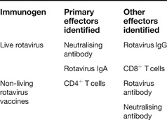
In spite of the many studies that have been conducted in humans and animal models, the effectors of protection generated after either live rotavirus infection or vaccination, or inoculation with non-living vaccines, remain only partially identified, as summarised in Table 2. Although it has not been necessary to have a full understanding of these mechanisms in order to generate effective vaccines, this understanding would greatly facilitate our ability to make rational improvements in current vaccines and treatment methods. For example, the question of the importance of neutralising antibodies in protection after live rotavirus infection or immunisation remains. If there are other effectors of protection, what are they and how do they work? Suggestions have been made, as already noted, but their credibility has limited support. Antibodies may play a reduced role after immunisation with certain non-living vaccines, but even this is uncertain and certainly not generally applicable to all non-living candidate vaccines or routes of immunisation. Thus, it can be safely stated that the knowledge gained until now has only scratched the surface of understanding the mechanisms of protection by any immunogen in any species administered by any route. There is much yet that needs attention.
Effectors of protection associated with rotavirus immunity

Acknowledgements
I thank my colleagues, collaborators and fellow investigators for their numerous published contributions that make reviews such as this possible. I also thank the three reviewers who anonymously provided helpful critiques that were used to improve this review.