The approach to patients with totally anomalous pulmonary venous connection has now become standardized, and surgical mortality has steadily declined. In patients with mixed totally anomalous pulmonary venous connection, in contrast, the small number of patients in individual series published thus far, along with incomplete ascertainment of the anatomy, and the morphologic heterogeneity, make it difficult to derive any conclusions regarding the best strategy for management, and the results of surgery. While various permutations and combinations of sites of drainage, the length of the confluent segment in the venous connections, the presence or absence of obstruction, and the surgical methods are all described in the setting of the “regular” forms of totally anomalous pulmonary venous connection, these details are largely lacking when the pulmonary veins all drain anomalously, but to mixed sites of drainage. With these deficiencies in mind, we have analyzed the published literature, using a MEDLINE search to identify the described instances of mixed totally anomalous pulmonary venous connection, the variations in anatomy with or without significant cardiac malformations, and the various sites of obstruction of the pulmonary venous pathway.Reference Delius, De Leval, Elliott and Stark1–Reference Rebensburg54 We have then synthesized all of these features with regard to their bearing on the strategies for surgical management.
Patients and methods
Criterions for selection of patients
As far as we could establish, there have been 386 cases of mixed totally anomalous pulmonary venous connection described in the English-language literature. We discovered these 386 patients of mixed totally anomalous pulmonary venous connection among 3772 patients described with totally anomalous pulmonary venous connections.Reference Delius, De Leval, Elliott and Stark1–Reference Rebensburg54
For the purposes of this review, we have selected 182 patients from 54 published series in whom an accurate anatomical description was provided that might possibly have a bearing on the natural history, diagnosis, surgical techniques, and peri-operative mortality.Reference Delius, De Leval, Elliott and Stark1–Reference Rebensburg54 In making this selection, we excluded 204 patients in whom we deemed the anatomical description of the patterns of pulmonary venous drainage to be inadequate for our purposes.
Incidence
The true incidence of mixed totally anomalous pulmonary venous connection is unknown. It varies in different published series from one-twentieth to one-tenth of the overall number of patients with totally anomalous pulmonary venous connection.Reference Delius, De Leval, Elliott and Stark1–Reference Rebensburg54 Our analysis revealed a cumulative incidence of 182 patients with mixed sites of drainage in 1,826 patients with totally anomalous pulmonary venous connection. Out of these patients, we identified 130 patients who underwent surgery.Reference Delius, De Leval, Elliott and Stark1–Reference Dickinson, Parmelazhagan and Tweedic13, Reference Muller19–Reference Winter21, Reference Watanabe, Murashita, Oka, Hatta, Imamura and Yasuda23–Reference El Said, Mullins and Mc Namara25, Reference Arbona, Kilman and Van Aman27, Reference Klint, Weldon, Hartmann, Schad, Hernandez and Goldring29, Reference Hawkins, Clark and Doty30, Reference Serraf, Belli, Roux, Sousa-Uva, Lacour-Gayet and Planche32–Reference Baumann34, Reference Neligan, O’Malley and Blake36–Reference Ando, Takahashi and Kikuchi44, Reference Chowdhury, Kothari and Airan46, Reference Boggs and Reed48–Reference Shimazaki, Nakano and Kato53 The pattern of pulmonary venous drainage was documented at autopsy in a further 28 patients,Reference Blake, Hall and Manion18, Reference Delisle, Ando and Calder22, Reference Carter, Capriles and Noe28, Reference Rubino, VanPraagh, Kadoba, Pessotto and Van Praagh31, Reference Gott, Lester, Lillehei and Varco35, Reference Nakib, Moller, Kanjuh and Edwards45, Reference Rebensburg54 while in 24 patients only an echocardiographic diagnosis had been provided.Reference Goswami, Shrivastava, Saxena and Dev14–Reference Chin, Sanders, Sherman, Lang, Norwood and Castaneda17, Reference Sreeram and Walsh26, Reference Lee, Wang, Wu, Chu and Lue47
Demographics and preoperative evaluation
The age at operation ranged from 12 hours to 24 years, with the median age in most series being less than 1 year.Reference Delius, De Leval, Elliott and Stark1–Reference Rebensburg54 Weight ranged from 2 to 50 kilograms, with the median weight in most series being less than 4 kilograms.Reference Delius, De Leval, Elliott and Stark1–Reference Rebensburg54 An obstructive pattern of drainage involving one or more veins is described in 108 (59.3%) of the patients.Reference Delius, De Leval, Elliott and Stark1–Reference Rebensburg54 The clinical picture varies considerably, from cyanotic newborns dying in the first few weeks after birth to the non-cyanotic adult with little disability.
Studies of the chest radiograph showed signs of asymmetric pulmonary venous congestion confined either to one lung or one lobe.Reference Delius, De Leval, Elliott and Stark1, Reference Oelert, Schafers, Stegmann, Kallfelz and Borst3–Reference Ward, Mullins, Huhta, Nihill, McNamara and Cooley8, Reference de Leval, Stark and Waterston10, Reference Fujimatsu, Tsunemoto and Shimada11, Reference Watanabe, Murashita, Oka, Hatta, Imamura and Yasuda23, Reference Snellen, Van Ingen and Hoefsmit24, Reference Neligan, O’Malley and Blake36, Reference Kanjuh, Katkov, Singh, Franciosi, Helseth and Edwards40, Reference Nakib, Moller, Kanjuh and Edwards45 Diagnosis of the mixed pattern may be difficult on echocardiography alone in many patients, and some sites of drainage may be missed. The indications of pre-operative catheterization in most studies were the anatomy being unresolved by echocardiography, the need to characterize pulmonary venous obstruction, or the exclusion of major associated cardiac anomalies.Reference Delius, De Leval, Elliott and Stark1–Reference Chin, Sanders, Sherman, Lang, Norwood and Castaneda17, Reference Muller19–Reference Winter21, Reference Watanabe, Murashita, Oka, Hatta, Imamura and Yasuda23–Reference Arbona, Kilman and Van Aman27, Reference Klint, Weldon, Hartmann, Schad, Hernandez and Goldring29, Reference Hawkins, Clark and Doty30, Reference Serraf, Belli, Roux, Sousa-Uva, Lacour-Gayet and Planche32–Reference Baumann34, Reference Neligan, O’Malley and Blake36–Reference Ando, Takahashi and Kikuchi44, Reference Chowdhury, Kothari and Airan46–Reference Shimazaki, Nakano and Kato53
Cross-sectional echocardiography and cardiac catheterization provided the necessary diagnostic information, and defined the anatomy before surgery, in 139 patients. Echocardiography was the only diagnostic study in 43 patients. Magnetic resonance imaging and computerized tomographic angiography have been used for further clarification of the pulmonary venous anatomy in selected cases in more recent series.Reference Choe, Lee, Kim, Ko, Kim and Han55, Reference Wang, Li and Chiu56 Balloon atrial septostomy was mostly performed at cardiac catheterization in patients with complex cardiac anomalies requiring palliation.Reference Delius, De Leval, Elliott and Stark1–Reference Chin, Sanders, Sherman, Lang, Norwood and Castaneda17, Reference Muller19–Reference Winter21, Reference Watanabe, Murashita, Oka, Hatta, Imamura and Yasuda23–Reference Arbona, Kilman and Van Aman27, Reference Klint, Weldon, Hartmann, Schad, Hernandez and Goldring29, Reference Hawkins, Clark and Doty30, Reference Serraf, Belli, Roux, Sousa-Uva, Lacour-Gayet and Planche32–Reference Baumann34, Reference Neligan, O’Malley and Blake36–Reference Ando, Takahashi and Kikuchi44, Reference Chowdhury, Kothari and Airan46, Reference Lee, Wang, Wu, Chu and Lue47–Reference Shimazaki, Nakano and Kato53
For patients who underwent pre-operative cardiac catheterization, systolic pulmonary arterial pressure ranged from 20 to 110 millimetres of mercury, with a mean of 53, and standard deviation of 18.9 millimetres of mercury. The ratio of systolic pulmonary arterial to systemic arterial pressure ranged from 0.22 to 1.6, with a mean of 0.70, and standard deviation of 0.04. Preoperative systemic saturations of oxygen ranged from 65% to 96%, with a mean of 75%, and standard deviation of 8.2%.
Definitions
We considered the mixed form of totally anomalous pulmonary venous connection to be isolated or simple if the patient had the anomalous pulmonary venous connections in association with an atrial septal defect, a patent arterial duct, or both. Patients were considered to have univentricular or biventricular physiology according to whether their anatomy was deemed suitable for univentricular or biventricular repair. Patients were defined as having heterotaxy syndrome when associated with complex anomalies of visceral-cardiac relationships along with complex intra-cardiac pathology.
Morphology
We used the descriptions of echocardiographic, angiographic, surgical, and autopsy findings of the 182 patients to stratify the various cases. We focused on the number of the veins draining to a confluent site, the pattern of drainage of the individual pulmonary veins, the presence or absence of a pulmonary venous confluence, the gross appearance of the confluence, the site or sites of pulmonary venous obstruction if present, the presence of diffuse stenosis within the channels of drainage, and the presence or absence of a plexus of pulmonary veins.
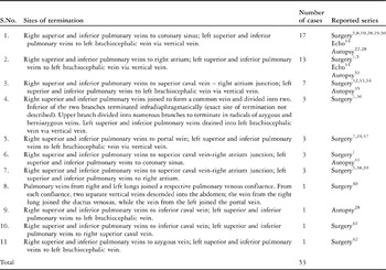
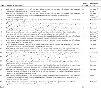
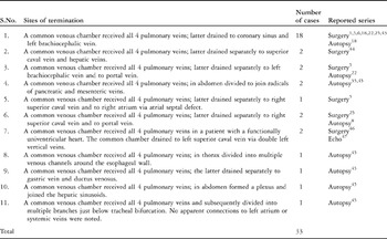
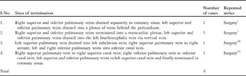
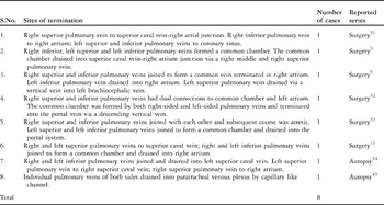
The anatomical variations, and frequency, of the entire spectrum of cases have been tabulated in Tables 1–5. We categorized the patients into several groups.
Group of patients with separate anomalous connection of veins from each lung.

Group of patients with isolated drainage of a solitary pulmonary vein at different body levels and drainage of the other 3 pulmonary veins to a common site.

Group of patients with drainage of all pulmonary veins through a common venous confluence but at various levels, namely, supracardiac, cardiac or infracardiac.

Group of patients with drainage at different levels through individual pulmonary veins.

Group of patients with bizarre anatomical variants.

• Bilateral and symmetrical connections. In this group, there were separate anomalous connections of all the veins from each lung, with the veins of each side joining to form confluences that then connected with the systemic veins at separate sites. Thus, the pattern produced a discontinuity of the right-sided and left-sided drainage. Within this group, we identified 11 different anatomic combinations among 53, or 29.1% of the total number of patients.Reference Delius, De Leval, Elliott and Stark1, Reference Choudhary, Bhan and Sharma5, Reference Raisher, Grant, Martin, Strauss and Spray7, Reference Ward, Mullins, Huhta, Nihill, McNamara and Cooley8, Reference de Leval, Stark and Waterston10, Reference Goswami, Shrivastava, Saxena and Dev14, Reference Delisle, Ando and Calder22, Reference Snellen, Van Ingen and Hoefsmit24, Reference Carter, Capriles and Noe28–Reference Bochdalek42
• Bilateral but asymmetrical connections. In this group, there was isolated drainage of a solitary pulmonary vein from one lung at different levels, with drainage of the other 3 pulmonary veins to a common site via a confluence. We found 17 different combinations in a total of 84 cases, accounting for 46.2% of the entire series.Reference Delius, De Leval, Elliott and Stark1–Reference Arbona, Kilman and Van Aman27
• Drainage through a confluence. In this group, pulmonary veins from both lungs drained through a common venous confluence, but the confluence itself then drained to the systemic veins at various levels, which could be supracardiac, cardiac, or infracardiac. We identified 11 different combinations of this type of mixed drainage in 33 described patients, accounting for 18.1% of the overall series.Reference Delius, De Leval, Elliott and Stark1, Reference Snellen, Van Ingen and Hoefsmit24, Reference Boggs and Reed48
• Individual drainage at different levels. In this group, we included 4 patients in whom the individual pulmonary veins from both lungs drained at different levels, which again could be supracardiac, cardiac, or infracardiac. These patients accounted for 2.2% of the total series.Reference Delius, De Leval, Elliott and Stark1, Reference Snellen, Van Ingen and Hoefsmit24, Reference Boggs and Reed48
• In our final group, we placed patients with pulmonary veins from both lungs draining in different combinations at different levels, but which did not permit categorization into any of the previous groups. There were 8 (4.4%) such patients found within the published literature. The anatomical variants were bizarre, and required pulmonary venous rechannelling using individualized surgical techniques.Reference Choudhary, Bhan and Sharma5, Reference Dickinson, Parmelazhagan and Tweedic13, Reference Nakib, Moller, Kanjuh and Edwards45, Reference Van Son, Hambsh and Mohr51–Reference Rebensburg54
Associated cardiac lesions
Due to the generalization of data, it is not possible to quote the exact incidence of associated anomalies with the various forms of mixed connection. Specific reference has been made previously, nonetheless, to the presence of heterotaxy syndrome, or isomerism of the atrial appendages, which includes absence of the coronary sinus in the setting of right isomerism, bilateral superior caval veins, double inferior caval veins, obstruction of the right and left ventricular outflow tracts, coarctation of the aorta, interrupted aortic arch, a functionally univentricular heart, perimembranous ventricular septal defect, pulmonary stenosis, atrioventricular septal defect with common atrioventricular junction, tetralogy of Fallot, double outlet right ventricle, mitral atresia, tricuspid atresia, discordant ventriculo-arterial connections, congenitally corrected transposition, double outlet left ventricle, pulmonary atresia, and hypoplastic left heart syndrome.Reference Delius, De Leval, Elliott and Stark1, Reference Oelert, Schafers, Stegmann, Kallfelz and Borst3–Reference Fujimatsu, Tsunemoto and Shimada11, Reference Dickinson, Parmelazhagan and Tweedic13–Reference Saxena, Reddy, Kothari, Juneja, Venugopal and Shrivastava16, Reference Winter21–Reference Snellen, Van Ingen and Hoefsmit24, Reference Carter, Capriles and Noe28, Reference Rubino, VanPraagh, Kadoba, Pessotto and Van Praagh31, Reference Willi and Gasser33, Reference Gott, Lester, Lillehei and Varco35, Reference Chowdhury, Kothari and Airan46–Reference Boggs and Reed48
Surgical approach and management
Surgical management has evolved with time. Depending on the anatomical locations, and the preference of the surgeon, the lesions have been approached through a left thoracotomy with incision across the sternum, right thoracotomy, or bilateral thoracotomies, mostly in older series. At present, most surgeons would prefer to employ a median sternotomy.Reference Delius, De Leval, Elliott and Stark1–Reference Dickinson, Parmelazhagan and Tweedic13, Reference Muller19–Reference Winter21, Reference Watanabe, Murashita, Oka, Hatta, Imamura and Yasuda23–Reference El Said, Mullins and Mc Namara25, Reference Arbona, Kilman and Van Aman27, Reference Klint, Weldon, Hartmann, Schad, Hernandez and Goldring29, Reference Hawkins, Clark and Doty30, Reference Serraf, Belli, Roux, Sousa-Uva, Lacour-Gayet and Planche32–Reference Baumann34, Reference Neligan, O’Malley and Blake36–Reference Ando, Takahashi and Kikuchi44, Reference Chowdhury, Kothari and Airan46, Reference Boggs and Reed48–Reference Shimazaki, Nakano and Kato53
Intra-operative management has also evolved in the current era. Early in the experience, hypothermia was accomplished by a combination of surface cooling and cardiopulmonary bypass. Since 1982, cardiopulmonary bypass alone has been used most frequently for core cooling. Patients are usually cooled to 18 to 28 degrees centigrade. Intermittent ischaemic arrest was used before 1980. Cardioplegic arrest is now used in most centers.Reference Delius, De Leval, Elliott and Stark1–Reference Dickinson, Parmelazhagan and Tweedic13, Reference Muller19–Reference Winter21, Reference Watanabe, Murashita, Oka, Hatta, Imamura and Yasuda23–Reference El Said, Mullins and Mc Namara25, Reference Arbona, Kilman and Van Aman27, Reference Klint, Weldon, Hartmann, Schad, Hernandez and Goldring29, Reference Hawkins, Clark and Doty30, Reference Serraf, Belli, Roux, Sousa-Uva, Lacour-Gayet and Planche32–Reference Baumann34, Reference Neligan, O’Malley and Blake36–Reference Ando, Takahashi and Kikuchi44, Reference Chowdhury, Kothari and Airan46, Reference Boggs and Reed48–Reference Shimazaki, Nakano and Kato53 Repair under deep hypothermia and circulatory arrest was used at the beginning of the experience in some centres.Reference Oelert, Schafers, Stegmann, Kallfelz and Borst3, Reference Whight, Barratt-Boyes, Calder, Neutze and Brandt4–Reference Ward, Mullins, Huhta, Nihill, McNamara and Cooley8, Reference Dickinson, Parmelazhagan and Tweedic13, Reference Berdat, Pfammattr, Genyk and Carrel20, Reference Serraf, Belli, Roux, Sousa-Uva, Lacour-Gayet and Planche32, Reference Gott, Lester, Lillehei and Varco35, Reference Thompson, McElhinney, Reddy, Jue and Hanley50 At present, routine hypothermic cardiopulmonary bypass with low flows, or a brief period of circulatory arrest, is preferred when permitted by the specific anatomy. The mean duration of circulatory arrest was 40 minutes, with a range of 20 to 60 minutes.
Surgical procedures for various anatomic types and subtypes
Group of patients with separate but symmetrical anomalous connection of veins from each lung
These patients can be approached conceptually as having bilateral totally anomalous pulmonary venous drainage. Several options have been suggested, although each option may be tailored to the individual patient and surgeon:
• Techniques for establishment of connection between right pulmonary veins and left atrium have been well described. Most techniques of connecting the left-sided veins involve an anastomosis to the left atrial appendage.Reference Choudhary, Bhan and Sharma5, Reference Ward, Mullins, Huhta, Nihill, McNamara and Cooley8, Reference de Leval, Stark and Waterston10, Reference Carter, Capriles and Noe28–Reference Hawkins, Clark and Doty30, Reference Willi and Gasser33–Reference Sepulveda, Lukas and Steinberg39, Reference Saxena, Fong, Lamb, Monro, Shore and Keeton41, Reference Bochdalek42
• Kanjuh and associatesReference Kanjuh, Katkov, Singh, Franciosi, Helseth and Edwards40 described an alternative technique in which the individual right and left pulmonary venous confluences were anastomosed to each other, and the resulting common pulmonary venous confluence was anastomosed to the left atrium.Reference Kanjuh, Katkov, Singh, Franciosi, Helseth and Edwards40 These authors described this operation on a patient in whom the pulmonary veins from each lung joined a respective pulmonary venous confluence. From each confluence, a vein descended into the abdomen, with the vein from the right lung joining the venous duct, while the vein from the left joined the portal vein.
• Serraf and colleaguesReference Serraf, Belli, Roux, Sousa-Uva, Lacour-Gayet and Planche32 introduced a modified superior approach by dividing the ascending aorta, which had the advantages of excellent exposure in the retroaortic space for access to the common veins, thus avoiding circulatory arrest, exposing the common vein and left atrium in the same plane and thereby reducing the risk of kinking at the anastomotic site. Anastomosis of the left-sided pulmonary veins to the roof of left atrium was preferred by these authors over the more accepted direct anastomosis of the veins to left atrial appendage, because the appendage is not always sufficiently developed to allow for a large anastomosis.Reference Serraf, Belli, Roux, Sousa-Uva, Lacour-Gayet and Planche32
• In the subset of patients with right pulmonary venous drainage to the superior caval vein, and left pulmonary venous drainage to the brachiocephalic vein, a combination of the modified superior approach suggested by Serraf and colleagues,Reference Serraf, Belli, Roux, Sousa-Uva, Lacour-Gayet and Planche32 and the classic technique for repair of the sinus venosus defect, allowed a satisfactory outcome in all patients, including neonates.
Group of patients with isolated drainage of a solitary pulmonary vein, and drainage of the other 3 pulmonary veins to a common site
• For those patients with a horizontal confluence of the three pulmonary veins with drainage to a vertical vein, or at infradiaphramatic level, the apex of the heart can be elevated over to the right side into the right pleural cavity as described by us earlier.Reference Choudhary, Bhan and Sharma5 A large anastomosis, as large or larger than the calculated area of the mitral valvar orifice, is then made externally between the left atrium and the common pulmonary venous chamber, using a continuous 6-0 or 7-0 polypropylene suture. In patients with a large atrial septal defect, a dacron or pericardial patch is used to close the atrial septum, extending it to the right of the anastomosis of the pulmonary venous confluence. This serves not only to enhance the capacity of left atrium, but also ensures an adequate communication between the pulmonary veins and the left atrium.
• For those patients with drainage of the three pulmonary veins into the coronary sinus, rechannelling can be accomplished by one of the three techniques, namely, unroofing of the coronary sinus and patch closure of the atrial septal defect, the technique suggested by Van Praagh and colleagues, or that proposed by Mee and his associates.Reference Delius, De Leval, Elliott and Stark1–Reference Dickinson, Parmelazhagan and Tweedic13, Reference Snellen, Van Ingen and Hoefsmit24
• For patients with three pulmonary veins draining at or above the junction of the superior caval vein and the right atrium, or if the right superior pulmonary vein drains cephalad into superior caval vein and the other 3 pulmonary veins drain directly into right atrium, rechannelling can be accomplished by modifying the technique proposed by Van Son and his associates.Reference Choudhary, Bhan and Sharma5, Reference Imoto, Kado, Asou, Shiokawa, Tominaga and Yasui6, 9, Reference Winter21, Reference El Said, Mullins and Mc Namara25, Reference Van Son, Hambsh and Mohr51
• For patients with a mixture of supracardiac and cardiac drainage, with right upper and right middle pulmonary veins draining directly into superior caval vein, rechannelling was accomplished by the technique described by Berdat and colleagues.Reference Berdat, Pfammattr, Genyk and Carrel20
In all of the above groups, the following options have been described for surgical management of the remaining pulmonary vein:
• An obstructed isolated pulmonary vein requires rechannelling.Reference Delius, De Leval, Elliott and Stark1–Reference Dickinson, Parmelazhagan and Tweedic13, Reference Muller19–Reference Winter21, Reference Watanabe, Murashita, Oka, Hatta, Imamura and Yasuda23–Reference El Said, Mullins and Mc Namara25, Reference Arbona, Kilman and Van Aman27 For patients with the isolated pulmonary vein draining via the vertical vein into the left brachiocephalic vein, the vertical vein can be transected at its junction with the brachiocephalic vein and reimplanted to the left atrial appendage, making as large an anastomosis as possible whilst avoiding tension or torsion.
• If the vein is not obstructed, the following options are available:
• It can be left uncorrected at the time of initial repair.Reference Delius, De Leval, Elliott and Stark1–Reference Dickinson, Parmelazhagan and Tweedic13, Reference Muller19, Reference Berdat, Pfammattr, Genyk and Carrel20, Reference Watanabe, Murashita, Oka, Hatta, Imamura and Yasuda23, Reference Snellen, Van Ingen and Hoefsmit24, Reference Kraybill, Luca, Emmanouilides, Reimenschneider, Allen and Gutgesell57 As postoperative pulmonary venous stenosis is less frequent with a large pulmonary vein, repair of the residual anomalous drainage in the second operation may be performed safely if needed.Reference Jaumin, Rubay and Moulin58, Reference Jenkins, Sanders, Coleman, Mayer and Colan59
• The lobe that is anomalously connected can be removed.Reference Brantigan60
Group of patients with drainage of all pulmonary veins through a common venous confluence, but with drainage at various levels
• In this category, only 14 out of 33 patients underwent surgery. The pulmonary venous patterns of 18 patients were established at autopsy, and in one patient were documented on echocardiography. Rechannelling was accomplished by unroofing of the coronary sinus, and closure using a pericardial patch as indicated. The vertical vein was ligated at its junction with the left brachiocephalic vein.Reference Delius, De Leval, Elliott and Stark1, Reference Choudhary, Bhan and Sharma5, Reference Imoto, Kado, Asou, Shiokawa, Tominaga and Yasui6, Reference Blake, Hall and Manion18, Reference Berdat, Pfammattr, Genyk and Carrel20, Reference Delisle, Ando and Calder22, Reference El Said, Mullins and Mc Namara25, Reference Arciprete, McKay, Watson, Hamilton, Wilkinson and Arnold43, Reference Ando, Takahashi and Kikuchi44, Reference Chowdhury, Kothari and Airan46
• One patient with drainage of the common pulmonary venous chamber into the right superior caval vein and portal vein underwent anastomosis of the common chamber to the left atrium, with ligation of the vertical vein at its junction with the right superior caval vein, leaving the infradiaphragmatic connection of the vertical vein unligated.Reference El Said, Mullins and Mc Namara25
• Another patient with a functionally univentricular heart and double left vertical veins draining into the left superior caval vein underwent bi-directional Glenn anastomoses, with ligation of one vertical vein proximal to its junction with the left superior caval vein. The overlooked additional vertical vein was ligated at a second operation.Reference Chowdhury, Kothari and Airan46
Group of patients with drainage at different levels through individual pulmonary veins
The four patients making up this group, who had terminations at supracardiac, cardiac, and infracardiac levels, have been managed surgically on an individualized basis.Reference Delius, De Leval, Elliott and Stark1, Reference Snellen, Van Ingen and Hoefsmit24, Reference Boggs and Reed48
Delius and associatesReference Delius, De Leval, Elliott and Stark1 reported one patient in whom right pulmonary veins drained into the coronary sinus, and left pulmonary veins drained into a plexus of veins behind the pericardium. The right pulmonary veins were rechannelled into left atrium by unroofing the coronary sinus and using a patch to close the atrial septal defect. For the left pulmonary veins, the pericardium was opened and the largest vein in the plexus was anastomosed to the posterior aspect of the left atrium.Reference Delius, De Leval, Elliott and Stark1
In the second patient, reported by the same authors, the largest vein in the plexus was opened, but extensive thrombus was found. A thrombectomy was attempted unsuccessfully. No patent veins of a satisfactory size could be identified in the plexus. The junction of the vertical and brachiocephalic veins was transected, and a large unrestricted anastomosis was performed to the left atrial appendage.Reference Delius, De Leval, Elliott and Stark1
For the remaining patients, the right-sided pulmonary veins were managed by conventional techniques. The vein draining into the superior caval vein was connected to the left atrium by a baffle. Patients with a descending vein had the vein widely mobilized and anastomosed to the back of left atrium with a running polypropylene suture. The distal descending vein was divided in 2 patients.Reference Snellen, Van Ingen and Hoefsmit24, Reference Boggs and Reed48
Group of patients with bizarre anatomical variants, which could not be categorized into any of the other groups
• One patient had the left pulmonary venous system draining to the coronary sinus, with the right superior pulmonary veins draining to the junction of the superior caval vein and the right atrium, with the right inferior pulmonary vein draining directly to the right atrium. He was managed by unroofing of the coronary sinus combined with Warden’s technique.Reference Van Son, Hambsh and Mohr51
• Another patient had the right inferior, left superior and left inferior pulmonary veins draining to a confluence, which then drained into the junction of the superior caval vein and the right atrium via the right middle and right superior pulmonary veins. During correction, the confluence was connected to left atrial appendage, the ridge between right superior pulmonary vein and the atrial septal defect was resected, and a large Dacron patch was used to close the atrial septal defect and baffle the right superior pulmonary vein.Reference Choudhary, Bhan and Sharma5
• This patient had right superior and inferior pulmonary veins joining to form a right common pulmonary vein. The right pulmonary artery was looping under these right-sided pulmonary veins, causing kinking and obstruction. The left inferior pulmonary vein drained directly into the right atrium, while the left upper pulmonary vein drained via a vertical vein into the left brachiocephalic vein. Under circulatory arrest, the right pulmonary vein was transected and anastomosed to left atrium. The atrial septal defect was closed using a fenestrated dacron patch which also served to redirect the left inferior pulmonary vein to the left atrium. The left superior pulmonary vein, along with vertical vein, was transected and anastomosed to the left atrial appendage. The sternum was left open for 48 hours.Reference Choudhary, Bhan and Sharma5
• In this patient, the right-sided pulmonary veins had dual connections to a confluence, and also to the left atrium. The confluence, formed by both right and left-sided pulmonary veins, terminated in the portal vein via a descending vertical vein. Under cardiopulmonary bypass and cold cardioplegia, the descending vertical vein was transected at the level of the diaphragm and was anastomosed to left atrial appendage. The atrial septal defect was closed through the right atrium using a dacron patch.Reference Mohiuddin, Levin, Runco and Booth52
• This patient had confluent right superior and right inferior pulmonary veins, but the subsequent channel was atretic. The left superior and left inferior pulmonary veins joined to form a confluence that drained into the portal vein. Under circulatory arrest, the left descending vertical vein was transected above the diaphragm, mobilized, and anastomosed to left atrium, the right pulmonary vein being anastomosed to the right atrium.Reference Shimazaki, Nakano and Kato53
• In this patient, right and left superior pulmonary veins drained into the superior caval vein, while the right and left inferior pulmonary veins joined to form a confluence that drained into the right atrium. Rechannelling was accomplished under circulatory arrest.Reference Dickinson, Parmelazhagan and Tweedic13
• The patterns of drainage for the final 2 patients were obtained at autopsy examination.Reference Nakib, Moller, Kanjuh and Edwards45, Reference Rebensburg54
Surgical Results
The overall reported mortality for patients undergoing rechannelling of mixed totally anomalous pulmonary venous connection is 22.9 percent (25 of 109) in those patients with specified pulmonary venous anatomy. The outcome of surgical procedures was not clearly described in 21 patients. These 25 patients died of diverse causes, including low cardiac output, intractable pulmonary hypertensive crises, pulmonary infection, multiple organ failure, massive pulmonary haemorrhage, refractory ventricular arrhythmias, and sepsis.Reference Delius, De Leval, Elliott and Stark1–Reference Dickinson, Parmelazhagan and Tweedic13, Reference Blake, Hall and Manion18–Reference El Said, Mullins and Mc Namara25, Reference Arbona, Kilman and Van Aman27–Reference Klint, Weldon, Hartmann, Schad, Hernandez and Goldring29, Reference Hawkins, Clark and Doty30–Reference Serraf, Belli, Roux, Sousa-Uva, Lacour-Gayet and Planche32, Reference Baumann34, Reference Ando, Takahashi and Kikuchi44–Reference Chowdhury, Kothari and Airan46, Reference Boggs and Reed48–Reference Rebensburg54
Discussion
The mixed variant of totally anomalous pulmonary venous connection is a rare congenital entity, but one that has both clinical and surgical fascination. That we were able to identify only 182 patients amongst 1,826 described as having repair of totally anomalous pulmonary venous connection underscores the rarity of this lesion, and the need for a high index of suspicion. Our review of the pertinent literature reveals a wide spectrum of anatomical and pathological presentation, many different technical aspects of surgical repair, and an increased operative risk when multiple terminations are present than when there is single termination.Reference Delius, De Leval, Elliott and Stark1, Reference Wilson, Ilbawi and DeLeon2, Reference Choudhary, Bhan and Sharma5–Reference Raisher, Grant, Martin, Strauss and Spray7, Reference Rodriguez-Collado, Attie and Zabal12, Reference Berdat, Pfammattr, Genyk and Carrel20, Reference Arbona, Kilman and Van Aman27, Reference Serraf, Belli, Roux, Sousa-Uva, Lacour-Gayet and Planche32, Reference Thompson, McElhinney, Reddy, Jue and Hanley50, Reference Van Son, Hambsh and Mohr51
The correct pre-operative diagnosis, and accurate anatomical description, constitutes the essential background for planning any surgical treatment. No defined strategy for investigation of these patients has been presented in the literature. The difficulties of the diagnosis are inherently associated with the highly variable anatomy. This is especially true in neonates, in whom identifying these variants during operation may be difficult.Reference Delius, De Leval, Elliott and Stark1, Reference Oelert, Schafers, Stegmann, Kallfelz and Borst3, Reference Whight, Barratt-Boyes, Calder, Neutze and Brandt4, Reference Choudhary, Bhan and Sharma5, Reference Raisher, Grant, Martin, Strauss and Spray7, Reference de Leval, Stark and Waterston10, Reference Fujimatsu, Tsunemoto and Shimada11, Reference Delisle, Ando and Calder22, Reference Carter, Capriles and Noe28
Radiological appearances of obstructive pulmonary hypertension are usually bilateral and symmetrical. Differences in signs of pulmonary venous obstruction between the right and left lungs signals the presence of more than one site of pulmonary venous termination or the hypoplastic state of the pulmonary veins draining one or other of the lungs.Reference Delius, De Leval, Elliott and Stark1, Reference Oelert, Schafers, Stegmann, Kallfelz and Borst3, Reference Choudhary, Bhan and Sharma5–Reference Ward, Mullins, Huhta, Nihill, McNamara and Cooley8, Reference Watanabe, Murashita, Oka, Hatta, Imamura and Yasuda23, Reference Snellen, Van Ingen and Hoefsmit24, Reference Klint, Weldon, Hartmann, Schad, Hernandez and Goldring29, Reference Krishnan, Airan and Iyer49
In most cases of totally anomalous pulmonary venous connection, echocardiography is sufficient for diagnosis, and is preferable to cardiac catheterization. Some authors have stated that cardiac catheterization may be unnecessary if three veins are well visualized, and there is no clinical evidence of obstruction. If, however, fewer than three pulmonary veins are identified by echocardiography, and in cases where there is difficulty in quantifying the presence or absence of obstruction, cardiac catheterization and angiocardiography is recommended to delineate the anatomic details.Reference Delius, De Leval, Elliott and Stark1, Reference Oelert, Schafers, Stegmann, Kallfelz and Borst3–Reference Raisher, Grant, Martin, Strauss and Spray7, Reference Goswami, Shrivastava, Saxena and Dev14–Reference Chin, Sanders, Sherman, Lang, Norwood and Castaneda17, Reference Delisle, Ando and Calder22, Reference Sreeram and Walsh26, Reference Rubino, VanPraagh, Kadoba, Pessotto and Van Praagh31, Reference Chowdhury, Kothari and Airan46, Reference Lee, Wang, Wu, Chu and Lue47, Reference Shimazaki, Nakano and Kato53 The literature documents a sensitivity of twothirds for diagnosis with echocardiography alone, whereas the sensitivity by catheterization was 100 percent.Reference Delius, De Leval, Elliott and Stark1–Reference Chin, Sanders, Sherman, Lang, Norwood and Castaneda17, Reference Muller19–Reference Winter21, Reference Watanabe, Murashita, Oka, Hatta, Imamura and Yasuda23–Reference Arbona, Kilman and Van Aman27, Reference Klint, Weldon, Hartmann, Schad, Hernandez and Goldring29, Reference Hawkins, Clark and Doty30, Reference Serraf, Belli, Roux, Sousa-Uva, Lacour-Gayet and Planche32–Reference Baumann34, Reference Neligan, O’Malley and Blake36–Reference Ando, Takahashi and Kikuchi44, Reference Chowdhury, Kothari and Airan46–Reference Shimazaki, Nakano and Kato53 Balloon atrial septostomy was once popular when repair was to be delayed, but is now reserved for cases with complex anatomy requiring palliation.Reference Delius, De Leval, Elliott and Stark1, Reference Oelert, Schafers, Stegmann, Kallfelz and Borst3–Reference Imoto, Kado, Asou, Shiokawa, Tominaga and Yasui6, Reference Raisher, Grant, Martin, Strauss and Spray7, Reference de Leval, Stark and Waterston10, Reference Fujimatsu, Tsunemoto and Shimada11, Reference Dickinson, Parmelazhagan and Tweedic13, Reference Goswami, Shrivastava, Saxena and Dev14–Reference Chin, Sanders, Sherman, Lang, Norwood and Castaneda17, Reference Sreeram and Walsh26
With the availability of Doppler echocardiography, transoesophageal imaging, 3-D analysis, and improved technical expertise, echocardiographers are getting better in identifying all four pulmonary veins, and their exact points of connection. The decision to proceed to cardiac catheterization, therefore, should now be made individually. Some patients remain moribund before operation, and it would be difficult to perform a catheterization in these patients. Specific indications for preoperative cardiac catheterization are to delineate the pulmonary venous anatomy in patients with suspected asymmetric pulmonary venous obstruction, to determine the aetiology of systemic desaturation, to confirm any diagnosis that is unresolved by echocardiography, to assess the presence of suspected major associated cardiac anomalies, and to measure the pulmonary arterial pressure in patients who present late. Magnetic resonance imaging and computerized tomographic angiography are other imaging modalities that may provide an accurate description of pulmonary venous drainage; early reports are promising but the clinical experience to date has been limited.Reference Choe, Lee, Kim, Ko, Kim and Han55, Reference Wang, Li and Chiu56 In general, when the mixed type of totally anomalous pulmonary venous connection is suspected by echocardiography, cardiac catheterization is preferable if the patient has stable haemodynamics. It is, of course, mandatory to identify all four pulmonary veins during the operation.
Anatomic variations and morphological classifications
In 1956, Neill, based on embryological speculation, stratified totally anomalous pulmonary venous connection as draining to the right atrium, the right cardinal system, the left cardinal system, or the umbilico-vitelline system.Reference Neill61 In 1959, Darling and associatesReference Darling, Rothurney and Craig62 proposed a classification based on anatomy. Within their system was a group in which some pulmonary veins terminated at one level, which could be supracardiac, cardiac or infracardiac, while the remainder terminated at another level. This classification accounts for most of the variants we encountered for mixed totally anomalous pulmonary venous connection. Kanjuh and colleaguesReference Kanjuh, Katkov, Singh, Franciosi, Helseth and Edwards40 then suggested another classification, based termination at one or multiple levels by one or multiple trunks, but included multiple connections at the same level as being “mixed”, which is not strictly the case. Burroughs and colleaguesReference Burroughs and Edwards63, and Smith and associatesReference Smith, Frye and Newton64, proposed a classification based on relative prognosis, taking into account the length of the anomalous channel, be it long, intermediate, or short, and the presence or absence of pulmonary venous obstruction. None of these classifications have not taken into account the entire spectrum of mixed totally anomalous pulmonary venous connection reported in the world literature, and the marked variations in the prognosis despite similar anatomy have served to cause some of these systems to fall into disuse.
We have categorized the identified cases into various groups, the differences between the groups having a strong bearing on the operative mortality, morbidity, techniques, and the outcome of surgical treatment.
Anatomical, physiological and surgical issues
The principles of management and surgical repair of this subset of patients have been difficult to define because of the small number of patients in most surgical series. Surgical repair has been associated with high mortality, although better outcomes have been obtained recently as a result of more accurate diagnostic techniques, early surgical intervention, and more precise surgical correction.Reference Delius, De Leval, Elliott and Stark1–Reference Dickinson, Parmelazhagan and Tweedic13, Reference Muller19–Reference Winter21, Reference Watanabe, Murashita, Oka, Hatta, Imamura and Yasuda23–Reference El Said, Mullins and Mc Namara25, Reference Arbona, Kilman and Van Aman27, Reference Klint, Weldon, Hartmann, Schad, Hernandez and Goldring29, Reference Hawkins, Clark and Doty30, Reference Serraf, Belli, Roux, Sousa-Uva, Lacour-Gayet and Planche32–Reference Baumann34, Reference Neligan, O’Malley and Blake36–Reference Ando, Takahashi and Kikuchi44, Reference Chowdhury, Kothari and Airan46, Reference Boggs and Reed48–Reference Shimazaki, Nakano and Kato53
The goal of any surgical technique for the repair of totally anomalous pulmonary venous connection is to achieve a widely patent anastomosis between the pulmonary venous chamber and left atrium, thus avoiding obstruction and the possibility of residual pulmonary hypertension. Several techniques have been devised to meet these demands. Some of them require too many atrial incisions, with the potential for atrial arrhythmias, and others do not offer adequate exposure.Reference Delius, De Leval, Elliott and Stark1–Reference Dickinson, Parmelazhagan and Tweedic13, Reference Muller19–Reference Winter21, Reference Watanabe, Murashita, Oka, Hatta, Imamura and Yasuda23–Reference El Said, Mullins and Mc Namara25, Reference Arbona, Kilman and Van Aman27, Reference Klint, Weldon, Hartmann, Schad, Hernandez and Goldring29, Reference Hawkins, Clark and Doty30, Reference Serraf, Belli, Roux, Sousa-Uva, Lacour-Gayet and Planche32–Reference Baumann34, Reference Neligan, O’Malley and Blake36–Reference Ando, Takahashi and Kikuchi44, Reference Chowdhury, Kothari and Airan46, Reference Boggs and Reed48–Reference Shimazaki, Nakano and Kato53 Still, the applied technique in these cases has a high content of individual adaptation of different techniques. Analyzing the outcome following surgery, the complexity of the anatomical findings mandates individualized surgical management. The technique of repair, however, has not been correlated with the incidence of secondary pulmonary venous obstruction.
The surgical approach to patients with mixed totally anomalous pulmonary venous connection can be individualized by classifying patients as having separate anomalous connection of veins from each lung, isolated drainage of a solitary pulmonary vein with drainage of the other 3 veins to a common site, or more bizarre combinations (Tables 1–5). Although standard techniques of rechannelling of totally anomalous pulmonary venous connection have produced satisfactory results, a substantial number of cases is documented in which these techniques have not provided optimum exposure, nor has complete rechannelling been achieved for all the anomalous pulmonary veins.
The commonest anatomic type encountered in variety with 3 veins draining via a confluence is for the confluence to drain into the coronary sinus. Although rechannelling of the confluence can easily be performed when it drains to the coronary sinus or to a vertical vein, the management of the remaining pulmonary vein must be individually tailored. Deslisle and co-authorsReference Delisle, Ando and Calder22 described the horizontal venous confluence as “the surgeon’s friend”, since it can readily be anastomosed to the back of left atrium.Reference Delisle, Ando and Calder22 Rechannelling of drainage of the confluence cephalad into superior caval vein risks the development of pulmonary venous obstruction, and junctional arrhythmias. Several investigators have described ingenious techniques in order to eliminate those drawbacks.Reference Choudhary, Bhan and Sharma5, Reference Berdat, Pfammattr, Genyk and Carrel20, Reference Winter21
An obstructed isolated anomalous pulmonary vein mandates rechannelling into the left atrium. Isolated unobstructed anomalous pulmonary venous drainage may be left uncorrected, or can be corrected during second surgery if needed, or may require removal of the anomalously connected lobe.Reference Delius, De Leval, Elliott and Stark1–Reference Dickinson, Parmelazhagan and Tweedic13, Reference Muller19–Reference Winter21, Reference Watanabe, Murashita, Oka, Hatta, Imamura and Yasuda23–Reference El Said, Mullins and Mc Namara25, Reference Arbona, Kilman and Van Aman27, Reference Brantigan60, Reference Babb, McGlynn, Pierce and Kirkman65–Reference Van Meter, LeBlanc, Culpepper and Ochsner68 Although patients with an uncorrected isolated pulmonary vein have tolerated the operation with an excellent long-term outcome, there have been isolated reports of new pulmonary vascular obstructive disease developing as a consequence of the left-to-right shunt present in patients with a single anomalous pulmonary vein.Reference Babb, McGlynn, Pierce and Kirkman65, Reference Saalouke, Shapiro, Perry and Scott LP67
Patients with separate anomalous connection of veins from each lung have been conceptually approached as having bilateral totally anomalous pulmonary venous drainage. Techniques for repair of these variants are well established, albeit that concerns have arisen regarding the patency of an anastomosis to the left atrial appendage, as the appendage is heavily pectinated, and the long-term patency of this anastomosis has not yet been established.Reference Choudhary, Bhan and Sharma5, Reference Ward, Mullins, Huhta, Nihill, McNamara and Cooley8, Reference de Leval, Stark and Waterston10, Reference Carter, Capriles and Noe28, Reference Klint, Weldon, Hartmann, Schad, Hernandez and Goldring29, Reference Hawkins, Clark and Doty30, Reference Serraf, Belli, Roux, Sousa-Uva, Lacour-Gayet and Planche32–Reference Baumann34, Reference Ramsbotham37–Reference Sepulveda, Lukas and Steinberg39, Reference Saxena, Fong, Lamb, Monro, Shore and Keeton41, Reference Bochdalek42
In 2 patients with a specified pulmonary venous anatomy, there was an extremely unusual pattern of mixed totally anomalous pulmonary venous connection, with a retrocardiac plexus behind pericardium draining the right lung in one case and the left lung in the other.Reference Delius, De Leval, Elliott and Stark1 Thus, the precise technique adopted in an individual patient must necessarily depend on the pattern of anomalous drainage. An individualized algorithm for management would be helpful, perhaps specified for each anatomic type.
Conclusions
Mixed totally anomalous pulmonary venous connection is an uncommon cardiac anomaly, with wide anatomic variations. The diagnostic accuracy is improving with the use of echocardiography, computerized tomographic angiography and careful intraoperative examination. We have reviewed the various reported patterns, stratifying them into several categories. Management, however, requires an individualized approach. Characterization of mixed totally anomalous pulmonary venous connection will hopefully increase awareness of its existence and its management.
Acknowledgement
The authors thank Prof. Robert H. Anderson, B.Sc., MD, MRCPath, for his constructive criticism and guidance in preparation of this manuscript.