INTRODUCTION
In the last decade, significant progress has been reported on the role of adjuvant radiotherapy, usually combined with chemotherapy, in improving the treatment outcomes in surgically treated gastric cancer patients.Reference Jansen, Boot, Verheij and van de Velde1, Reference Macdonald2, Reference Xiong, Gunderson, Yao and Ajani3 For a diagnosed patient with localised cancer, surgery remains the primary modality. However, post-surgical loco regional recurrences remain high and range from 25 to 55%, and unfortunately most patients will die with in 1–2 years. Efforts to improve survival and disease control with chemotherapy, either as neoadjuvant or adjuvant to surgery, did not show uniform effects or benefits.Reference Macdonald2, Reference Falcone4 Positive improvements have come from addition of radiotherapy to chemotherapy and surgery in suitable subjects, since locoregional adjuvant irradiation should result in improving the disease control.Reference Macdonald, Smalley and Benedetti5
The role of preoperative radiochemotherapy in gastric cancer is still under investigation. Preoperative radiochemotherapy is given in the hope that tumour might be diminished and resected thereafter. Radiochemotherapy may downstage the disease extent, reduce the likelihood of surgical contamination and hence result in reduced local recurrence, as well as the incidence of regional metastases and peritoneal metastases.Reference Ajani, Mansfield and Janjan6, Reference Lowy, Feig, Janjan, Rich, Pisters, Ajani and Mansfield7, Reference Balandraud, Moutardier, Giovannini, Giovannini, Lelong, Guiramand, Magnin, Houvenaeghel and Delpero8
During the last few years, more and more gastric cancer patients are being referred to radiation oncologists, and the familiarity to implement a safe radiotherapy technique is lacking in general. Since the New England Journal of Medicine report in 2001, there are a few literatures which have described the radiotherapy protocols and techniques.Reference Smalley, Gunderson, Tepper, Martenson, Minsky, Willett and Rich9, Reference Tepper and Gunderson10 The recommended dose of 45 Gy in 25 fractions over 5 weeks to tumour and locoregional nodal regions is now accepted protocol with ±5–7% deviation.Reference Stroom and Heijmen11 However the stated techniques of anteroposterior two-fields coverageReference Macdonald, Smalley and Benedetti5 of the desired target entails risking the kidneys, spinal cord and liver to long-term toxicities. If the current objectives are to achieve 3-year survivorship in 50% of patients treated with chemoradiotherapy, time–dose–volume relationships for these critical organs within the irradiated volume require improved radiotherapy techniques.Reference Jansen, Boot, Verheij and van de Velde1, Reference Falcone4 Addition of chemotherapy concurrent to irradiation course still lack accurate details of organ threshold dose levels and current estimates for any 3-D organ dose is based on radiotherapy protocols alone.Reference Emami, Lyman, Brown, Coia, Goitein, Munzenrider, Shank, Solin and Wesson12 There are some publications which have worked in improving the techniques by multiple fields of conventional, conformal or intensity-modulated radiotherapy (IMRT) practices.Reference Chung, Shakespeare, Wynne, Lu, Mukherjee and Back13–Reference Ringash, Perkins, Brierley, Lockwood, Islam, Catton, Cummings, Kim, Wong and Dawson16 So, where do we stand as far as acceptable treatment techniques for adjuvant or neoadjuvant practices of radiotherapy for gastric cancer?
According to estimates of cancer incidences, nearly 66% of all stomach cancers occur in less developed countries, that is 619235/933937 cases.17 The available radiotherapy infrastructures in these countries remain far from optimal, and execution of a complex radiotherapy practice for gastric cancer will be a challenge for most radiotherapists. This report is aimed to provide an appraisal of radiation techniques and parameters, based upon existing informations, so that a radiation oncologist can workout a suitable practice for a given gastric cancer patient, more so in a setting of the less developed countries.
OBJECTIVES
The broad objectives of managing a diagnosed stomach cancer patient are to assess the suitability of surgery. Current standard of therapy is to offer primary surgery as partial gastrectomy (for tumour ablative surgery often a total gastrectomy may be justified) along with D1/D2 nodal dissection,Reference Macdonald2, Reference Bonenkamp, Hermans and Sasako18, Reference Brennan19 followed by post-operative chemoradiotherapy. In this situation, two to three cycles of chemotherapy regimens are delivered (as systemic/radiosensitizer agents) before, after and more importantly concurrent with irradiation to locoregional tumour bed region.Reference Xiong, Gunderson, Yao and Ajani3, Reference Macdonald, Smalley and Benedetti5 One to two cycles are delivered before radiotherapy, two cycles concurrently and in some studies two more cycles after irradiation. Tumour bed radiotherapy is in the range of 40–50 Gy at 180–200 cGy per fraction, five daily fractions per week.Reference Macdonald, Smalley and Benedetti5, Reference Caudry, Escarmant, Maire, Demeaux, Guichard and Azaloux14, Reference Leong, Willis, Joon, Condron, Hui and Ngan15 Preoperative regimen is more limited in clinical practice, delivering two cycles of chemotherapy concomitant with radiation dose of same magnitude, that is 45 Gy.Reference Ajani, Mansfield and Janjan6–Reference Balandraud, Moutardier, Giovannini, Giovannini, Lelong, Guiramand, Magnin, Houvenaeghel and Delpero8 The present therapy outcomes anticipate a survival at 3–5 years in a range of 35–50 %.Reference Jansen, Boot, Verheij and van de Velde1–Reference Smalley, Gunderson, Tepper, Martenson, Minsky, Willett and Rich9
RADIOTHERAPY PRACTICES
Certain guidelines and codes of practice are imperative to deliver the pre- or post-operative radiotherapy in uniform and optimal manner. These include staging/imaging data, and patient’s surgical and pathology records. Pre-irradiation evaluations can be categorised as follows:
Preoperative radiochemotherapy
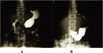
Pretreatment staging should include (1) complete oesophagogastroscopy with biopsy proof, (2) barium study of upper gastrointestinal (GI) tract in a prone and in a standing position (Figure 1), (3) computed tomography (CT) of the abdomen. The other routine evaluations include a chest x-ray, complete blood count and serum biochemistry for liver and renal profiles. The value of a baseline tumour-marker record of carcinoembryonic antigen and carbohydrate antigen 19.9 still remain debatable. Endoscopic ultrasonography and laparoscopy are not yet available in many institutions and should not be considered as mandatory.
Preoperative barium study of stomach region in a prone (A) and in a standing (B) position showed organ movement. In a standing position the lower border of stomach is situated one and half of vertebrae below than in a prone position.

Post-operative radiochemotherapy
Post-operative evaluation is more complex to define the treatment volume and all possible care is necessary before planning radiotherapy : (1) preoperative oesophagogastroscopy, (2) preoperative and post-operative diagnostic CT scans should be compared to delineate the tumour bed and anastomosis region with margin, (3) post-operative barium study to show post-operative gastric remnant and site of anastomosis, (4) surgical report and pathological report, (5) surgical clips if surgery is R1 or R2.
RADIOTHERAPY TECHNIQUES
Irradiation volumes and organs at risk of therapeutic injury
In recent times it has been understood that post-surgical adjuvant or currently explored neoadjuvant radiotherapy for stomach cancer is a complex practice from clinical and technical standpoints.Reference Emami, Lyman, Brown, Coia, Goitein, Munzenrider, Shank, Solin and Wesson12 The critical organs which surround the stomach region and can show adverse late effects on normal tissues, due to inclusion in irradiated volume, are: both kidneys, liver, spinal cord, small and large intestines, pancreas, lungs and heart.
Deciding the target volumes [clinical target volume (CTV), planning target volume (PTV)] is done in several ways at present,Reference Smalley, Gunderson, Tepper, Martenson, Minsky, Willett and Rich9, Reference Leong, Willis, Joon, Condron, Hui and Ngan15, Reference Willet, Gunderson, Perez, Brady, Halperin and Schmidt-Ullrich20 starting from simple 2-D estimation to complex 3-D contouring. The CTV or more broadly the PTV is designed to encompass, (1) the stomach or post-operative remnant stomach with anastomoses ends, plus a minimum of 3–5 cm margins, (2) regional nodes of perigastric, portahepatis, celiac regions as minimum and (3) if necessary the pancreaticodeodenal, splenic hilar, local periaortic and lower periesophageal nodal stations.
Precise definitions of regions of interest for tumour dose delivery and at same time organs at risk (OAR) for late radiation morbidities are to be set before the actual planning process begins. These requirements should be followed for both preoperative or post-operative irradiation practice.
Radiotherapy planning and delivery requisites
—Clinical evaluation of patient’s performance and nutritional status is done carefully so that a patient is suitable to undergo the rigours of chemoradiotherapy course which may range from 5–8 weeks for neoadjuvant approach to 8–16 weeks for post-operative adjuvant therapy.Reference Macdonald, Smalley and Benedetti5, Reference Ajani, Mansfield and Janjan6 As far as possible, maintaining enteral nutrition is the preferred choice. A Karnofsky Performance Score of ≥70 is ideal.
—Simulator planning is used to design the radiation portals and shielding of OAR. This may be adequate, when it is combined with barium fluroscopy and intravenous urography, for the 2-D planning for antero-posterior or three/four fields fixed dose delivery.
—CT scan planning. It is now realised that the complex anatomical region of upper abdomen require a CT scan–based planning for optimised radiotherapy delivery by the 3-D planning and dosimetry.
—Shields. Custom-made shields for kidneys, liver, spinal cord and optionally for spleen and heart will be needed for fixed fields. However, multi-leaf collimator (MLC)-based linac delivery, if available, can obviate these requirements to a great extent.
—Teletherapy machines. Modern linac with energies of 6–20 MV photons is ideal choice and facilities for conformal radiotherapy (CRT) and IMRT will improve the options in future. In its absence, isocentric cobalt can deliver the irradiation, but with higher risks of normal tissue and organ injury.
—Computerised treatment planning. Dosimetry evaluations for both 2-D and 3-D deliveries are becoming the standards and this step is critical to assessment of radiotherapy practice of an institution. Dosimetry records are to be kept for both online and hardcopy analysis.
—Manpower. Radiation oncologists, physicists, technologists and nursing personnel are all involved in the chemo-irradiation course of therapy and they should have familiarity in managing these patients. Anatomical and imaging knowledge is essential for planning and dosimetry, changes in clinical status need attentions, and morbidity assessment is part of quality health care delivery in radiotherapy.
Radiotherapy planning
All data of the patient related to disease extent, imaging, endoscopy, pathology and prior treatments by surgery and/or chemotherapy are scrutinised at this stage. The planning of radiation portals is done serially by simulation, CT scans and then reconstructed by digitally reconstructed radiograph (DRRs), if both facilities are present. All the OAR right from thorax to lower abdomen are imaged at these planning steps (simulator/CT scans), so that proper contouring and shielding can be achieved. The target region delineation and contouring will depend upon whether simulator fields only or simulator combined with CT scan portal planning are generated. Then the physicist, dosimetrist and radiation oncologist will combine together for CTV, PTV, OARs contouring, field arrangements and dose planning etc. prior to final approval. The field extents will differ according to pre- or post-operative status, tumour location in stomach, surgical pathology report of tumour-node involvements and preoperative and post-operative CT scan findings.
Although CTV is a concept for optimal target region definitions, yet such clear-cut delineations will not be easy in the post-operative situation, even with barium fluoro-simulation (for anastomosis site) and CT scan–based planning.Reference Smalley, Gunderson, Tepper, Martenson, Minsky, Willett and Rich9, Reference Leong, Willis, Joon, Condron, Hui and Ngan15 One has to fall back on anatomic landmarks of generated images to define the target region. In addition, PTV as 1–2 cm margin around CTV can, in some situations, adversely include larger volume of OARs and a critical judgement is called for so that unnecessary irradiation is avoided.
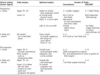
Differing field extents for radiation portals are described in Table 1. This can be treated as a broad guideline only.
As per above descriptions of radiation portals, it is intended to treat the stomach and locoregional target volumes by more than two fields. Although Intergroup trial and subsequent adjuvant radiotherapy consensus report have recommended post-operative adjuvant radiotherapy by antero-posterior opposed radiation fields,Reference Macdonald, Smalley and Benedetti5, Reference Smalley, Gunderson, Tepper, Martenson, Minsky, Willett and Rich9 even meticulous planning with opposed fields can deliver higher than late morbidity threshold doses to OARs in most clinical situations. Modern imaging, simulation and CT scan–based plannings can be time consuming to design three or more fields. However it would be clinically justifiable to move away from antero-posterior two-field irradiation practice, since more and more patients are going to live longer.Reference Chung, Shakespeare, Wynne, Lu, Mukherjee and Back13, Reference Leong, Willis, Joon, Condron, Hui and Ngan15, Reference Ringash, Perkins, Brierley, Lockwood, Islam, Catton, Cummings, Kim, Wong and Dawson16
Guidelines for radiotherapy portals

Note: (1) Simulation field planning is done with barium(for anastomosis/stomach contour)and iv contrast(for kidneys); (2) for conventional plan: antero-posterior (A-P) fields 12–15 cm width and 12–18 cm lengths are usual dimensions, and lateral (lat) fields would be 10–12 cm width and same length as A-P; (3) CRT/IMRT fields are planned on multi-segmental CT scan contouring, so that initial and boost field sizes are tailored at different stages of RT course; (4) not more than one-third volumes of liver and kidneys are included within radiation portals, so that total liver dose is <30 Gy and total dose for each kidney is <20 Gy, spinal cord dose is kept at <40 Gy; (5) shieldings are placed at different stages to shield kidneys(after 20 Gy) and liver(after 30 Gy) in conventional RT course; (6) whenever possible target volume and OARs contouring are done to generate 3-D dosimetry and dose–volume histograms, otherwise manual dose plottings are to be done for conventional RT evaluations.
CRT, conformal radiotherapy; IMRT, intensity-modulated radiotherapy; LN region, lymphnode region; RT, radiotherapy.
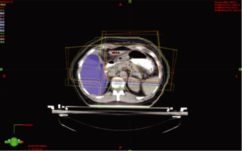
Preoperative radiotherapy planning may be easier, because the endoscopy and CT scan findings of intact stomach will be helpful to generate the stomach and locoregional extents.Reference Ajani, Mansfield and Janjan6 The delivery of preoperative radiotherapy to a patient of gastric cancer by CT scan–based 3-D contouring, with placement of three fields and wedges, is shown in Figure 2.
Preoperative CT scan–based contours and three-fields technique, which minimize the dose to the kidneys and to the spinal cord.

For the post-operative patient, the adjuvant radiotherapy (RT) is aimed at tumour bed, anastomosis site with margins, and the locoregional lymphatics. All pre- and post-operative CT scans should be evaluated, along with surgical and histopathological reports. If possible a barium study and repeat upper GI endoscopy should be carried out before radiotherapy planning. All these informations will be helpful to mark out the locoregional target volumes for initial and boost fields while doing the simulator field placements and CT scan–based planning. The simulator barium-fluoro demarcation of stomach outlines (preoperative) and anastomotic junctions plus remnant stomach (post-operative) can be combined with planning CT to derive DRRs for conventional 2-D/3-D dosimetry or multi-slice contouring for CRT/IMRT-directed radiation deliveries.
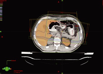
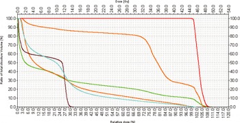
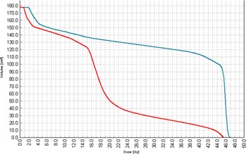
The three-fields planning for conventional RT can generate satisfactory isodose coverage, that is anterior plus two lateral for cardia and fundus tumours or, antero-posterior plus left lateral for body and antral tumors. Wedges would be helpful to tailor the dose gradients. The four-fields box technique would be tried in a situation where entire stomach with locoregional boundaries are treated en bloc. Whenever possible, the lateral fields may be angled anteriorly to avoid the spinal cord and kidneys. The CT scan–based 3-D planning for a post-operative gastric cancer patient is shown in Figure 3. It can be appreciated from the dose–volume histograms of this three-fields technique (Figure 4), when compared with antero-posterior two-fields (Figure 5), that the OARs will receive much lesser dose.
Post-operative treatment planning and isodose coverage for three-fields technique.

Representative dose–volume histograms (DVHs) for PTV (red line), liver (higher orange line), body (green line), left kidney (lower orange line), right kidney (turquoise line) and spinal cord (brown line).

Dose–volume histogram (DVH) for left kidney using three-fields technique (red line) and anteroposterior-posteroanterior (AP-PA) technique (blue line).

Alternatively when CRT/IMRT are available then multi-segmental contouring with single isocentre for upper and lower halves of the fields or split-fields arrangements are generated. OARs are defined alongside the regions of interest. Critical to the dose planning and isodose evaluations would be a broad agreement between radiation oncologist, physicist and dosimetrist a priori to select dose ranges they would like to deliver to target region and OARs.
DISCUSSION
The results of randomized adjuvant trials in gastric cancer show increase in disease control and survival rates, when chemoradiotherapy is added to surgery.Reference Jansen, Boot, Verheij and van de Velde1–Reference Smalley, Gunderson, Tepper, Martenson, Minsky, Willett and Rich9 As more and more gastric cancer patients will be treated by radiotherapy, usually combined with chemotherapy, the successful therapy will depend upon patient tolerance. It is observed that nearly 20–30% of all patients planned for pre- or post-operative chemoradiotherapy could not complete the intended therapy protocols and nearly 40–50% patients had treatment-related acute toxicities compromising the treatment outcomes.Reference Macdonald, Smalley and Benedetti5–Reference Lowy, Feig, Janjan, Rich, Pisters, Ajani and Mansfield7 The reported trials have generally used the antero-posterior two-fields in radiotherapy practiceReference Macdonald, Smalley and Benedetti5, Reference Smalley, Gunderson, Tepper, Martenson, Minsky, Willett and Rich9 and this is likely to cause both acute and late morbidities. Hence, the improvements in radiation therapy techniques and parameters are necessary.
As per the cancer burden described in the introduction, nearly two-thirds or more of all gastric cancer patients will be treated in less developed countries with varied radiotherapy infrastructures. The present report is intended to help the radiation oncology team in implementing a safe radiotherapy protocol of techniques and practices. The radiotherapy for gastric cancer, either pre- or post-operative, should be based upon 3-D dose definition (and recording) technology, with very accurate definition of GTV, CTV, PTV and normal OAR (liver, kindeys, spinal cord, bowel, pancreas, heart, lungs). Multiple-field conformal techniques with shielding by multi-leaf collimator allow to protect normal tissues and reduce the risk of radiation injuries.Reference Caudry, Escarmant, Maire, Demeaux, Guichard and Azaloux14, Reference Leong, Willis, Joon, Condron, Hui and Ngan15 Wherever feasible, the institution should shift from conventional to CRT/IMRT practices, although these can be manpower and technology intensive.
We have resisted from describing in detail the currently reported heterogenous chemotherapy and surgical practices, which often range from conservative to aggressive, affecting the radiotherapy morbidities. The impact of other anticancer modalities can be minimized by making changes in radiation therapy techniques. The practice of radiation therapy by antero-posterior fields will deliver higher dose to critical organs and will encompass higher irradiated tissue volumes, as compared to the multiple-fields delivery. This is likely to have greater consequential late effects (CLEs) due to the acute radiation morbidities inflicted.Reference Dorr and Hendry21, 22 In currently practiced aggressive radiotherapy or chemoradiotherapy protocols, the acute reactions become intense and these are prominent in critical organs where the acute response disrupts the mechanical and chemical barriers. This can cause additional trauma seen as CLE. Such effects can be pronounced in early responding organs of the GI system leading to higher than currently estimated late effects.
Our argument will be to incorporate all informations of imaging and clinical data, in order to treat these patients with multi-field techniques in any available radiotherapy setting.
SUMMARY
Recent studies have found improved outcomes in surgically treated gastric cancer when chemoradiotherapy is delivered in a scheduled adjuvant or neoadjuvant manner. Radiation oncology community has the new responsibility to treat these patients in such a way that target region receives the intended 45 Gy and the surrounding critical organs are spared from acute, consequential and long-term late effects. The intergroup trial and subsequent consensus reports recommend elaborate plannings for opposed antero-posterior two-fields radiation delivery. There is concern and reluctance from radiation oncologists to treat all gastric cancer patients by antero-posterior fields which has inherent limitations in delivering higher doses to OAR and to treated volumes. The descriptions of planning requirements, radiation field plans, isodose contouring for target vs. OARs would be useful guides. For all future studies, the multi-field conventional or conformal/IMRT radiotherapy will be the direction to move ahead and evaluate its benefits in terms of locoregional control, critical organ effects and long-term survivals.
ACKNOWLEDGEMENT
The data collection and preparation of manuscript were done during the professional visit of Dr. B.K. Mohanti from his assignment at Muscat to Gliwice, Poland.