Introduction
In recent years, organophosphate pesticides have been widely used, especially in the field of agriculture. The O,O-diethyl o-3,5,6-trichloropyridin-2-yl phosphorothioate, which is also known as chlorpyrifos (CPF), is one of the organophosphates that is extensively used to protect agricultural products from insects. However, one concern with the use of pesticides is their toxicity to aquatic ecosystems and the direct effects of agricultural discharges on human life.[ Reference Freitas, Singer, Muller and Schwarzenbach 1 – Reference Castillo, Ruepert and Solis 3 ] CPF is moderately toxic by reaction with a serine residue on acetylcholinesterase (AChE), which inhibits the enzymatic reaction of AChE with acetylcholine chloride. This inhibition directly affects brain development, especially in children.[ Reference Eaton, Daroff, Autrup, Bridges, Buffler, Costa, Coyle, McKhann, Mobley, Nadel, Neubert, Hermann and Spencer 4 ] Accordingly, this issue of CPF contamination has aroused interest from many research fields such as environment, medicine, chemical, biology, among others. Hence, numerous scientific approaches to detect CPF have been developed.[ Reference Zou, Du, Wang, Smith, Timchalk, Li and Lin 5 , Reference Uygun and Dilgin 6 ]
Standard analytical methods can be used for efficient identification of CPF, such as gas chromatography with mass spectroscopy[ Reference Oliva, Navarro, Barba and Navarro 7 , Reference Goncalves and Alpendurada 8 ] or high-performance liquid chromatography.[ Reference Harshit, Charmy and Nrupesh 9 – Reference Abu-Qare and Abou-Donia 12 ] However, these methods are known to require highly skilled operators and to be expensive and time-consuming. In addition to these techniques, surface plasmon resonance (SPR) and electrochemical immunosensing, which operate based on enzyme/antibody immobilization, can be used as pesticide sensors with high selectivity and sensitivity. These techniques have been implemented using the AChE enzyme as a biomarker to directly detect biologic interaction with CPF.[ Reference Mauriz, Calle, Lechuga, Quintana, Montoya and Manclus 13 , Reference Yao, Liang, Huang, Wang and Qiu 14 ] Nevertheless, these methods required many material preparation steps and long-duration incubation for immobilization with the enzyme. Moreover, optimal environmental conditions were necessary to prevent damage to the enzyme from acidic or basic conditions, organic solvents, high temperatures, and so forth.
Recently, photoelectrochemical (PEC) measurements, which leverage the synergetic advantages of optical and electrochemical sensing, have been considered to be an alternative strategy for detection of CPF.[ Reference Li, Zheng, Liu, Zhao and Liu 15 , Reference Gong, Wang, Li and Wang 16 ] One challenge associated with PEC sensing is to amplify the photocurrent signal. To address this need, titanium dioxide (TiO2) has been widely used for its high photocatalytic activity and non-toxicity. However, photoabsorption of TiO2 is limited to the ultraviolet (UV) range, but only approximately 10% of the radiant solar energy is in the UV range.[ Reference Fu 17 ] To extend the photoresponse of the electrode to include light in the visible range, conjugated polymers have been extensively employed as energy providers in photovoltaic devices for their high charge-carrying capacity and high absorption efficiency.[ Reference Sapp, Elliott, Contado, Caramori and Bignozzi 18 – Reference Li, Shrotriya, Huang, Yao, Moriarty, Emery and Yang 20 ] Poly(3-hexylthiophene) (P3HT), which can be used as an electron donor material, has an absorption peak of approximately 1.8 eV,[ Reference Soci, Hwang, Moses, Zhu, Waller, Gaudiana, Brabec and Heeger 21 ] offering the possibility of band gap alignment and compatibility with TiO2. P3HT–functionalized TiO2 has been used in many applications such as solar cells and sensors.[ Reference Chen, Nakahara, Wei, Nordlund and Russell 22 – Reference Tiwari, Singh, Joshi, Chakrabarti, Takashima, Kaneto and Prakash 27 ] Recently, a PEC sensor for CPF detection was developed, which could effectively detect the CPF molecules at zero potential under visible light irradiation.[ Reference Li, Li, Xu and Hu 28 ] Use of this method resulted in a very weak photocurrent signal (of the order of nA), but the detection limit was still high (0.01 µM) in comparison with the standard instruments.
Herein, we propose a novel approach to enhance the photocurrent generated in PEC measurements and apply this method for sensitive detection of CPF. This approach leverages hybrid excitation of grating-coupled[ Reference Baba, Wakatsuki, Shinbo, Kato and Kaneko 29 , Reference Baba, Aoki, Shinbo, Kato and Kaneko 30 ] and localized surface plasmons[ Reference Pangdam, Nootchanat, Ishikaw, Shinbo, Kato, Kaneko, Thammacharoen, Ekgasit and Baba 31 ] on the photoelectrode. As a result of the synergistic effects of both types of plasmons,[ Reference Lertvachirapaiboon, Supuntabut, Baba, Ekgasit, Thammacharoen, Shinbo, Kato and Kaneko 32 , Reference Nootchanat, Ninsonti, Baba, Ekgasit, Thammacharoen, Shinbo, Kato and Kaneko 33 ] the short-circuit potential was improved, thereby improving the CRF-detection limit to 7.5 nM. The grating structure (grating pitch Λ of 740 nm) was fabricated on the photoelectrode via nanoimprinting of a CYTOP fluoropolymer with a recordable digital versatile disk (DVD-R) template, followed by gold thin-film deposition. Then, gold nanoparticles (AuNPs) were deposited on the P3HT–TiO2 functionalized gold-grating layer. The SPR excitation of this structure was characterized by measuring the SPR reflectivity under p-polarized light irradiation. The results indicated that the wavelength of the excited propagating surface plasmon corresponded with the absorption peak of the fabricated photoelectrode. CPF detection using the fabricated system was demonstrated for a range of CPF concentrations. We demonstrated that the novel plasmonic PEC sensor can be used for the detection of CPF at zero potential based on the photovoltaic effect with an amplified detection signal and improved detection limit.
Experimental section
Chemicals and materials
TiO2 nanoparticles with diameters of 21 nm were obtained from AEROSIL. CYTOP (CTL-809M, with a glass transition temperature of 375 K) was purchased from ASAHI glass. P3HT, ethanol, 1,2-dichlorobenzene, CPF (analytical standard), and phosphate-buffered saline (PBS; pH 7.3) were purchased from Sigma-Aldrich.
Fabrication of sensing layers and experimental setup
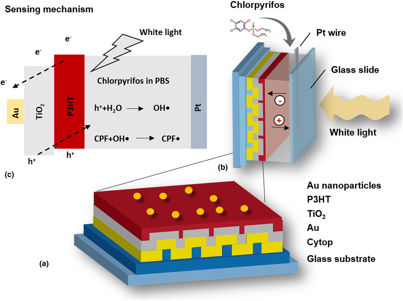
Figure 1(a) shows a schematic of the fabricated plasmonic photoelectrode. The photoelectrode was constructed by spin-coating CYTOP onto a BK-7 glass slide at 3000 rpm for 30 s, and then drying the CYTOP at 100 °C for 15 min to obtain a 400-nm-thick film. A DVD-R disk was used as a template for nanoimprinting.[ Reference Hara, Lertvachirapaiboon, Ishikawa, Ohdaira, Shinbo, Kato, Kaneko and Baba 34 , Reference Nootchanat, Pangdam, Ishikawa, Wongravee, Shinbo, Kato, Kaneko, Ekgasit and Baba 35 ] The template was cut into a size of 2.5 cm × 2.5 cm and then washed with nitric acid, ethanol, and deionized water. The grating structure was subsequently etched in a 1 : 4 acetone:ethanol solution for 15 min before rinsing with deionized water. The DVD-R mold with the grating structure was stamped onto the CYTOP film with a force of 25 MPa at 110 °C for 90 min. The grating-structured CYTOP/glass substrate was coated with approximately 3 nm of Cr and 60 nm of gold using vacuum evaporation. Next, TiO2 nanoparticles were dispersed in ethanol at a concentration of 3 mg/mL, spin-coated on the gold-coated grating structure at 3000 rpm for 30 s, and dried at 100 °C for 1 h. The sample was then annealed at 250 °C for 30 min, forming a 100-nm-thick TiO2 film. P3HT was dissolved in 1,2-dichlorobenzene at a concentration of 10 mg/mL and deposited on the TiO2 layer by spin-coating at 2000 rpm for 60 s prior to annealing at 120 °C for 30 min, thus producing a 50-nm-thick P3HT film. Finally, ~3-mm-diameter AuNPs were loaded onto the P3HT film by vacuum evaporation. A UV–vis spectrometer (V-650, JASCO) was used to record the absorption spectra of each layer during the preparation process. The surface morphology of the gold-coated grating substrate, gold-coated grating/TiO2/P3HT, and the gold-coated grating/TiO2/P3HT loaded with AuNPs were studied via atomic-force microscopy (SPM-9600 from Shimadzu Corporation). Results indicated that the grating pattern remained intact after coating with TiO2/P3HT and AuNPs (see Supplementary Data, Fig. S1).
Schematic diagrams of (a) the fabricated photoelectrode with a grating structure, (b) the PEC cell for CPF measurements, and (c) the CPF sensing mechanism.

A schematic diagram of the PEC measurements is shown in Fig. 1(b). A platinum wire was used as the counter electrode. A 0.1 M PBS solution was used as the electrolyte. In the PEC measurements, the fabricated cell with the active area of 10.17 mm2 was mounted on a goniometer. A solar simulator (HAL-C100 from ASAHI Spectra USA Inc.) with a light intensity of 75 mW/cm2 was used as the light source to illuminate the cell on its counter-electrode/glass-slide side.
The SPR reflectivity measurements were performed under p-polarized light illumination for excitation of the surface plasmons; s-polarized light was used for comparison. The white light was collimated by objective lenses and applied at a fixed incident light angle with varying wavelengths. A spectrometer was used to measure the reflected light.
Results and discussion
SPR properties
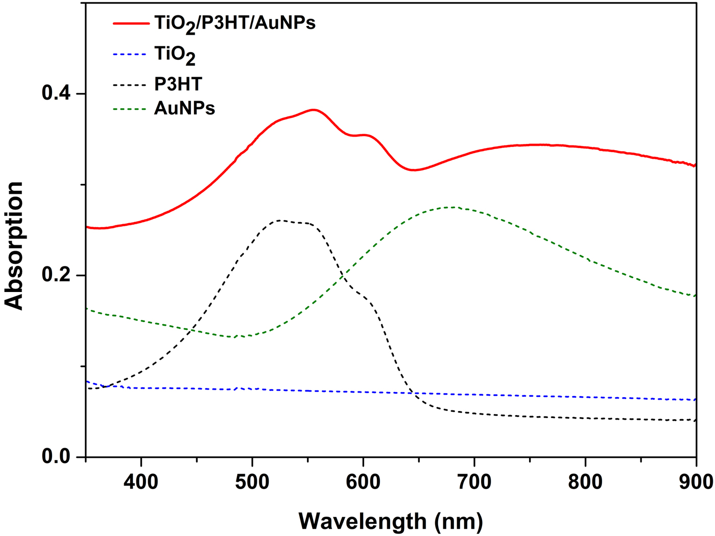
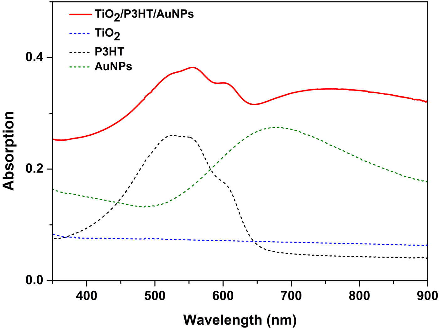
Figure 2 shows the UV–vis spectra of TiO2/P3HT/AuNPs, TiO2, P3HT, and AuNPs on glass slides to determine which peaks in the spectrum originated from which layer in the composite layered structure. A Q-band peak at 550 nm with a shoulder at 600 nm originating from the P3HT film and a broad shoulder at 720 nm originating from the AuNPs were observed in the multilayered film.
UV–vis spectra of TiO2/P3HT/AuNPs, TiO2, P3HT, and AuNPs.
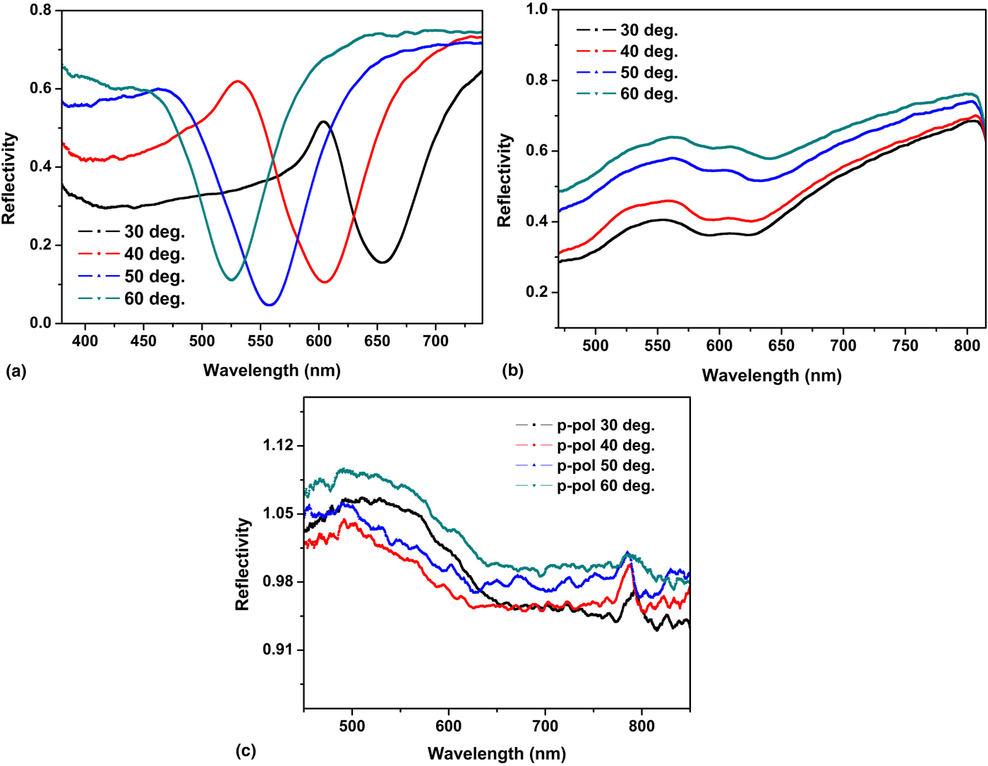
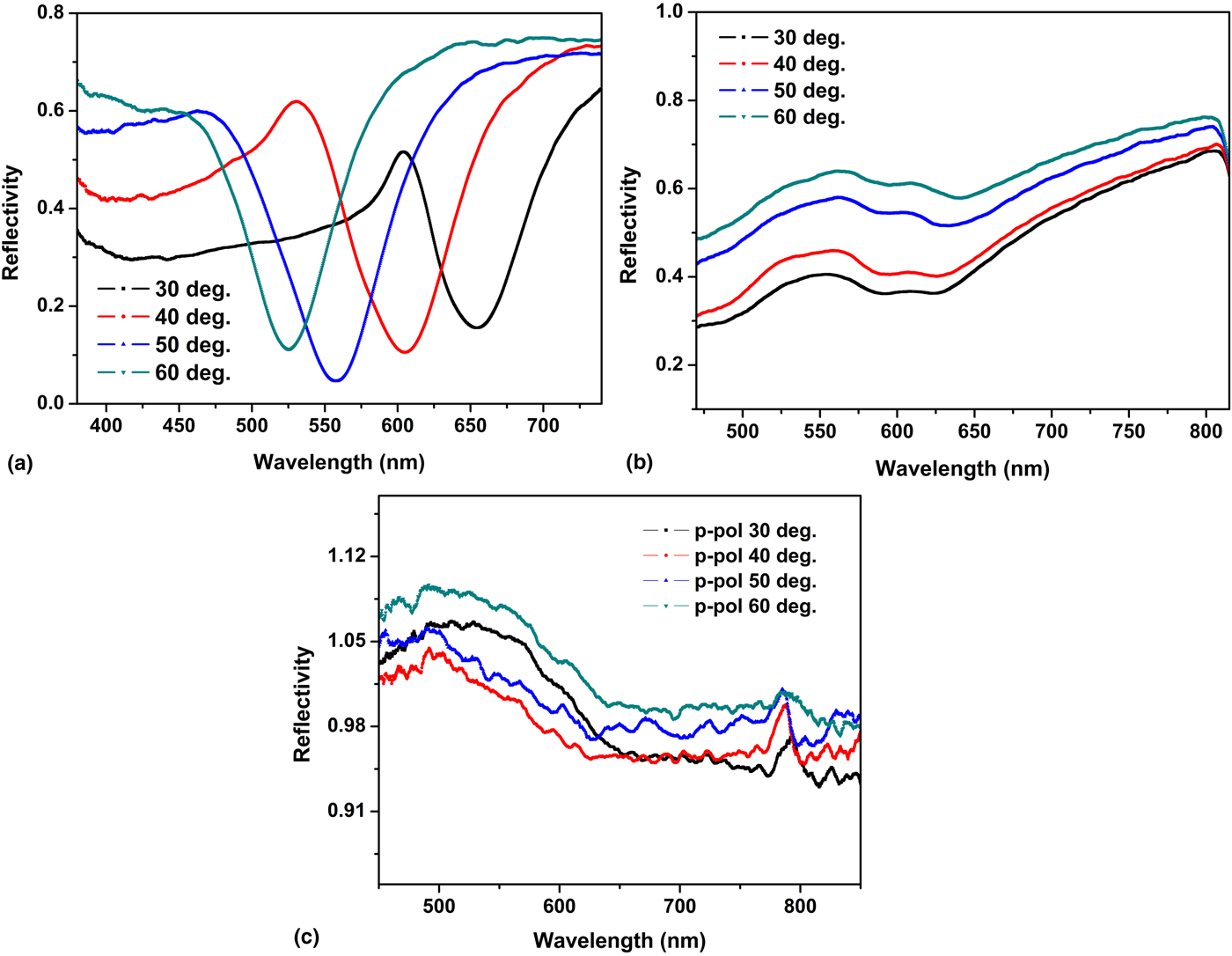
The excitation of SPR was studied by measuring the reflectivity curves as a function of wavelength from the fabricated AuNP-loaded photoelectrode cell in PBS. The ratio of the light reflected by the fabricated cell to those of a flat gold reference specimen was taken as the reflectivity. Figure 3(a) shows the SPR reflectivity curves of the fabricated cell with a grating structure measured at incident angles ranging from 30° to 60° with p-polarized light irradiation. The results show a resonance dip for each incident angle due to propagating surface plasmon excitation, indicating that enhanced electric fields could be obtained in the cell. The observed resonance dips shifted toward shorter wavelengths as the incident angle increased. The reflectivity curve of the fabricated cell with a grating structure under s-polarized light, as well as that of the cell without the grating structure under p-polarized light, were also measured and are shown in Figs. 3(b) and 3(c). In these conditions, the reflectivity dips did not occur due to lack of excitation of propagating SPR. Comparing the surface plasmon dips [Fig. 3(a)] with the absorption spectrum of the multilayered film (Fig. 2), we found that the wavelengths at which dips occur (around 40°−60°) corresponded to the main absorption peak wavelength of the PEC cell, which originated from the absorption of P3HT.
Reflectivity curves of the fabricated PEC cell obtained by illumination with (a) p-polarized light and (b) s-polarized light on the grating substrate, and (c) p-polarized light on a cell with a flat gold substrate, each measured at incident light angles ranging from 30° to 60°.
Short-circuit photocurrent measurements
To measure the electrical signal from the PEC sensor in the presence of CPF, the short-circuit current at 0 V was measured. The underlying mechanism that gives rise to the short-circuit current is that upon injection of CPF, P3HT generates exited electrons due to visible light irradiation, which are injected into the conduction band of TiO2, and subsequently to the gold electrode. Simultaneously, holes in the valence band of TiO2 migrate to the valence band of P3HT. The holes can react with PBS to generate hydroxyl radicals (•OH). This dissociation reacts with CPF, forming CPF•, which can amplify the sensor's response.
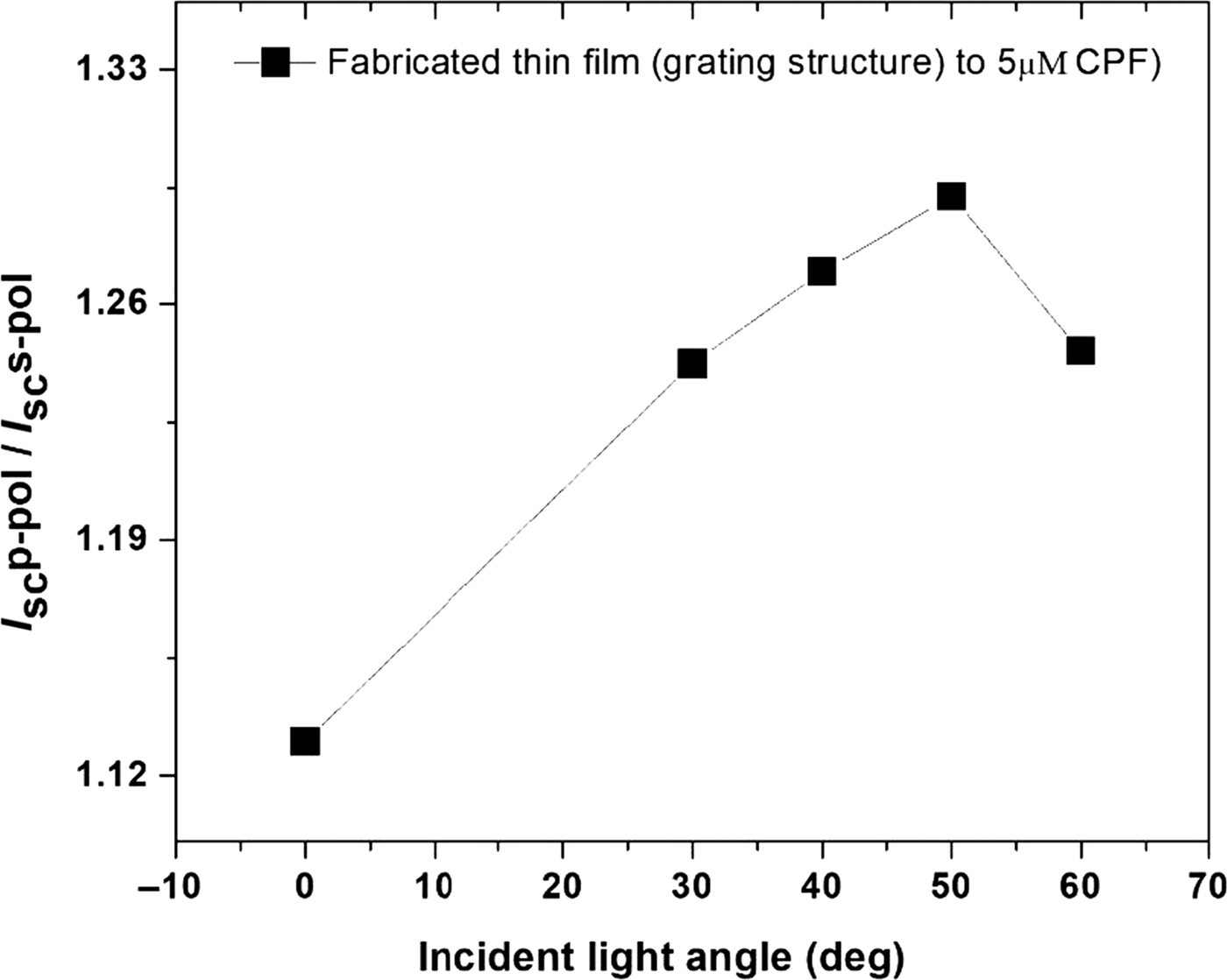
We first studied the effect of propagating surface plasmon excitation on the photocurrent at zero potential as a function of the incident light angle between 0° and 60°. It is noted that no current was observed under light irradiation in the absence of CPF. Figure 4 shows the enhancement factor of the short-circuit current in the fabricated cells with 5 µM CPF in PBS, where the enhancement factor is defined as the ratio of the current measured with propagating surface plasmon excitation (p-polarized light irradiation) to the current without surface plasmon excitation (s-polarized light irradiation). The enhancement factor gradually increased as the incident light angle increased from 0° to 40° and reached a peak at 50°, which coincides with the surface plasmon dip wavelength observed in the fabricated cell and with the absorption of the P3HT film. This result is reasonable because the light absorption of the P3HT film is enhanced by the strong electric field generated by the excitation of a propagating surface plasmon at the optimal incident angle.
Enhancement factor of the short-circuit current, defined as the ratio of the current with surface plasmon excitation (p-polarized light irradiation) to the current without surface plasmon excitation (s-polarized light irradiation) in the presence of 5 µM CPF.

Short-circuit photocurrent enhancement for CPF detection by hybrid surface plasmon excitation
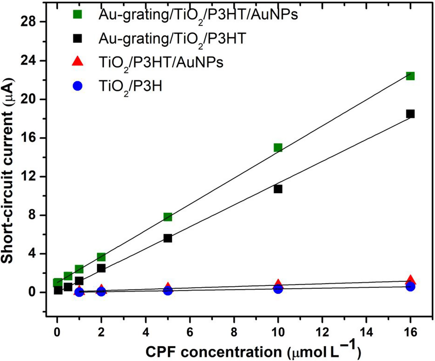
The short-circuit photocurrent signal enhancement by both propagating surface plasmon and localized surface plasmon excitations were studied. Photoelectrodes were prepared in four configurations: flat gold substrates and gold-coated grating substrates, each prepared with or without loaded AuNPs. The short-circuit photocurrent signal was measured as a function of the CPF concentration (0.01–16 µM), as shown in Fig. 5. The linearity of the signal as a function of the CPF concentration was observed in each photoelectrode sample. The flat gold-coated substrate provided a lower I SC signal than the gold-coated grating substrate. This is because the strong electric field intensity generated by the propagating surface plasmon on the grating gold surface increases the light absorption[ Reference Baba, Wakatsuki, Shinbo, Kato and Kaneko 29 , Reference Baba, Aoki, Shinbo, Kato and Kaneko 30 ] of P3HT, which subsequently increases the number of holes generated in P3HT, leading to more reactions with PBS to generate more •OH and CPF• radicals. When AuNPs were loaded on P3HT, the resulting signal was enhanced compared with that without AuNPs. This may be because the localized surface plasmon excited around the AuNPs enhances the electric field in the P3HT film.[ Reference Pangdam, Nootchanat, Ishikaw, Shinbo, Kato, Kaneko, Thammacharoen, Ekgasit and Baba 31 ]
Plots of the short-circuit photocurrent of the fabricated PEC cells with various compositions as a function of the CPF concentration. The short-circuit current was measured at an optimal incident angle of 50°.
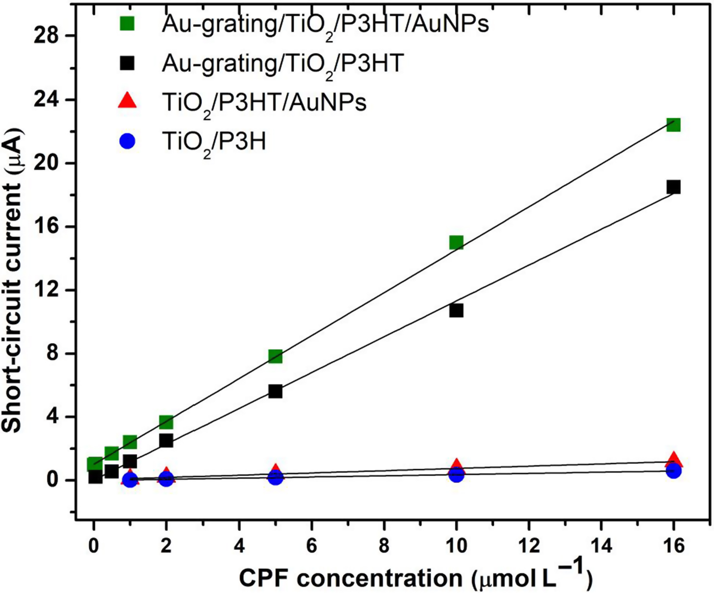
Due to the synergetic effect of propagating surface plasmon and localized surface plasmon, an extremely low limit of detection of 7.5 nM was achieved. Although the sensitivity was still one or two orders lower than that obtained by enzyme/antibody-based immunoassay systems with the electrochemical technique,[ Reference Sun, Wang, Wen, Gan, Lei and Liu 36 ] surface enhanced Raman,[ Reference Kim, Barcelo and Li 37 ] and magnetic platform[ Reference Pino, Ivandini, Nakata, Fujisjima, Merkoci and Einaga 38 ] or by magnetic-based molecularly imprinted technique with Kretschmann-based SPR,[ Reference Yao, Liang, Huang, Wang and Qiu 14 ] our technique exhibited high sensitivity without using complicated systems. Furthermore, the signal intensity was amplified to μA, producing nA-level signals, which are significantly more than that obtained using a previously reported method.[ Reference Li, Li, Xu and Hu 28 ] Based on our study, the advantages afforded by the PEC sensor using the gold-coated grating structure and loaded AuNPs were effectively demonstrated, including enhancement of the detection limit and amplification of the short-circuit current.
Conclusion
We developed a PEC CPF sensor with a photoelectrode composed of gold-coated grating substrate/TiO2/P3HT/AuNPs. The propagating SPR on the gold grating, which corresponded well with the absorption peak wavelength of the P3HT film, enhanced the short-circuit photocurrent for highly sensitive detection of CPF. Moreover, loading of AuNPs further enhanced the short-circuit photocurrent due to the localized surface plasmon effect. The modified hybrid plasmonic photoanode was enhanced compared with photoanodes having structures without surface plasmon excitation. By the synergetic effect of both types of plasmons, the short-circuit photocurrent was amplified to a μA scale, which is notably higher than the signal obtained using a previously reported method, by which several tens of nA-level signals were obtained in all CPF concentration. Moreover, CPF was detected at a very low concentration of 7.5 nM using this system. The demonstrated novel method offers a multifunctional photovoltaic effect which can be used in several PEC-based sensing applications.
Supplementary material
The supplementary material for this article can be found at https://doi.org/10.1557/mrc.2017.131.
Acknowledgments
This work was supported by Japan Society for the Promotion of Science (JSPS) KAKENHI Grant Numbers JP16K13662 and JP17H03231. T.T. acknowledges the support of “research and researchers for industries (RRI)” scholarship of the Thailand Research Fund.