Background
Persistent fifth aortic arch is a very rare congenital anomaly as the fifth aortic arch normally involutes in fetal life before birth, and the fourth aortic arch persists as the true arch.Reference Van Praagh and Van Praagh 1 Several studies have described the arch and its presentation and management.Reference Van Praagh and Van Praagh 1 – Reference Chiappa, Greco, Fainardi, Passantino, Serranti and Favilli 9 It becomes clinically relevant when it is associated with coarctation or interruption of the true aortic arch. Intra-operative discovery of the anomaly led to an unusual but successful approach for repair. We also describe the unusual appearance of the vessel’s wall, which was ductal in origin.
Case report
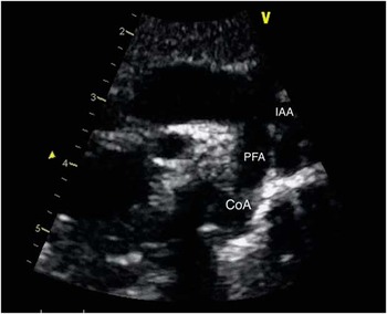
A baby girl was born at 39 weeks of gestation via a planned C-section, weighing 2.94 kg, as the baby had a prenatal diagnosis of a left congenital diaphragmatic hernia containing colon and liver that was confirmed by an MRI performed during pregnancy. Delivery was uneventful, and the baby had an echocardiogram on day of life 0, which showed an unobstructed left aortic arch with a large patent ductus arteriosus. She underwent a transabdominal repair for her left congenital diaphragmatic hernia on day of life 2. Around day of life 12, she was noted to be more tachypnoeic, and an echocardiogram revealed significant coarctation (peak velocity across the isthmus of 3.8 m/s) with moderate atrial septal defect and a left superior caval vein to the coronary sinus with no patent ductus arteriosus (Fig 1). Neither the interrupted aortic arch nor the persistent fifth aortic arch was recognised. Her four-extremity blood pressure gradient was significant for a 25-mmHg gradient. Prostaglandin infusion was initiated without clinical or echocardiographic improvement. Given the interpretation of the echocardiogram, an end-to-end anastomosis via left thoracotomy was planned on day of life 14.
Preoperative echocardiogram showing severe coarctation (CoA) of the persistent fifth arch (PFA), which was misinterpreted as the true aortic arch at its junction with the descending aorta. In retrospect, the interrupted aortic arch (IAA) can be seen on top of the PFA.

During dissection, we realised that the left subclavian artery and the descending aorta were discontinuous, and there appeared to be an aberrant right subclavian artery originating from the proximal descending aorta travelling posterior to the oesophagus. What we had assumed to be the aortic arch, into which the ligamentum arteriosus entered, had a distinctly different characteristic and appearance, which was different from the ascending aorta and the descending aorta (Fig 2a). Owing to severe hypoplasia of the true aortic arch, which gave rise to four head vessels with no connection to the descending aorta, an extended end-to-end repair was not possible. Other alternatives such as closing the incision and addressing this via sternotomy on bypass versus obtaining additional imaging were also considered. Ultimately, we decided to incorporate the fifth aortic arch into the repair, relieving the discrete juxtaductal coarctation. The ligamentum arteriosus was divided between suture ligatures of 5–0 prolene. Heparin was administered, and the coarctation segment was resected leaving the retro-oesophageal artery attached to the descending aorta. All ductal tissues were removed. Examination of the fifth aortic arch demonstrated an abnormal blood vessel, and where we had been previously manipulating the blood vessel there were multiple full-thickness lacerations. For this reason, the proximal clamp was moved onto the ascending aorta, and all of the fifth aortic arch was resected. An adequate-sized defect was created on the ascending aorta, and an end descending aorta-to-side ascending aortic anastomosis was accomplished with significant difficulty. Following repair, the cuff pressure in the lower extremity was slightly higher than that in the right radial. She was extubated to continuous positive airway pressure ventilation on postoperative day 3 and placed on room air a few days later. A CT angiogram was obtained before discharge, which showed mild narrowing at the anastomotic site (Fig 2b).
( a ) A diagram of the intra-operative abnormality showing type A interrupted aortic arch (IAA), type B persistent fifth aortic arch (PFAA) with severe coarctation, ligamentum arteriosum, four arch vessels, and an aberrant right subclavian artery (ARSA). Presence of two RSAs caused confusion intra-operatively but was suspected and then confirmed with CT angiography postoperatively (Printed with permission from Texas Children’s Hospital). ( b ) Postoperative CT angiogram with three-dimensional reconstruction of the repair showing mild narrowing at the anastomotic site.

Discussion
Van Praagh was the first to describe persistent fifth aortic arch or the so-called double aortic arch in 1969. He labelled the two arches as a superior channel, or fourth aortic arch, and an inferior channel, or fifth aortic arch.Reference Van Praagh and Van Praagh 1 Since then, several case reports have been published describing this anomaly.Reference Gerlis, Ho and Anderson 3 – Reference Zhao, Su, Liu, Cao and Ding 5 , Reference Valderrama, Álvarez, Ballesteros, Rodríguez and Zunzunegui 8 – Reference Khan and Nihill 10
The persistent fifth aortic arch can be either right sided or left sided and is subdivided into two major classes. The first one is a systemic-to-systemic connection (types A and B), which is the most common one. The second one is a systemic-to-pulmonary connection (type C). Type A is characterised by superior and inferior channels. The superior channel gives rise to the head vessels and is considered to be composed distally of the left fourth aortic arch. The inferior channel originates proximally, opposite the ostium of the innominate artery and re-joins the superior channel distally, proximal to the ligamentum arteriosum. This inferior channel represents a left persistent fifth aortic arch. Type B consists of interruption of the superior channel, with a patent inferior arch. Finally, type C is described as a systemic-to-pulmonary arterial connection arising proximal to the first brachiocephalic artery – fifth aortic arch – and joining the pulmonary artery – sixth aortic arch.Reference Gerlis, Ho and Anderson 3 , Reference Zhao, Su, Liu, Cao and Ding 5 According to this classification, we classified our case as type B persistent fifth aortic arch with coarctation.
This anomaly can be associated with other intracardiac defects such as pulmonary atresia, atrial septal defect, ventricular septal defect, truncus arteriosus,Reference Khan and Nihill 10 tricuspid atresia, transposition of the great arteries, tetralogy of Fallot, and patent ductus arteriosus.Reference Van Praagh and Van Praagh 1
It can be discovered using an echocardiogram, CT, MRI, or cardiac catheterisation. In our institution, the gold standard is echocardiography to diagnose aortic coarctation, and a CT scan will only be performed when the extent of the arch hypoplasia is questionable on the echocardiogram. Unfortunately, the echocardiogram for this patient was misinterpreted by both the cardiologist and the surgeon, and the correct diagnosis was missed. Fortunately, the baby did well. Performing a preoperative CT scan would have been ideal for such a patient who had a previous congenital diaphragmatic hernia and would have guided our surgical approach.
Its clinical presentation varies widely from being an incidental finding to causing haemodynamic collapse and significant cyanosis.Reference Van Praagh and Van Praagh 1 , Reference Khan and Nihill 10 Treatment of such anomaly depends on the associated lesions. In types A and B, the presence of coarctation mandates an invasive intervention. Several types of surgical and catheter-based interventions have been described in the literature.Reference Culham and Reed 2 , Reference Gibbin, Midgley, Potter and Martin 4 – Reference Tittel, Kaldararova, Olejnik, Tittelova and Masura 6 , Reference Valderrama, Álvarez, Ballesteros, Rodríguez and Zunzunegui 8 In type C, a systemic-to-pulmonary shunt is necessary to be able to come off prostaglandins.Reference Khan and Nihill 10 In our case, the discovery of the anomaly intra-operatively mandated our surgical approach. If this particular anatomy had been known preoperatively, our surgical approach would have been different. The approach would have been an arch advancement via median sternotomy on cardiopulmonary bypass and antegrade cerebral perfusion for brain protection. The principle of arch advancement procedure in both situations is the same, where the proximal descending aorta is advanced to the distal ascending aorta with an end-to-side anastomosis. The only difference is the utilisation of cardiopulmonary bypass, which makes median sternotomy a much safer operation than performing a thoracotomy for such anatomy. None of the previous reports have described the persistent fifth aortic arch being composed of ductal tissue. In our case, the whole fifth aortic arch was composed of ductal tissue. The final pathology report confirmed our finding, justifying excision of the whole fifth arch and performing an arch advancement. This was an interesting finding, as in usual aortic arch development the distal fourth aortic arch becomes the ductus arteriosus and not the fifth arch. Our only explanation for this finding is the possible migration of ductal tissue to the fifth arch. This would be similar to ductal tissue migration to the branched pulmonary arteries or subclavian artery causing significant narrowing of the vessel.
We believe that this case is instructive, because the persistent fifth aortic arch can lead to misinterpretation of the echocardiogram and failure to diagnose type A interrupted aortic arch. Discovery of this combination of anomalies was surprising intra-operatively and required a complete understanding of aortic arch embryology. Although aortic arch advancement via left thoracotomy was ultimately successful, it was potentially unsafe and underscores the importance of the correct diagnosis in preoperative planning. The decision to attempt using the fifth arch in the repair was taken because of a lack of understanding of its characteristics, guided by the fact that the ductus arteriosus had closed and the persistent fifth aortic arch remained patent.
Acknowledgements
The authors would like to thank Dr. Diego Lara for providing us with the echocardiography image used in this paper. Permission was granted by the parents to publish the case report.
Authors’ contributions: all authors participated in creating content for the manuscript, editing, and provided final approval for submission.
Financial Support
This research received no specific grant from any funding agency or from commercial or not-for-profit sectors.
Conflicts of Interest
The authors declare no potential conflicts of interest with respect to the study, authorship, and/or publication of this article.