




No CrossRef data available.
Article contents
The effect of colostrum on pigs pre-natally or post-natally exposed to Schistosoma japonicum
Published online by Cambridge University Press: 05 October 2004
Abstract
Pre-natal infection of Schistosoma japonicum in pigs may prove to be a useful model in shedding light on human pre-natal schistosomiasis. This study describes the effects of immune colostrum on worm burdens, tissue egg counts, liver pathology and crude worm or egg antigen-specific IgG and IgA responses, in groups of pigs pre-natally, pre-natally+post-natally or post-natally exposed to S. japonicum. Results suggest that pre-natal exposure and immune colostrum did not affect the establishment of a post-natal challenge infection. However, immune colostrum seemed to increase the levels of septal fibrosis in pre-natally exposed pigs. These findings indicate that further investigations will prove valuable, elucidating the influence of the parasitological and immunological status of the sow, on pre-natally exposed pigs, and on the ability of these pigs to develop resistance against S. japonicum later in life.
Information
- Type
- Research Article
- Information
- Copyright
- © 2004 Cambridge University Press
INTRODUCTION
Schistosomiasis japonica remains a major public health problem in regions of the People's Republic of China, the Philippines and in minor endemic foci in other Southeast Asian countries (Ross et al. 2001; Wu et al. 2002). Although the prevalence among human populations has decreased markedly during the last 50 years to about 1·4 million people infected today, 70–75 million people are still at risk of contracting the infection. The zoonotic component and vast transmission zones make schistosomiasis japonica control a difficult task (McGarvey et al. 1999; Zhou et al. 2002).
Pre-natal transmission of S. japonicum was first suggested as a possible route of infection a century ago (Narabayashi, 1914; Cort, 1921; Kikuchi, 1957; Wang, 1959). For several decades these studies did not receive much attention, but with the recent establishment of the pre-natal S. japonicum/porcine model, a number of studies have addressed the phenomenon of pre-natal schistosomiasis. Willingham et al. (1999) showed that S. japonicum readily establish in the pig foetus if the sow is infected during mid to late pregnancy. When sows were infected during early pregnancy a high percentage of stillbirths and neonatal deaths were observed. Similar results have been found for pre-natal transmission of S. mansoni and S. haematobium in mice (el Temsahi & el Gohari, 1999), implying that exposure to schistosomes during early pregnancy may induce abortion. Information on congenital schistosomiasis, the immune responses induced during infection and on abortion rates in humans from schistosome endemic areas compared to abortion rates in non-endemic areas is scarce and results obtained are inconclusive (Nour et al. 1990; Romia et al. 1992; Shi et al. 2001). Hence, further studies are needed in order to obtain a deeper understanding of the host–parasite relationship in pre-natal infections that might reveal important differences between immunological responses induced during pre-natal and post-natal schistosomiasis.
The pathological and immunopathological consequences of a pre-natally acquired S. japonicum infection in pigs have been touched upon by several authors (Willingham et al. 1999; Johansen et al. 2000, 2001; Iburg et al. 2002, personal communication), but to our knowledge previous studies have not addressed the impact of maternal transferred immunity on resistance to infection. This was done here through comparison of parasitological data and parasite antigen-specific IgG and IgA antibody levels between groups of pigs pre-natally and/or post-natally exposed to S. japonicum, receiving colostrum and milk from either infected or uninfected sows.
MATERIALS AND METHODS
Parasites
S. japonicum cercariae used in this study were of a Chinese mainland strain (Johansen et al. 2000), maintained in Oncomelania hupensis hupensis snails and female NMRI mice at The Danish Bilharziasis Laboratory. Cercariae were harvested as previously described (Moloney & Webbe, 1982).
Animals and experimental design
Twelve helminth-naïve and specific pathogen-free Danish Landrace/Yorkshire/Duroc and/or Hampshire cross-breed sows were inseminated, and 6 of these were infected by intramuscular injection of 10000 S. japonicum cercariae as described by Willingham et al. (1996) 10 weeks post-insemination. After synchronized delivery, a total of 53 piglets were allocated into 8 groups, of which some received colostrum and milk from infected sows, and others from uninfected sows (Table 1).

Pre-natally exposed piglets receiving colostrum from infected sows stayed with their mother, while pre-natally exposed piglets receiving colostrum from uninfected sows were moved from their mother to an uninfected sow immediately after birth. Similarly piglets born to uninfected mothers receiving colostrum from uninfected sows stayed with their mother, while unexposed piglets receiving colostrum from infected sows were moved. Hence cross-suckling only occurred in some groups (1, 2 and 6). Nine weeks post-delivery 2 groups of pre-natally infected piglets (1 and 3) and 2 groups of piglets born to uninfected sows (6 and 7) received a challenge infection of 1000 cercariae. Group 5 served as a congenitally infected, non-challenged control group and group 8 served as uninfected controls receiving an injection of plain suspension medium. Blood and faecal samples were collected from week 1 and thereafter once a month throughout the experiment. All pigs were killed by an overdose of pentobarbital 24–25 weeks after delivery except pigs in group 5, which were killed at the time of the challenge infection of groups 1, 3, 6 and 7. Animals were treated in accordance with protocols approved by the animal ethics committee of Denmark.
Parasitological techniques
Pigs were perfused as described by Bogh et al. (1997). S. japonicum worms were counted according to sex and maturity. Subsequent to perfusion, the intestinal tract of each pig was examined for residual worms, tissue samples were taken from the liver, and the mucosa was scraped off the caecum for tissue egg counts. The number of eggs in the liver and caecum was estimated according to the method described by Giver et al. (1999) and faecal egg counts were obtained according to the protocol of Willingham, Johansen & Barnes (1998). Fecundity of female worms was estimated by relating the number of eggs per gram (epg) of caecum mucosa to the number of worm pairs recovered. Gross pathology of the livers was independently evaluated by 2 scorers and classified into one of the following 4 categories; none=0, mild=1, moderate=2, or severe=3 depending on the degree of portal and septal fibrosis (Johansen, 1998; Johansen et al. 2000, 2001).
Measurement of antigen-specific antibodies
Crude worm antigen was prepared from adult worms recovered from mice as previously described (Smithers & Terry, 1967) and stored at −80 °C. Thawed pelleted worms were resuspended in phosphate-buffered saline inhibitor solution (PBS with phenylmethylsulphonylfluoride, L-1-tosylamide-phenylchloromethylketone and N-a-p-tosyl-L-lysine-chloromethylketone hydrochloride, all purchased from Sigma, Denmark) and sonicated for 20 s three times with 20 s intervals. After sonic disruption, the solution was kept at 4 °C overnight and the supernatant was centrifuged at 10000 g for 20 min at 4 °C. The supernatant was dialysed against PBS overnight at 4 °C in Slide-A-Lyzer cassettes (molecular weight cut-off value of 10000, Pierce, USA). The solution was sterile-filtered through a 0·45 μm CA-membrane (Bie & Berntsen, Denmark), and the protein concentration was determined using Coomassie Plus protein assay (Bio-Rad Laboratories, Denmark) with bovine serum albumin as the standard. To prepare soluble egg antigen, mouse livers were digested with 10 mg collagenase (Sigma, Denmark) and 200 mg trypsin (Sigma, Denmark) overnight. The solution was filtered through a series of sieves, sedimented and centrifuged as previously described (Willingham et al. 1998). The quality and number of eggs were estimated under the microscope, and crude egg antigen was made according to the procedure above. Four μl of crude worm antigen preparation (10 mg/ml) in 12 ml of 0·1 M Na2CO3 buffer (3 μg/ml), pH 9·6, were coated on polystyrene microtitre plates (Maxisorp F96, Nunc, Denmark) and kept at 4 °C overnight. After washing 3 times with 0·05% Tween-20 in PBS, wells were blocked with 0·5% bovine serum albumin (BSA, Sigma, Germany) in PBS for 2 h at room temperature, and 100 μl of a 1/1000 dilution of sera for IgG measurements or 1/300 dilution of sera for IgA measurements were allowed to react with worm antigen overnight at 4 °C. Wells were washed and horse-radish peroxidase-conjugated goat anti-pig IgG antibody (24-A100105P, Bethyl, The SMS-group, Denmark) in 0·5% BSA/PBS (1/3000) or 100 μl of HRP-conjugated goat anti-pig IgA-alpha chain antibody (24-A100102P, Bethyl, The SMS-group, Denmark) in 0·05% BSA/PBS (1/1000) were added and incubated for 1 h at room temperature. After further washing, o-phenylene diamine dihydrochloride (OPD peroxidase substrate, Dako, Denmark) was added according to the manufactor's protocol and the reaction was terminated with 1·0 M sulphuric acid after 8 min for IgG and 18 min for IgA measurements. OD values were measured at 490 nm with an ELISA reader (Microplate reader 550, Bio-Rad, USA). Specific IgG and IgA against crude S. japonicum egg antigen was measured by coating plates with 124 μl of crude egg antigen solution (1 μg/ml) in 12 ml of 0·1 M Na2CO3 buffer. Serum was added as described above. The incubation with substrate was terminated after 3 min for IgG and 7 min for IgA. As negative controls, non-Ag coated wells and wells with no primary antiserum were performed. All reactions were done in triplicate and data were only used when the standard deviation of the mean was <0·01. Positive reference sera were used to draw a standard curve, and OD values were adjusted for plate-to-plate variation by inference from this standard curve and expressed in U/ml (Rihet et al. 1991). By definition, 1 U/ml is the amount of specific IgG and IgA measured in a 1/1000 or a 1/300 dilution of the most reactive serum respectively. Data were analysed using Microplate Manager (Bio-Rad, version 4.0).
Statistical analysis
Data are presented as the group mean±S.D. Statistical differences between group means for worm burdens and faecal and tissue egg counts, were determined using One-way analysis of variance and post hoc comparisons were done with Scheffe's range test. Prior to analysis, caecum and liver egg counts were transformed to log10. The General Linear Model analysis for repeated measurements was used for IgG and IgA levels followed by One-way analysis of variance. Possible correlations between numbers of worm pairs, maximum faecal egg counts and tissue egg counts were assessed by using Pearson's correlation, as were the possible correlations between IgG or IgA and worm burden, faecal and tissue egg counts. The ranked degree of liver gross pathology was compared between different groups using Kruskal–Wallis test and Mann–Whitney U-test. All analyses were done using SPSS version 11.0. Values of P<0·05 were considered significant.
RESULTS
All sows exposed to cercariae were confirmed as infected by examination of the number of schistosome eggs excreted with faeces (data not shown). The gestation period lasted 115–116 days and the litter size ranged between 9 and 13 piglets per sow. No piglets showed clinical signs of schistosomiasis and no weight differences or differences in weight gain throughout the experiment were observed between piglets born to infected or uninfected sows (data not shown). Infection was demonstrated in 43 of the 46 piglets, the 3 piglets, in which no eggs or worms were recovered, belonging to groups 2 and 4.
The mean number of worms and the geometric mean counts of tissue eggs are presented in Table 2. Significant differences in the total number of worms were only seen between groups of pre-natally infected pigs and groups of pre-natally infected pigs receiving a post-natal challenge infection (P<0·01). The male to female worm ratio ranging from 1·4–1·6 was stable regardless of infection history.
Table 2. Schistosoma japonicum worm and tissue egg counts (Mean worm, liver and caecum egg counts and female worm fecundity in pigs pre-natally exposed to cercariae of S. japonicum at 10 weeks gestation and/or post-natally exposed 9 weeks post-delivery, ±S.D. (95% Confidence intervals).)

Numbers of faecal eggs excreted remained low throughout the experiment, and there were no significant differences at any time between the 7 groups due to the high variation within each group (data not shown).
The geometric mean number of tissue eggs in liver and caecum was comparable between the pre-natally+post-natally exposed groups (1 and 3) and the post-natally infected groups (6 and 7), both exceeding that of the pre-natally exposed groups (2 and 4) (P<0·01). No statistically significant differences in fecundity (caecum eggs/female) were observed between any of the groups.
At the time of perfusion, differences were seen in the level of septal and portal fibrosis between groups 2 and 4 harbouring low worm burdens, in comparison to groups 1, 3, 6 and 7 harbouring larger worm burdens (P<0·05). Indeed, a strong correlation was seen between portal fibrosis and total number of worms (r=0·512, n=43, P<0·01) and tissue eggs (liver r=0·374, caecum r=0·389, n=43, P<0·01) as well as between septal fibrosis and total number of worms (r=0·617, n=43, P<0·01) and tissue eggs (liver r=0·528, caecum r=0·532, n=43, P<0·01). The level of septal fibrosis was higher in pre-natally exposed pigs receiving colostrum from infected sows (group 4) in comparison to the level seen in pre-natally exposed pigs with similar worm and tissue egg counts but receiving colostrum from uninfected sows (group 2) (P<0·05). This was not observed between any other groups.
The specific IgG responses to S. japonicum worm and egg antigens are shown in Fig. 1. Five weeks post-delivery, equivalent to 11 weeks post-pre-natal exposure, a stable low IgG level was seen in all pre-natally infected groups. Levels of IgG in pre-natally exposed groups receiving a post-natal exposure (groups 1 and 3), and in groups only being post-natally exposed (groups 6 and 7) were significantly higher than the levels seen in pre-natally exposed pigs (groups 2 and 4) 20 weeks post-delivery (P<0·05).

Fig 1. Worm and egg-specific IgG responses in groups of pigs pre-natally, pre-natally+post-natally or post-natally exposed to Schistosoma japonicum. Groups 1, 2, 3, 4 and 5 were pre-natally exposed and groups 1, 3, 6 and 7 were post-natally exposed 9 weeks post-delivery. Groups 1, 2 and 7 received colostrum from uninfected sows while groups 3, 4, 5 and 6 received colostrum from infected sows. For full group description see Table 1.
No differences were observed at any time between groups of pre-natally+post-natally exposed pigs in comparison to post-natally exposed pigs. Pre-natally exposed pigs, receiving colostrum from uninfected sows (group 2) reached significantly higher worm- specific IgG levels at 21 weeks post-exposure, compared to the uninfected control group (group 8) (P<0·05). Throughout the study period, a tendency to higher IgG levels which was most pronounced 21 weeks post-exposure was observed in group 2 in comparison to group 4 (P=0·06). Six weeks after post-natal exposure a positive correlation between IgG levels and total number of worms at the time of perfusion was seen in all post-natally exposed groups (r=0·618, n=26, P<0·01), and 21 weeks after pre-natal exposure in group 2 (r=0·849, n=7, P<0·05).
A similar IgG response pattern was seen for crude egg antigen (Fig. 1), apart from a tendency to lower levels in pre-natally+post-natally exposed pigs receiving colostrum from infected sows (group 3), compared to pigs pre-natally+post-natally exposed, receiving colostrum from uninfected sows (group 1) (P=0·07). Only pre-natally+post-natally exposed pigs receiving colostrum from uninfected sows reached egg-specific IgG levels significantly higher than the uninfected control animals (group 8) (P<0·01), peaking at 11 weeks post-exposure. A positive correlation between egg antigen-specific IgG and tissue egg counts at the time of perfusion was seen in all post-natally exposed groups 11 weeks after the exposure (P<0·01).
All groups post-natally exposed showed increasing levels of worm and egg-specific IgA, reaching peak levels at 11 weeks post-exposure, significantly higher than the level of the control group (P<0·05) (Fig. 2). Levels of worm and egg-specific IgA in groups only pre-natally exposed did not reach levels significantly higher than the control. No significant differences were observed between worm-specific IgA levels in pre-natally+post-natally exposed groups compared to post-natally exposed groups irrespective of colostrum donor (groups 3, 6 and 7), but higher peak levels of egg specific IgA in pre-natally+post-natally pigs receiving colostrum from uninfected sows (groups 1 and 7) were observed (P<0·05). A positive correlation was seen between worm and egg-specific IgA and total number of worms and tissue eggs respectively 6 weeks after post-natal exposure (P<0·01).

Fig. 2. Worm and egg-specific IgA responses in groups of pigs pre-natally, pre-natally+post-natally or post-natally exposed to Schistosoma japonicum. Groups 1, 2, 3, 4 and 5 were pre-natally exposed and groups 1, 3, 6 and 7 were post-natally exposed 9 weeks post-delivery. Groups 1, 2 and 7 received colostrum from uninfected sows while groups 3, 4, 5 and 6 received colostrum from infected sows. For full group description see Table 1.
DISCUSSION
This study addressed some fundamental questions about the nature of the host–parasite relationship in pre-natally and post-natally S. japonicum-infected pigs. Post-natal exposure to S. japonicum cercariae resulted in infection levels of similar sizes in all exposed pigs, irrespective of a previous pre-natal exposure, in line with previous studies (Johansen et al. 2001; Willingham et al. 1997). This indicates that pre-natal exposure to the parasite does not induce resistance to a later challenge infection, in contrast to what is seen in pigs receiving the initial infection post-natally (Sorensen et al. 1999). This susceptibility to a post-natal challenge infection was recently questioned by Pedersen, Johansen & Ornbjerg (2003). Pre-natally exposed pigs generally harbour lower infection levels compared to pigs exposed post-natally, and hence the low level of the primary infection, and not the route of infection/antigen exposure, could be the cause of the observed susceptibility. However, in the study by Pedersen et al. (2003) an anti-fecundity effect in pigs receiving a low level primary infection was observed, indicating that there are indeed differences in pathology and immune responses induced in pre-natally exposed pigs and in low dose post-natally infected pigs. Thus, results suggest that pigs might develop partial tolerance towards S. japonicum after infection/antigen exposure in utero, rendering the pigs less able to mount anti-worm and/or anti-fecundity responses to a challenge infection, and unable to fight off infection following treatment (Johansen & Ornbjerg, 2003).
This corresponds to findings in children born to mothers infected with filarial parasites, showing higher susceptibility to the same infection later in life, and appearing to have a profound life-long tolerance to antigens from the microfilarial stage (Allen & Maizels, 1996; Steel et al. 1994; Lammie et al. 1991). A research group in Egypt have addressed the occurrence of transplacental transmission of S. mansoni in the human population and found positive antigen levels in 66% of newborns, implying that pre-natal exposure might play a role in the human host–parasite relationship as well as in the epidemiology, population biology and pathogenesis of schistosomiasis (Hassan et al. 1997).
In the present study we furthermore showed that the infection status of the sows, from which colostrum was received, did not influence the infection intensities in pigs pre-natally, pre-natally+post-natally or post-natally exposed. This implies that colostrum from an infected sow does not affect establishment and fecundity of the parasite.
The formation of both septal and portal fibrosis in post-natally exposed pigs correlated with levels of infection, irrespective of the infection status of the colostrum donor. Although not significant at group level, individual animals pre-natally+post-natally exposed showed less hepatic fibrosis compared to animals solely post-natally exposed. It has previously been reported that pigs infected pre-natally+post-natally show less pathology than post-natally infected pigs, in spite of significant challenge worm establishment and tissue egg deposition in all animals (Johansen et al. 2001).
Interestingly, the level of septal fibrosis in pre-natally exposed pigs, with similar infection intensities, was significantly higher in pigs receiving immune colostrum than in pigs receiving colostrum from uninfected sows. This indicates that septal fibrosis formation in response to an infection acquired in utero, might be affected by antibodies or immune cells passed on to the piglets during suckling, in line with previous studies (Iburg et al. 2002).
After post-natal exposure, IgG and IgA levels increased in all pigs irrespective of previous infection history, indicating that pre-natal exposure does not have an effect on the antigen specific IgG and IgA responses to a post-natal infection. Levels of worm and egg specific IgG and IgA, in pigs only pre-natally exposed, were significantly lower than the levels in post-natally exposed pigs. This could reflect immunosuppression following pre-natal exposure to parasite antigens or simply reflect differences in infection intensities between the groups of animals (Pedersen et al. 2003). Irrespective of infection intensities, immune colostrum seemed to have a negative effect on antibody levels, as groups receiving colostrum from uninfected sows reached higher levels of parasite-specific IgG and IgA compared to groups receiving immune colostrum. This suggests that immune colostrum might contain antibodies and/or T cells with anti-idiotypic specificity, or other factors playing an educational role, leading to suppression of the production of parasite specific antibodies later in the life of the pigs. Antibodies and T cells with anti-idiotypic specificity have been found in neonates born to schistosomiasis infected mothers, indicating that in utero idiotypic manipulation might occur (Butterworth, 1993; Eloi-Santos et al. 1989). Anti-idiotypic antibody molecules have also been reported to be expressed in chronic S. japonicum infections in mice at the time of reduced granulomatous inflammation (Kresina & Olds, 1986), and a potential role for auto-anti-idiotypic antibodies in the regulation of resistance to S. mansoni and S. japonicum infection has been suggested (Kresina et al. 1992; Phillips et al. 1988).
In summary, the present study confirmed our previous observations that pre-natal exposure to S. japonicum, induces responses different from responses induced during post-natal infections (Johansen & Ornbjerg, 2004; Johansen et al. 2001). Pre-natal exposure and immune colostrum did not affect the establishment of a post-natal challenge infection. However, immune colostrum did result in higher levels of septal fibrosis in pre-natally exposed pigs, and seemed to have a suppressive effect on parasite specific antibody levels, implying that immune colostrum may play a role in the pathogenesis observed in pre-natal infections. Due to differences in infection intensities between groups of pigs in this study, results obtained are inconclusive. Hence, further studies are required in order to address the influence of the parasitological and immunological status of the sow on pre-natally exposed pigs, and on the ability of those pigs to develop resistance later in life.
This study was financially supported by The Centre of Experimental Parasitology. Special thanks to Heidi M. Nielsen and Charlotte Raun Berholdt from the Danish Bilharziasis Laboratory for technical assistance. Valuable discussions with Claus Reimert and Henry Madsen were greatly appreciated.
References
REFERENCES
ALLEN, J. E. &
MAIZELS, R. M. (
1996).
Immunology of human helminth infection.
International Archives of Allergy and Immunology
109,
3–10.CrossRefGoogle Scholar
BOGH, H. O.,
WILLINGHAM, A. L.,
JOHANSEN, M. V.,
ERIKSEN, L. &
CHRISTENSEN, N. O. (
1997).
Recovery of Schistosoma japonicum from experimentally infected pigs by perfusion of liver and mesenteric veins.
Acta Veterinaria Scandinavica
38,
147–155.Google Scholar
BUTTERWORTH, A. E. (
1993).
Immunology of schistosomiasis. In
Human Schistosomiasis (
ed. Jordan, P., Webbe, G. & Sturrock, R. F.), pp.
331–336.
CAB International,
Wallingford.
CORT, W. W. (
1921).
Prenatal infestation with parasitic worms.
Journal of the American Medical Association
76,
170–171.Google Scholar
EL TEMSAHI, M. M. &
EL GOHARI, S. H. (
1999).
The possibility of transplacental transmission of experimental schistosomiasis.
Journal of the Egyptian Society of Parasitology
29,
599–608.Google Scholar
ELOI-SANTOS, S. M.,
NOVATO-SILVA, E.,
MASELLI, V. M.,
GAZZINELLI, G.,
COLLEY, D. G. &
CORREA-OLIVEIRA, R. (
1989).
Idiotypic sensitization in utero of children born to mothers with schistosomiasis or Chagas' disease.
Journal of Clinical Investigation
84,
1028–1031.CrossRefGoogle Scholar
GIVER, H.,
JOHANSEN, M. V.,
CHRISTENSEN, N. O. &
NANSEN, P. (
1999).
Schistosoma japonicum in the pig: a new technique for estimation of intestinal tissue egg counts.
Southeast Asian Journal of Tropical Medicine and Public Health
30,
664–669.Google Scholar
HASSAN, M. M.,
HASSOUNAH, O. A.,
HEGAB, M.,
SALAH, K.,
EL MAHROUKY, L. &
GALAL, N. (
1997).
Transmission of circulating schistosomal antigens from infected mothers to their newborns.
Journal of the Egyptian Society of Parasitology
27,
773–780.Google Scholar
IBURG, T.,
BALEMBA, O. B.,
DANIZER, V.,
LEIFSSON, P. S. &
JOHANSEN, M. V. (
2002).
Pathogenesis of congenital infection with Schistosoma japonicum in pigs.
Journal of Parasitology
88,
1021–1024.CrossRefGoogle Scholar
JOHANSEN, M. V. (
1998).
Effect of praziquantel treatment on experimental porcine Schistosoma japonicum infection.
Parasitology
116,
519–524.CrossRefGoogle Scholar
JOHANSEN, M. V.,
BOGH, H. O.,
NANSEN, P. &
CHRISTENSEN, N. O. (
2000).
Schistosoma japonicum infection in the pig as a model for human schistosomiasis japonica.
Acta Tropica
76,
85–99.CrossRefGoogle Scholar
JOHANSEN, M. V.,
IBURG, T.,
BOGH, H. O. &
CHRISTENSEN, N. O. (
2001).
Postnatal challenge infections of congenitally Schistosoma japonicum-infected piglets.
Journal of Parasitology
87,
813–815.Google Scholar
JOHANSEN, M. V. &
ORNBJERG, N. (
2004).
Prenatal Schistosoma japonicum infection in piglets: effect of repeated exposure of the dams on treatment efficacy and susceptibility to challenge infections.
Journal of Parasitology
90,
392–396.CrossRefGoogle Scholar
KIKUCHI, S. (
1957).
Studies on placental infection of Schistosoma japonicum.
Sogo Igaku
14,
537–554.Google Scholar
KRESINA, T. F.,
CHEEVER, L. W.,
CHIREAU, M.,
JOHNSON, J.,
RAMIREZ, B.,
PETERS, P. &
OLDS, G. R. (
1992).
Human EBV-transformed lymphocytes of patients with Schistosoma japonicum infection secrete idiotypically related immunoregulatory antibodies.
Clinical Immunology and Immunopathology
65,
325–329.CrossRefGoogle Scholar
KRESINA, T. F. &
OLDS, G. R. (
1986).
Concomitant cellular and humoral expression of a regulatory cross-reactive idiotype in acute Schistosoma japonicum infection.
Infection and Immunity
53,
90–94.Google Scholar
LAMMIE, P. J.,
HITCH, W. L.,
WALKER ALLEN, E. M.,
HIGHTOWER, W. &
EBERHARD, M. L. (
1991).
Maternal filarial infection as risk factor for infection in children.
Lancet
337,
1005–1006.CrossRefGoogle Scholar
McGARVEY, S. T.,
ZHOU, X. N.,
WILLINGHAM, A. L.,
FENG, Z. &
OLVEDA, R. (
1999).
The epidemiology and host-parasite relationships of Schistosoma japonicum in definitive hosts.
Parasitology Today
15,
214–215.CrossRefGoogle Scholar
MOLONEY, N. A. &
WEBBE, G. (
1982).
A rapid method for the infection of laboratory mice with Schistosoma japonicum.
Transactions of the Royal Society of Tropical Medicine and Hygiene
76,
200–203.CrossRefGoogle Scholar
NARABAYASHI, H. (
1914).
Demonstration of specimens of Schistosoma japonicum: congenital infection and its route of infection (in Japanese).
Kyoto Igaku Zasshi
11,
2–3.Google Scholar
NOUR, N. M.,
FAYED, M. A.,
HUSSEIN, S. M.,
SHAHEEN, H. I. &
ABDEL-RAHEAM, R. S. (
1990).
Prevalence of morbidity of schistosomiasis among preschool children in Bani-Suef governorate.
Journal of the Egyptian Society of Parasitology
20,
793–803.Google Scholar
PEDERSEN, I. T.,
JOHANSEN, M. V. &
ORNBJERG, N. (
2003).
The effect of a low level primary Schistosoma japonicum infection on establishment of a challenge infection in pigs.
Parasitology
127,
121–130.CrossRefGoogle Scholar
PHILLIPS, S. M.,
PERRIN, P. J.,
WALKER, D. J.,
FATHELBAB, N. G.,
LINETTE, G. P. &
IDRIS, M. A. (
1988).
The regulation of resistance to Schistosoma mansoni by auto-anti-idiotypic immunity.
Journal of Immunology
141,
1728–1733.Google Scholar
RIHET, P.,
DEMEURE, C. E.,
BOURGOIS, A.,
PRATA, A. &
DESSEIN, A. J. (
1991).
Evidence for an association between human resistance to Schistosoma mansoni and high anti-larval IgE levels.
European Journal of Immunology
21,
2679–2686.CrossRefGoogle Scholar
ROMIA, S. A.,
HANDOUSSA, A. E.,
YOUSEFF, S. A. &
EL ZAYAT, M. M. (
1992).
Transplacental transfer of schistosomal antigens and antibodies.
Journal of the Egyptian Society of Parasitology
22,
575–582.Google Scholar
ROSS, A. G.,
SLEIGH, A. C.,
LI, Y.,
DAVIS, G. M.,
WILLIAMS, G. M.,
JIANG, Z.,
FENG, Z. &
McMANUS, D. P. (
2001).
Schistosomiasis in the People's Republic of China: prospects and challenges for the 21st century.
Clinical Microbiology Reviews
14,
270–295.CrossRefGoogle Scholar
SHI, Y. E.,
JOHANSEN, M. V.,
LI, F. R.,
WILLINGHAM, A. L.,
BOGH, H. O.,
LIAO, L. G.,
HAN, J. J. &
NING, C. X. (
2001).
An epidemiological investigation of congenital Schistosoma japonicum transmission in Hubei Province, PR China.
Southeast Asian Journal of Tropical Medicine and Public Health
32,
323–325.Google Scholar
SMITHERS, R. S. &
TERRY, R. J. (
1967).
Naturally acquired resistance to experimental infection of Schistosoma mansoni in the rhesus monkey (Macaca mulatta).
Parasitology
55,
701–710.Google Scholar
SORENSEN, E.,
JOHANSEN, M. V.,
WILSON, S. &
BOGH, H. O. (
1999).
Elucidation of Schistosoma japonicum population dynamics in pigs using PCR-based identification of individuals representing distinct cohorts.
International Journal for Parasitology
29,
1907–1915.CrossRefGoogle Scholar
STEEL, C.,
GUINEA, A.,
McCARTHY, J. S. &
OTTESEN, E. A. (
1994).
Long-term effect of prenatal exposure to maternal microfilaraemia on immune responsiveness to filarial parasite antigens.
Lancet
343,
890–893.CrossRefGoogle Scholar
WANG, X. Y. (
1959).
Abortion in cattle (in Japanese). In
Control of Domestic Animal Schistosomiasis, pp.
141–151.
Ministry of Health, People's Republic of China (ed.).
Shanghai Science and Technology Press,
Shanghai,
China.
WILLINGHAM, A. L.,
BOGH, H. O.,
JOHANSEN, M. V.,
CHRISTENSEN, N. O. &
NANSEN, P. (
1997).
Schistosoma japonicum infection in the pig: the effect of a patent primary infection on a challenge infection.
Acta Tropica
66,
51–59.CrossRefGoogle Scholar
WILLINGHAM, A. L.,
BOGH, H. O.,
VENNERVALD, B. J.,
JOHANSEN, M. V.,
ERIKSEN, L.,
CHRISTENSEN, N. O. &
NANSEN, P. (
1996).
Worm establishment and egg production of Schistosoma japonicum in pigs infected by percutaneous methods or intramuscular injection.
Veterinary Parasitology
61,
157–163.CrossRefGoogle Scholar
WILLINGHAM, A. L.,
JOHANSEN, M. V. &
BARNES, E. H. (
1998).
A new technic for counting Schistosoma japonicum eggs in pig feces.
Southeast Asian Journal of Tropical Medicine and Public Health
29,
128–130.Google Scholar
WILLINGHAM, A. L.,
JOHANSEN, M. V.,
BOGH, H. O.,
ITO, A.,
ANDREASSEN, J.,
LINDBERG, R.,
CHRISTENSEN, N. O. &
NANSEN, P. (
1999).
Congenital transmission of Schistosoma japonicum in pigs.
American Journal of Tropical Medicine and Hygiene
60,
311–312.CrossRefGoogle Scholar
WU, X. H.,
WANG, T. P.,
LU, D. B.,
HU, H. T.,
GAO, Z. B.,
ZHU, C. G.,
FANG, G. R.,
HE, Y. C.,
MEI, Q. J.,
WU, W. D.,
GE, J. H. &
ZHENG, J. (
2002).
Studies of impact on physical fitness and working capacity of patients with advanced Schistosomiasis japonica in Susong County, Anhui Province.
Acta Tropica
82,
247–252.CrossRefGoogle Scholar
ZHOU, X.,
MINGGANG, C.,
McMANUS, D. &
BERGQUIST, R. (
2002).
Schistosomiasis control in the 21st century. Proceedings of the International Symposium on Schistosomiasis, Shanghai, 4–6 July 2001.
Acta Tropica
82,
95–114.Google Scholar

Table 1. Experimental design

Table 2. Schistosoma japonicum worm and tissue egg counts

Fig 1. Worm and egg-specific IgG responses in groups of pigs pre-natally, pre-natally+post-natally or post-natally exposed to Schistosoma japonicum. Groups 1, 2, 3, 4 and 5 were pre-natally exposed and groups 1, 3, 6 and 7 were post-natally exposed 9 weeks post-delivery. Groups 1, 2 and 7 received colostrum from uninfected sows while groups 3, 4, 5 and 6 received colostrum from infected sows. For full group description see Table 1.
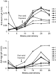
Fig. 2. Worm and egg-specific IgA responses in groups of pigs pre-natally, pre-natally+post-natally or post-natally exposed to Schistosoma japonicum. Groups 1, 2, 3, 4 and 5 were pre-natally exposed and groups 1, 3, 6 and 7 were post-natally exposed 9 weeks post-delivery. Groups 1, 2 and 7 received colostrum from uninfected sows while groups 3, 4, 5 and 6 received colostrum from infected sows. For full group description see Table 1.
 You have
Access
- 4
- Cited by