





1. Introduction
The developing brain seems to have a remarkable ability to adapt to environmental changes. Many theoretical frameworks, such as the interactive specialization theory (Johnson, Reference Johnson2011) posit that there is a constant interaction between the environment and brain maturation. Bilingualism provides a unique opportunity to examine whether dual language exposure and use modify the functional and structural architecture of the brain.
It has been suggested that the sustained need to monitor languages, to shift between them, and to suppress one while speaking another would enhance cognitive control (Abutalebi & Green, Reference Abutalebi and Green2016; Blumenfeld & Marian, Reference Blumenfeld and Marian2011). Specifically, skills such as inhibition, shifting, selective attention, and updating (executive functions, Blom et al., Reference Blom, Boerma, Bosma, Cornips and Everaert2017; Yurtsever et al., Reference Yurtsever, Anderson and Grundy2023) are likely to be impacted by bilingualism. Yet this “bilingual effect” on executive functions (EFs) has not always been found in children, especially once socioeconomic factors are considered (Clayden et al., Reference Clayden, Hope, Argyri, Goksan, Stefani, Wei and Liegeois2023; Dick et al., Reference Dick, Garcia, Pruden, Thompson, Hawes, Sutherland, Riedel, Laird and Gonzalez2019; Naeem et al., Reference Naeem, Filippi, Periche-Tomas, Papageorgiou and Bright2018). Recent reviews have concluded that effects may be specific to certain tasks (such as inhibition, attention switching, monitoring; Giovannoli et al., Reference Giovannoli, Martella, Federico, Pirchio and Casagrande2020), show small effect sizes, and are prone to publication bias (see reviews in Gunnerud et al., Reference Gunnerud, Ten Braak, Reikerås, Donolato and Melby-Lervåg2020; Paap et al., Reference Paap, Johnson and Sawi2015; Ware et al., Reference Ware, Kirkovski and Lum2020). A more critical question is to what extent domain-general networks are sensitive to bilingual use and exposure. Yet whether bilingualism impacts the connectivity of brain networks involved in EFs is still poorly understood, especially in childhood, when the brain is particularly amenable to adaptation.
1.1. Bilingualism effects on the adult brain
The neural signature of bilingualism in children remains elusive, with most research examining effects on brain connectivity in adulthood. Functional MRI studies examining EF networks in adults indicate bilingual effects both during tasks and at rest (review in Pliatsikas & Luk, Reference Pliatsikas and Luk2016). At rest, enhanced functional connectivity in lifelong bilinguals has been reported in widespread language-related networks that include the inferior frontal gyrus (fronto-parietal, fronto-frontal, and interhemispheric networks (Berken et al., Reference Berken, Chai, Chen, Gracco and Klein2016; X. Fan et al., Reference Fan, Liao, Lei, Zhao, Xia, Men, Wang, Hu, Liu, Qin, Tan, Gao, Dong, Tao and He2021; see Luk et al., Reference Luk, Green, Abutalebi and Grady2011 for a review) and in temporo-parietal networks (Marin-Marin et al., Reference Marin-Marin, Palomar-García, Miró-Padilla, Adrián-Ventura, Aguirre, Villar-Rodríguez and Costumero2021). A recent resting-state fMRI study showed that adult bilinguals had increased connectivity compared to monolinguals, particularly evident between the cerebellum and EF-related networks such as attentional and default-mode networks (DMN). Global efficiency was also higher in early and simultaneous bilinguals (Gracia-Tabuenca et al., Reference Gracia-Tabuenca, Barbeau, Kousaie, Chen, Chai and Klein2024). Another study focusing on EF networks at rest showed that bilingual adults had stronger functional connectivity than monolinguals in a subnetwork within the cingulo-opercular network (Cai et al., Reference Cai, Xu, Fan, Ma, Fan, Wang, Wu, Pan, Yin and Li2021). Enhanced functional connectivity has also been reported as a result of lifelong bilingualism in the DMN (Grady et al., Reference Grady, Luk, Craik and Bialystok2015; Stevens et al., Reference Stevens, Khan, Anderson, Grady and Bialystok2023), an intrinsic domain-general network. Regarding structural connectivity, bilingual effects have been observed in white matter integrity as measured by increased fractional anisotropy (FA), for example in the inferior longitudinal fasciculus (ILF) and corpus callosum in adulthood (Luk et al., Reference Luk, Bialystok, Craik and Grady2011; Pliatsikas et al., Reference Pliatsikas, Moschopoulou and Saddy2015). Findings have, however, been mixed regarding both the direction and anatomical location of differences between bilinguals and monolinguals (Tao et al., Reference Tao, Wang, Zhu and Cai2021). As EFs and their underlying networks follow a protracted development into adulthood (Crone & Steinbeis, Reference Crone and Steinbeis2017), it is unclear whether the effects of bilingualism on domain-general control networks can already be observed in childhood.
1.2. Bilingualism as a continuum
It is now widely accepted that examining the bilingual experience as a continuum is critical when investigating neural adaptations (DeLuca et al., Reference DeLuca, Rothman, Bialystok and Pliatsikas2019; Kremin & Byers-Heinlein, Reference Kremin and Byers-Heinlein2021; Li et al., Reference Li, Legault and Litcofsky2014). Indeed, bilingual individuals can vary widely in their language experiences, such as patterns of exposure to and use of their languages in different types of settings. Duration and extent of bilingual language use, second language (L2) age of acquisition, active use of L2, and quality of input in both languages are some of the language experience factors reported as critical brain modifiers (DeLuca et al., Reference DeLuca, Rothman, Bialystok and Pliatsikas2019). For instance, in adult studies, increased bilingual proficiency has been linked to thinner cortex in EF-related regions (anterior prefrontal and cingulate cortices, Claussenius-Kalman et al., Reference Claussenius-Kalman, Vaughn, Archila-Suerte and Hernandez2021) and to density changes in inferior frontal language regions (Stein et al., Reference Stein, Federspiel, Koenig, Wirth, Strik, Wiest, Brandeis and Dierks2012). L2 age of acquisition predicts white matter structure in thalamocortical radiations and in several language-related tracts, such as the inferior longitudinal fasciculus (ILF), inferior fronto-occipital fasciculus (IFOF), uncinate fasciculus (UF; Rossi et al., Reference Rossi, Cheng, Kroll, Diaz and Newman2017) and left arcuate fasciculus (AF; Hämäläinen et al., Reference Hämäläinen, Sairanen, Leminen and Lehtonen2017). Similarly, grey matter effects of L2 age of acquisition are widespread and depend on metrics (Claussenius-Kalman et al., Reference Claussenius-Kalman, Vaughn, Archila-Suerte and Hernandez2020). In addition to structural changes, L2 proficiency and L2 age of acquisition modulate brain functional connectivity at rest in both language and EF-related networks (Berken et al., Reference Berken, Chai, Chen, Gracco and Klein2016; Dash et al., Reference Dash, Joanette and Ansaldo2022; Sulpizio et al., Reference Sulpizio, Del Maschio, Del Mauro, Fedeli and Abutalebi2020). For example, a resting-state fMRI study showed that earlier L2 age of acquisition and greater diversity of language use correlated with higher interhemispheric frontal functional connectivity and greater cingulate-putamen connectivity, respectively (Gullifer et al., Reference Gullifer, Chai, Whitford, Pivneva, Baum, Klein and Titone2018). Altogether, the neuroimaging literature indicates that both quality and quantity of bilingual language use/exposure impact a range of brain structures and networks in adulthood, but findings may differ between studies (Li et al., Reference Li, Legault and Litcofsky2014; see Tao et al., Reference Tao, Wang, Zhu and Cai2021 for a review). Given the dynamic interaction between environmental factors and brain maturation during development (Johnson, Reference Johnson2011), there is a need to characterize bilingualism effects on the brain before adulthood.
1.3. Bilingualism effects on the child’s brain
Few studies have examined bilingual effects on the developing brain (see review in Tao et al., Reference Tao, Wang, Zhu and Cai2021). A large-scale whole-brain study (over 600 individuals aged 3 to 21 years; Pliatsikas et al., Reference Pliatsikas, Meteyard, Veríssimo, DeLuca, Shattuck and Ullman2020) reported that cortical thinning trajectories differ between monolinguals and bilinguals across 12 cortical regions. Bilingual individuals demonstrated less grey matter loss, especially in frontal and parietal regions, from late childhood onwards. Importantly, as seen in adults, paediatric studies highlight that experience-related factors modulate many brain effects. For instance, how “balanced” (proficient in both languages) children are impacts thickness in left temporal and frontal regions as well as putamen size (Archila-Suerte et al., Reference Archila-Suerte, Woods, Chiarello and Hernandez2018). An analysis of the Adolescent Brain Cognitive Development (ABCD) study cohort (Vaughn et al., Reference Vaughn, Nguyen, Ronderos and Hernandez2021) identified cortical thickness differences between bilingual and monolingual children aged 9–10 years. Within the bilingual group, higher English use correlated with thicker frontal and parietal cortices, whereas greater English vocabulary was associated with greater frontal and temporal cortical thickness (Vaughn et al., Reference Vaughn, Nguyen, Ronderos and Hernandez2021). Regarding white matter, one study (Mohades et al., Reference Mohades, Struys, Van Schuerbeek, Mondt, Van De Craen and Luypaert2012) revealed that bilingual children exposed to two languages from birth had higher FA in the left IFOF than later bilinguals, but lower FA in anterior callosal tracts relative to monolinguals. The aforementioned large-scale study (Pliatsikas et al., Reference Pliatsikas, Meteyard, Veríssimo, DeLuca, Shattuck and Ullman2020) found that only the fronto-striatal tracts followed different maturational trajectories in monolinguals and bilinguals, out of 20 tracts examined. Paediatric studies of functional connectivity are scarcer. One study in young children (aged 3–5 years) revealed that children growing up in multilingual environments show a combination of increased fronto-parietal and decreased fronto-temporal connectivity relative to a monolingual group (Thieba et al., Reference Thieba, Long, Dewey and Lebel2019). A recent study of the ABCD cohort (n = 335) showed that occipital-prefrontal functional connectivity at rest was significantly higher in 9 to 10 year-old multilinguals than their monolingual peers (Kwon et al., Reference Kwon, Yoo, Nguyen, Jeong and Chun2021). Altogether, the brains of monolingual and bilingual children appear to undergo different growth trajectories, yet little is known about how experience factors affect brain structural and intrinsic functional connectivity in language- and EF-related networks.
1.4. The present study
Here we report results from a pre-registered study (Goksan et al., Reference Goksan, Argyri, Clayden, Liegeois and Wei2020) in school-aged children that combines structural and resting-state MRI with extensive linguistic information on bilingual children’s experience in acquiring English and Greek. Resting-state connectivity allows us to examine intrinsic adaptations in the efficiency of brain networks that may not be detected using structural imaging. Our main research question is: How do language-related experience factors modulate brain connectivity in bilingual children?
Our registered protocol also stated that we aimed to examine whether structural connectivity and resting-state functional connectivity differ between bilinguals from birth (“simultaneous”), later bilinguals (“sequential”), and monolinguals. Given recent proposals in the field to move beyond categorical distinctions such as “early” versus “late” bilinguals, and to conceptualise bilingualism as a continuous variable (e.g., Chung-Fat-Yim et al., Reference Chung-Fat-Yim, Sorge and Bialystok2020), these group comparisons are presented as secondary (see Supplementary Introduction for justification).
Here we aimed to characterise how age at systematic exposure to the L2, language use, and bilingual language proficiency modulate brain connectivity. We examined language networks (AF and IFOF microstructure; fronto-temporal functional connectivity) as well as EF-related domain general networks (SLF microstructure; default mode network, DMN; salience network, SLN; fronto-parietal control network, FCN; striatal functional connectivity). We did not predict bilingualism to modulate corticospinal tract microstructure or motor and visual thalamo-cortical functional connectivity, and therefore these are referred to as “control” visual and motor networks.
Children in our sample attended English-speaking schools in an English-speaking country. We therefore hypothesised that increased proficiency and usage of Greek, their heritage language, would be associated with enhanced connectivity in attention (SLN) and language (fronto-temporal) networks as a reflection of increased attentional control (Menon & Uddin, Reference Menon and Uddin2010) and linguistic demands, respectively.
2. Method
2.1. Rationale for participant recruitment
Studies have suggested an impact of linguistic distance on bilingual cognitive performance (Gallo et al., Reference Gallo, Myachykov, Nelyubina, Shtyrov, Kubiak, Terekhina and Abutalebi2023; Morrison & Taler, Reference Morrison and Taler2023) and language outcomes in bilinguals (Bialystok et al., Reference Bialystok, Luk and Kwan2005; Blom et al., Reference Blom, Boerma, Bosma, Cornips, van den Heuij and Timmermeister2020). To reduce potential variability due to languages within the bilingual group, we examined this question using a single language pair, namely English and Greek. Bilingual participants were in full-time mainstream education in the UK (English as societal/school language or L2). They were exposed to Greek from birth and had had systematic exposure to English at different ages. Data on race/ethnicity was not collected.
2.2. Participants
Typically developing English-speaking monolingual and English–Greek bilingual children aged 8–10 years were recruited. The registered protocol divided the bilingual group into simultaneous and sequential groups, and therefore we followed the original inclusion criteria, namely: (i) monolingual participants had to be exclusively exposed to English at home since birth, (ii) simultaneous bilinguals had to be exposed to Greek and English from birth, and (iii) sequential bilinguals had to be exposed to Greek from birth and to English between 3 and 5 years old, i.e., they should have been born in Greece and arrived in the UK between 3 and 5 years of age. Due to difficulties recruiting children in the sequential bilingual group, we had to widen our inclusion criteria and thus recruited two additional children who arrived in the UK between one and two years of age.
Children were not included in the study if (i) they had early systematic exposure to languages other than English and Greek, (ii) their Greek-speaking parents were born and/or had lived in the UK for most of their lives, (iii) they were not attending mainstream schools, (iv) they had any MRI contraindications (e.g., metal implants or braces). Additionally, we excluded participants if they had conditions that may have affected language development, such as (i) a history of hearing impairment (ii) a diagnosis of language learning difficulties, developmental conditions (e.g., ADHD, autism, dyslexia), neurological conditions (e.g., epilepsy, cerebral palsy), severe chronic medical conditions, or were born extremely premature (earlier than 33 weeks gestational age), or (iii) had non-verbal intelligence below 70.
Seventeen monolinguals and 36 bilinguals aged 8–10 years were recruited via local English–Greek bilingual networks, local advertising, and Greek complementary schools. All participants were given a thank-you voucher.
The authors assert that all procedures contributing to this work comply with the ethical standards of the relevant national and institutional committees on human experimentation and with the Declaration of Helsinki of 1975, as revised in 2008. Children gave informed assent and parents/guardians informed consent. The research project was approved by the UCL Institute of Education Research Ethics Committee (approval number: 1080).
Standardised tests were used to measure the participants’ linguistic and non-linguistic abilities and were administered in person, either in a research room or at the participants’ homes. The bilingual children’s parents completed a detailed language background questionnaire. Moreover, monolingual and bilingual children were invited to an MRI brain scanning session where structural (T1- and diffusion-weighted) and functional (resting-state) images were acquired.
2.3. Background variables
2.3.1. Fluid intelligence and working memory
We measured non-verbal fluid intelligence and verbal working memory as background variables (see protocol in Goksan et al., Reference Goksan, Argyri, Clayden, Liegeois and Wei2020 for justification). Non-verbal ability was assessed using the Raven’s Progressive Matrices (CPM) test (Raven, Reference Raven2008) while verbal working memory (“number repetition”) was assessed using a digit span task (forward and backward repetition of a series of numbers (Semel et al., Reference Semel, Wiig and Secord2006)). Standard scores range from 1 to 19 (mean = 10, standard deviation = 3).
2.3.2. Linguistic proficiency: Expressive vocabulary
Verbal ability in both English and Greek was tested using the Raven’s Crichton Vocabulary Scales (CVS) test (Raven, Reference Raven2008). The CVS is a standardised expressive vocabulary test which is developed for use alongside the aforementioned non-verbal ability CPM test. Children are asked to describe the meaning of words in a list (Maximum score is 80, i.e., the total number of words). Bilinguals were tested on English and Greek vocabulary, whereas monolinguals were assessed only in English.
2.3.3. Executive function (EF)
EF skills were also tested focusing on the selective, sustained and switching aspects of attention. Four subtests from TEA-Ch 2 (Manly et al., Reference Manly, Anderson, Crawford, George and Robertson2016), a standardised computer-based test, were used. Attention switching was measured using a subtest (“Red and blues, bags and shoes” or RBBS) that involves sorting four objects according to two predetermined categories (bags and shoes are sorted for either their colour or whether they pair with a hand or feet). Selective attention was measured using a subtest that involves target detection amongst distractors (“Hecuba visual search”, HVS). Sustained attention was measured by two tasks. In the first task (“Cerberus”), children are required to detect target sounds (dog barks) while ignoring distractors (other animal noises). The final score is weighted for accuracy on all trials. In the second task, children’s “simple” reaction time (SRT) was measured during a 5-minute subtest where they needed to press a button in response to the appearance of a blue blob on the screen.
It should be noted that the attention-switching task may invoke inhibitory control in the form of inhibition of a prepotent response. Specifically, participants need to ignore goal-irrelevant stimuli (e.g., whether the object is red or blue) to produce a goal-relevant response (i.e., whether the object pairs with a hand or feet). The mean reaction time is calculated from the average response to the ‘switch trials’ only, that is, the first response where the rule has switched from either a red/blue decision to a hand/feet decision or vice versa.
The TEA-Ch 2 assessment produces standard scores (either population-based or education-referenced) for each subtest (mean = 10, SD = 3).
2.4. Bilingual experience measures
Bilingual children’s parents were asked to complete a detailed questionnaire, i.e., the Alberta Language Environment Questionnaire (ALEQ Heritage) adapted by Daskalaki and colleagues (Daskalaki et al., Reference Daskalaki, Chondrogianni, Blom, Argyri and Paradis2019). The questionnaire collects information on family demographics, the child’s length and pattern of exposure to English and Greek since birth, home language use, as well as bilingual exposure and use in contexts outside the home, extracurricular activities, literacy practices, time of arrival to the UK, and parents’ education background. Socioeconomic status was calculated based on years of maternal education. The original ALEQ (Paradis, Reference Paradis2011) measures current English language use, whereas the adapted version used in the present study (ALEQ Heritage) measures current Greek language use. The 5-point Likert rating scale for all relevant items reflects this adaptation: 0-English always/Greek never, 1-English usually/Greek seldom, 2-English 50%/Greek 50%, 3-English seldom/Greek usually, 4-English never/Greek always. We focus on the following outcome measures:
2.4.1. Language use at home
Language use at home is calculated by averaging: (i) Greek/English input and (ii) Greek/English output, ranging from 0 to 1. This score is a measure of the amount of English/Greek input and output the child is exposed to and directs to other interlocutors. Higher language use scores (>0.5) indicate a higher relative Greek language use at home, whereas lower scores (<0.5) indicate a higher relative English language.
2.4.2. Richness score
English and Greek richness scores are a measure of English/Greek language use during various activities (e.g., English/Greek language use with friends, literacy activities, music lessons, sports) inside and outside the home, ranging from 0 to 1. The calculation of richness scores in the adapted version of the questionnaire is different from the original ALEQ because it includes more questions (e.g., more activities have been added to capture more settings in which bilingual children socialise). Our protocol (Goksan et al., Reference Goksan, Argyri, Clayden, Liegeois and Wei2020) provides a more detailed explanation of the key differences between the two versions of ALEQ.
2.4.3. Combined Greek score
A combined score was calculated and used to indicate Greek language use (range 0–2). The calculation involves adding together children’s scores for language use at home and Greek richness. The combined score will more accurately reflect children’s amount of input and Greek language use in various settings inside and outside the home.
2.4.4. Age at systematic exposure to L2
For the purposes of this study, age at systematic exposure to L2 is defined as the age at which children started having consistent and significant interaction with English speakers (all bilingual participants were exposed to Greek from birth). For example, a simultaneous English–Greek bilingual child looked after by an English-speaking caregiver from the age of 6 months, or starting nursery at age 12 months, would be considered to have systematic exposure at ages 6 and 12 months, respectively. ALEQ includes a question on the age bilingual children started receiving consistent and significant exposure to English (L2), such as entering an English-language nursery and so forth We therefore included this information in our statistical analysis as this measure captures more accurately the onset of systematic exposure to English as an L2.
2.5. MRI data collection and analysis
We collected (i) high resolution T1-weighted structural datasets (TR = 2,300 ms, echo time (TE) = 2.74 ms, flip angle = 8°, 1 mm isotropic voxels, field of view = 256 × 256 mm), (ii) diffusion-weighted datasets, whose parameters are detailed below, and (iii) resting-state functional MRI (T2* BOLD-weighted, flip angle = 75°, TE = 26 ms, TR = 1,240 ms, field of view = 200 × 200 mm, 2.5 mm isotropic voxels, multi-band accelerator factor 2) on a 3 T Magnetom Prisma scanner (Siemens), using a 32-channel head coil.
3D diffusion-weighted MRI scans were collected using a single-shot multi-shell diffusion MRI sequence. The images are encoded along 60 independent directions with b-values of 1,000 and 2,200 s/mm2 (TR = 3,050 ms, TE = 60 ms, FA = 90°, TA = 7 min 16 s, FOV = 220 × 220 mm, matrix size = 110 × 110, voxel size = 2 × 2 × 2 mm, number of contiguous axial slices = 66, slice thickness = 2 mm, distance factor = 10%, slice acquisition = interleaved, PAT = GRAPPA, multi-band acceleration factor = 2, fat suppression = on). We also acquired 13 b0 images and one reverse phase-encoded b0.
Children watched a movie of their choice during the acquisition of structural data (around 15 min), and were asked to stare at a fixation cross on a screen for the duration of the resting-state fMRI acquisition period (around 6 minutes), with no specific instructions regarding blinking or swallowing. Detailed acquisition parameters and scanning preparation for children are reported in our published protocol (Goksan et al., Reference Goksan, Argyri, Clayden, Liegeois and Wei2020).
Each scanning session lasted approximately 45 min. This included the time taken to set up and settle each participant, followed by collection of the structural, diffusion and functional resting-state images (total acquisition time approximately 22 min).
2.5.1. MRI preprocessing
MRI data were analysed using specific pre-processing steps described in our protocol paper and at https://github.com/sgoksan/paed_mri_preprocessing, following quality control.
Briefly, T1-weighted image preprocessing steps included brain extraction, correction of RF inhomogeneity, and segmentation into distinct brain tissue types. Diffusion-weighted data were corrected for susceptibility-induced distortions, eddy currents and head movements. For functional MRI data, steps included motion correction, distortion correction, brain extraction and grand mean scaling. A single-band reference was acquired at the start of the functional scan. It was used as a baseline for motion correction and to co-register fMRI images to the T1-weighted image. MELODIC (model-free fMRI analysis using probabilistic independent component analysis, ICA) was used to decompose functional data into spatially independent components. These components were then automatically labelled as either signal or noise (i.e., motion or physiological artefact) using ICA-AROMA (Automatic Removal of Motion Artifacts). After automatically removing ICA components that depict physiological noise or movement, we applied a high-pass temporal filter at 0.01 Hz (100 s period) following quality control. No global regression was performed. Motion in diffusion data was handled by FSL-EDDY. For fMRI data, we used FSL-MCFLIRT (via FEAT, with the bias-corrected SBref image used as a reference). Standard parameters for the FSL diffusion and functional pipelines were applied, and results were manually checked. A graphical representation of the MRI analysis pipeline is available in our protocol publication (Goksan et al., Reference Goksan, Argyri, Clayden, Liegeois and Wei2020).
In addition, total grey matter, intracranial volume and subcortical volume metrics were generated from T1-weighted structural images using FreeSurfer (version 7.2.0, Desikan et al., Reference Desikan, Ségonne, Fischl, Quinn, Dickerson, Blacker, Buckner, Dale, Maguire, Hyman, Albert and Killiany2006).
2.5.2. Structural connectivity
Anatomically constrained tractography was performed using MRtrix version 3.0.2 to generate all tracts of interest (Smith et al., Reference Smith, Tournier, Calamante and Connelly2012; Tournier et al., Reference Tournier, Smith, Raffelt, Tabbara, Dhollander, Pietsch, Christiaens, Jeurissen, Yeh and Connelly2019). Seed and target regions were drawn using each participant’s eigenvector maps, modulated by fractional anisotropy, and overlaid onto individual structural images. All tracts were seeded and generated separately per hemisphere, with a midline sagittal plane used as an exclusion region. The language tracts, AF (dorsal language pathway) and IFOF (ventral language pathway) were generated using one seed and two inclusion regions (adapted from Morgan et al., Reference Morgan, Su, Reilly, Conti-Ramsden, Connelly and Liégeois2018). The seed region for AF was inferior to the central sulcus, with one inclusion region within the arch of AF and a second, small inclusion region anterior to the seed, below the precentral sulcus. The seed region for IFOF was within the extreme/anterior floor of the external capsule. Anterior and posterior inclusion planes were drawn for IFOF, which took cross sections of frontal and posterior occipital fibres. The fronto-parietal tract (or superior longitudinal fasciculus, SLF) was generated using the same seed and anterior inclusion region as AF. Moreover, a second inclusion region, anterior to the seed, was specified, near the angular gyrus and anterior to the arch of the AF tract. Finally, the SLF required an additional exclusion plane which encompassed a cross-section of the arch of the AF and stopped fibres entering the CST via the internal capsule. Lastly, for the CST, a seed was placed close to the hand nodule in the precentral gyrus, and voxels within the pons, surrounded by transverse pontine fibres and the middle cerebellar peduncle, were selected to create a ventral inclusion and stop region (Liégeois et al., Reference Liégeois, Tournier, Pigdon, Connelly and Morgan2013). The tractography reconstruction algorithm was instructed to generate streamlines until 3,000 were selected per tract. Subsequently, a threshold of 10% was applied to remove voxels in which fewer than 30 streamlines had been selected. (Liégeois et al., Reference Liégeois, Tournier, Pigdon, Connelly and Morgan2013). We used the default probabilistic tractography algorithm in MRtrix3, iFOD2, whereby path segments are sampled from fibre orientation distributions. We kept the default parameters (45 degrees curvature limit, step width of half a voxel). In MRtrix3, streamlines are generated repeatedly until the threshold of 3,000 streamlines is reached, after discarding any that do not meet anatomical constraints.
2.5.3. Functional connectivity
Resting-state functional data were analysed as per our published protocol (Goksan et al., Reference Goksan, Argyri, Clayden, Liegeois and Wei2020). Briefly, regions of interest for language networks were the inferior frontal gyrus and posterior superior temporal gyrus. For EF-related networks, we examined the frontoparietal control network (FCN: middle frontal gyrus and inferior/superior parietal lobule), the salience network (SLN: anterior insula and anterior cingulate gyrus), the default mode network (DMN: posterior cingulate gyrus, ventromedial prefrontal cortex, and inferior parietal lobule/angular gyrus), and subcortical control region networks (putamen and caudate). To mirror our structural connectivity analysis, we extracted intra-hemispheric networks separately in the left and right hemispheres.
Regions of interest for control networks were the primary visual cortex and lateral geniculate nucleus in the thalamus for the visual network, and the bilateral precentral gyrus (primary motor cortex region) and ventral lateral thalamic nucleus for the motor network (Power et al., Reference Power, Cohen, Nelson, Wig, Barnes, Church, Vogel, Laumann, Miezin, Schlaggar and Petersen2011).
The cortical regions of interest (ROIs) were derived from the FreeSurfer (version 7.2.0) atlas.
2.6. Statistical analysis
2.6.1. Descriptive data and background variables
For background variables, we report on the three groups separately as they were recruited with distinct criteria. We compared groups using one-way ANOVAs or non-parametric equivalents for continuous variables, and chi-square for categorical variables. Where ANOVAs showed a significant difference by group, post-hoc analyses were conducted by calculating the estimated marginal means for each group using the “emmeans” package for R (DOI 10.32614/CRAN.package.emmeans), and then obtaining p-values for pairwise contrasts between those marginal means with Tukey correction for multiple comparisons.
We included fluid intelligence and number repetition scores as confounding factors in the connectivity analysis and when comparing attention scores between groups (see protocol in Goksan et al., Reference Goksan, Argyri, Clayden, Liegeois and Wei2020, for justification).
2.6.2. Connectivity metrics
For the white matter analysis, outcome variables were weighted FA values from tracts of interest. Weighted mean FA was calculated by weighting the FA value per voxel by the number of streamlines from the tract of interest passing through that voxel, which generated the mean FA measure per tract. For the functional connectivity analysis, the outcome variable was the mean absolute edge weight within each network, where each edge was weighted by the Pearson correlation between the relevant pair of cortical time series.
Finally, we also carried out unplanned analyses on subcortical volumes (caudate and putamen) extracted using FreeSurfer (Desikan et al., Reference Desikan, Ségonne, Fischl, Quinn, Dickerson, Blacker, Buckner, Dale, Maguire, Hyman, Albert and Killiany2006), given recent evidence of effects of bilingualism experience on striatal metabolism (Pliatsikas et al., Reference Pliatsikas, Pereira Soares, Voits, Deluca and Rothman2021).
We examined effects in the two hemispheres separately. We also subsequently averaged left and right hemisphere metrics to examine bilateral effects, as we had no a priori hypothesis about lateralised effects of bilingualism on control networks in our protocol. All brain metrics were transformed by regressing out nuisance variables, namely gender, age, maternal education, total intracranial volume, fluid intelligence and number repetition standard scores.
When we compared metrics across the three groups, we used ANOVAs and ran post-hoc tests where main group effects were detected.
2.6.3. Associations between connectivity metrics and bilingual variables
We examined the effects of bilingual experience on brain metrics within the bilingual group (combined sequential and simultaneous bilingual participants) using multiple linear regressions, with the following predictors: age at systematic exposure to English, Greek language use, Greek vocabulary score, English vocabulary score, Greek Richness score.
All analyses were conducted with an alpha significance level of 0.05 (two-tailed). We report all significant findings without correcting for multiple comparisons across networks to ensure that all results from the analyses planned in our protocol are reported.
3. Results
3.1. Sample characteristics
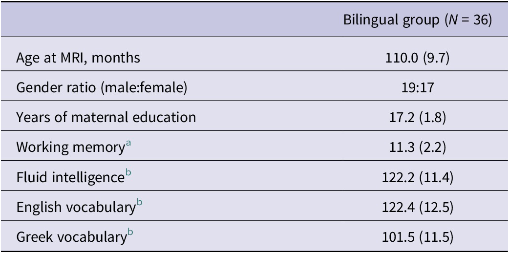
Most bilingual children had mothers who held a university degree and had intelligence scores one standard deviation above the mean (Table 1). Demographic characteristics for the three groups can be found in Supplementary Material (Tables S1 and S2).
Demographic and cognitive characteristics of the bilingual group

Table 1. Long description
The table consists of two columns. The first column lists the characteristics, and the second column provides the data for the Bilingual group (N = 36). Continuous variables are presented as mean with standard deviation in parentheses.
* Age at M R I, months: 110.0 (9.7)
* Gender ratio (male:female): 19:17
* Years of maternal education: 17.2 (1.8)
* Working memory: 11.3 (2.2). This is a standard score with a normative mean of 10 and standard deviation of 3.
* Fluid intelligence: 122.2 (11.4). This is a standard score with a normative mean of 100 and standard deviation of 15.
* English vocabulary: 122.4 (12.5). This is a standard score with a normative mean of 100 and standard deviation of 15.
* Greek vocabulary: 101.5 (11.5). This is a standard score with a normative mean of 100 and standard deviation of 15.
Note: Continuous variables are expressed as mean (standard deviation) per group.
a Standard score with normative mean = 10 (SD = 3)
b Standard score with normative M = 100 (SD = 15).
After preprocessing, all MRI scans were of usable quality, with no major motion or other artefacts that could not be corrected.
Greek input and use at home were higher for the sequential than the simultaneous bilingual group, but these two groups did not differ on English or Greek richness scores (Supplementary Table S3).
MRI analyses revealed that there were no group differences in global grey matter (F 2,50 = 2.23, p = 0.118) or intracranial (F 2,50 = 0.62, p = 0.545) volumes.
3.2. Bilingual experience: associations with brain connectivity
In the combined bilingual group, the effects of bilingualism variables were observed in domain-general (cognitive control) but not in language networks.
3.2.1. EF-related (cognitive control) networks
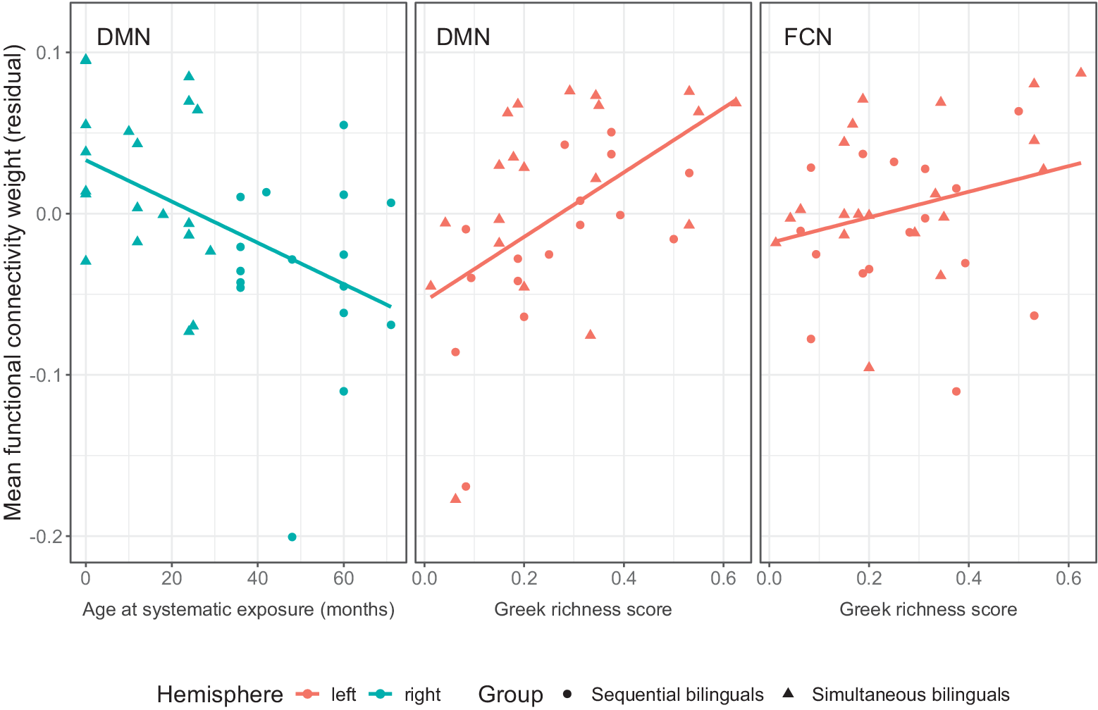
Functional connectivity: Increased DMN connectivity was associated with higher Greek richness scores (t 30 = 2.78, p = 0.0092). No other associations between connectivity and age of second language onset, usage, language ability, or age at systematic exposure to English were observed.
When examining hemisphere-specific effects, increased left hemisphere DMN connectivity was associated with higher Greek richness scores (t 30 = 3.46, p = 0.0016; Figure 1). In contrast, higher right DMN connectivity was associated with younger age at systematic exposure to English (t 30 = −2.23, p = 0.033, Figure 1). No other significant association with language usage or language ability was observed for the left or right DMN.
Relationships between age at systematic exposure (in months), Greek richness scores and executive control network functional connectivity in bilingual children, after correcting for nuisance variables. Note: Simultaneous (triangles) and sequential (circles) bilinguals were combined in this analysis. DMNQ = Default Mode Network, FCNQ = Fronto-Parietal Control Network. Left hemisphere, red; right hemisphere, blue.

Figure 1. Long description
A three-panel scatter plot with a shared Y axis labeled Mean functional connectivity weight (residual) ranging from minus 0.2 to 0.1.
Panel 1 (Left): Titled D M N. The X axis is Age at systematic exposure (months) from 0 to 60. Data points are teal, representing the right hemisphere. A linear regression line shows a negative correlation. Markers include triangles for simultaneous bilinguals and circles for sequential bilinguals, concentrated between 0 and 40 months.
Panel 2 (Center): Titled D M N. The X axis is Greek richness score from 0.0 to 0.6. Data points are salmon-red, representing the left hemisphere. A linear regression line shows a positive correlation. Markers are scattered across the score range with a higher density of triangles above the line.
Panel 3 (Right): Titled F C N. The X axis is Greek richness score from 0.0 to 0.6. Data points are salmon-red for the left hemisphere. A linear regression line shows a slight positive correlation, though more horizontal than the center panel. Markers are widely dispersed.
A legend at the bottom indicates Hemisphere: left is salmon-red, right is teal. Group: circles are sequential bilinguals, triangles are simultaneous bilinguals.
Increased left hemisphere FCN connectivity was associated with increased Greek richness score (t 30 = 2.14, p = 0.040).
Structural connectivity: Earlier age at systematic exposure to English was associated with higher FA in the left SLF (t 30 = −2.08, p = 0.047).
Subcortical volumes: Higher Greek richness scores were also associated with smaller right putamen volumes (t 30 = −2.24, p = 0.033).
3.2.2. Language-related networks
No significant association was detected between language network metrics and bilingual experience.
3.2.3. Motor and visual networks
There were no significant relationships with bilingual experience-related factors regarding functional connectivity in the motor or visual networks, or in the weighted FA of the corticospinal tracts.
3.3. Group differences in connectivity
We report results from the comparison of the three recruited groups. Of note, an exploratory analysis revealed no group difference between the monolingual and bilingual (combined) groups in any network examined.
3.3.1. EF-related (cognitive control) networks
Functional connectivity: Mean adjusted functional connectivity weight (i.e., average edgewise functional connectivity weight after correcting for nuisance variables) differed between groups in the default mode network (DMN) only, when left and right metrics were averaged (F 2,49 = 3.86, p = 0.028; F 2,49 = 1.61, p = 0.21 for the salience network; F 2,49 = 1.27, p = 0.29 for the FCN network; F 2,49 = 0.77, p = 0.47 for the subcortical control network). The greatest difference in DMN connectivity was observed between the simultaneous and sequential bilingual groups (t 49 = −2.69, p = 0.026 after correcting for multiple comparisons).
When examining hemisphere-specific effects, group differences were detected in the right default mode network only (F 2,49 = 4.90, p = 0.012; Supplementary Figure S2). Again, the greatest difference was observed between the simultaneous and sequential bilingual groups (t 49 = −2.95, p = 0.013 after correcting for multiple comparisons), with the difference between monolinguals and sequential bilinguals also significant (t 49 = 2.44, p = 0.048).
Negative functional weights were vanishingly rare in the data, with only one negative value in one subject across the networks of interest, so their impact on the results was negligible.
Structural connectivity: After correcting for sex, age at scan, intracranial volume, fluid intelligence and working memory scores, the three groups did not differ with respect to mean FA in the fronto-parietal tracts.
Language-related networks: There was no difference between the groups with respect to functional connectivity of the language network (F 2,49 = 1.06, p = 0.36). Similarly, neither the arcuate fasciculi nor the inferior fronto-occipital fasciculi differed between the groups with respect to weighted mean FA, after correcting for the covariates.
3.3.2. Subcortical volumes
There were no group differences in caudate or putamen volumes.
3.4. Results overview
In summary (Figure 2), the strongest bilingualism effects in our study were seen in the default mode network (DMN), with additional adaptations in other brain networks (fronto-parietal, subcortical). Greek richness score was the experience-related variable that showed the most associations with connectivity, impacting three networks (DMN, FCN, putamen volume).
Illustrative summary of findings: impact of bilingualism on network connectivity in middle childhood. Note: Positive correlations, +; negative correlations, −. Right hemisphere is on the right. Effects on the FCN and superior longitudinal fasciculus (SLF) were only detected in the left hemisphere. We did not detect any bilingualism effects on language networks, namely on the microstructure of the arcuate fasciculus (AF) and inferior fronto-occipital fasciculus (IFOF), or on the fronto-temporal network at rest. Created with Biorender.com.

Figure 2. Long description
The diagram is organized into two main vertical sections. On the left are three colored boxes representing bilingualism factors. On the right are five brain illustrations categorized into Language networks and Domain general networks.
* Language networks section at the top shows a brain with connections labeled A F and I F O F. No arrows point to this section.
* Domain general networks section below contains four brain illustrations.
* The first factor box, Younger age at systematic exposure to L 2, has two blue arrows. One points to the S L F brain with a green plus symbol. The second points to the Default Mode D M N brain with a green plus symbol.
* The second factor box, Birth versus childhood onset bilingualism, has a green arrow pointing to the Default Mode D M N brain with a green plus symbol.
* The third factor box, Richness of bilingual environment, has three brown arrows. One points to the Default Mode D M N brain with a green plus symbol. The second points to the Frontoparietal F C N brain with a green plus symbol. The third points to a cross-section of the Putamen with a red minus symbol.
* A green plus symbol indicates a positive correlation and a red minus symbol indicates a negative correlation.
4. Discussion
The current study aimed to examine the role of age at systematic exposure to English as an L2, language use, and bilingual language proficiency in modulating brain connectivity. Our findings uncovered a complex picture relating to the effects of bilingual experience-related factors on brain connectivity.
4.1. Associations between bilingual experience and connectivity in the default mode network
Younger age at systematic exposure to English as the L2/societal language was associated with higher connectivity in the right DMN and left frontoparietal tracts. In contrast, effects on the right putamen, left FCN and left DMN were seen for higher Greek “richness score”, corresponding to the active use of the heritage language in a variety of settings in and outside the home. This complexity highlights the widespread impact of early bilingual experiences on brain networks that are involved in language use, learning, and monitoring.
The DMN is typically activated during internal cognitive processes such as mind wandering, autobiographical time travelling, and self-focus. In contrast, its activity reduces during engagement in externally focused and attention-demanding tasks (Menon, Reference Menon2023). Between childhood and adolescence, the DMN’s internal connectivity increases (Fan et al., Reference Fan, Liao, Lei, Zhao, Xia, Men, Wang, Hu, Liu, Qin, Tan, Gao, Dong, Tao and He2021; Sherman et al., Reference Sherman, Rudie, Pfeifer, Masten, McNealy and Dapretto2014), but few studies have examined how the environment impacts this network.
We observed lateralised associations concerning this network, whereby a richer bilingual environment was associated with greater connectivity in the left DMN, and younger age at systematic exposure to the societal language (English) was associated with greater right-sided connectivity. Further studies should explore whether these lateralised effects on the DMN are confirmed. No effect of proficiency (vocabulary) was detected on the DMN, however, suggesting crystallised knowledge is not reflected in this network. These results contrast with the association between vocabulary and frontal and temporal cortical thickness (Vaughn et al., Reference Vaughn, Nguyen, Ronderos and Hernandez2021). We also did not replicate the positive association between diverse language use (i.e., the Greek richness score) and functional connectivity in networks involving the anterior cingulate (such as the SLN) or putamen reported in proficient bilingual adults (Gullifer et al., Reference Gullifer, Chai, Whitford, Pivneva, Baum, Klein and Titone2018). Overall, our results suggest that early and active use of two languages in a wide variety of settings is a key contributor to DMN adaptations in childhood.
4.2. Associations between bilingual experience and connectivity in other domain-general networks
We additionally observed associations between the FCN and bilingual experience. The positive association between Greek richness and left FCN, alongside left DMN connectivity, suggests a strengthening effect of rich and varied bilingual use and exposure. This expands on evidence of stronger frontoparietal resting-state connectivity in younger children (aged 3–5 years) growing up in multilingual environments (Thieba et al., Reference Thieba, Long, Dewey and Lebel2019). As we also observed that earlier age at bilingual exposure is associated with stronger structural connectivity of the left SLF, our childhood findings confirm that both age at onset and quality of bilingual exposure impact left frontoparietal networks, possibly reflecting language control or executive control adaptations.
4.3. Associations between bilingual experience and connectivity in language networks
Paradoxically, our data revealed several experience-related effects on connectivity in childhood, but not on language networks. In addition, these effects were associated with bilingual experience, but not language use or vocabulary. Richness effects were found on the left DMN, left FCN, and right putamen, suggesting that EF development may be particularly sensitive to the quality and variety of bilingual experience. For subcortical structures, we however, did not replicate adult findings such as group differences in striatal volumes (Burgaleta et al., Reference Burgaleta, Sanjuán, Ventura-Campos, Sebastian-Galles and Ávila2016; Pliatsikas et al., Reference Pliatsikas, DeLuca, Moschopoulou and Saddy2017) or positive relationships between putamen and amount of bilingual experience (Korenar et al., Reference Korenar, Treffers-Daller and Pliatsikas2023). We therefore hypothesise that subcortical adaptations differ in children and adults, and may take years to become detectable. Overall, our “Greek richness score” findings highlight that diverse bilingual immersion and engagement are critical for both subcortical and cortical adaptations to occur in the childhood period.
4.4. Group differences in connectivity: bilingualism as a categorical variable
Our analysis of group differences revealed that after 8–10 years of bilingual exposure from birth, the brain has already undergone adaptations manifesting as increased default mode network (DMN) connectivity at rest relative to later bilinguals, but not to monolinguals (as seen in adults; see review in Tao et al., 2021). Exposure to two languages from birth therefore shapes brain functional connectivity at rest in a domain-general EF-related network, namely the default-mode network (DMN). We had hypothesised enhanced DMN resting-state connectivity in both simultaneous and sequential bilingual children relative to monolinguals, based on a study in older adults who had decades of bilingual experience (Grady et al., Reference Grady, Luk, Craik and Bialystok2015). Here we did not observe this effect in simultaneous bilinguals and observed reduced connectivity in late bilinguals. These results indicate that extensive cumulative experience spanning more than a decade may be necessary to enhance DMN connectivity.
We most consistently observed reduced connectivity in sequential bilinguals, relative to simultaneous bilinguals (bilateral effects) and monolinguals (right hemisphere effects). It is noteworthy that group effects in DMN connectivity were mainly due to differences between the sequential bilinguals and the other two groups. We observed no differences between monolinguals and simultaneous bilinguals. As these were unexpected findings, our interpretations can only be speculative at this stage (see Supplementary Discussion). We observed no group differences in classical language networks, either relating to structural (arcuate and inferior fronto-occipital fasciculus) or functional connectivity (Pliatsikas et al., Reference Pliatsikas, Moschopoulou and Saddy2015; Tao et al., Reference Tao, Wang, Zhu and Cai2021). The fact that we controlled for many nuisance variables and compared carefully matched groups could explain the lack of replication of previous studies.
4.5. Theoretical implications
Overall, our study results inform theoretical accounts of bilingual development. The dynamic restructuring model, DRM (Pliatsikas, Reference Pliatsikas2019), proposes that bilingualism impacts grey and white matter structure in three stages, and that adaptations depend on the quality and quantity of bilingual experience. We add that bilingual experience also impacts the brain’s intrinsic connectivity (Figure 2). The complex relationship we observed between bilingual factors and brain subnetworks is consistent with the general premise of the adaptive control hypothesis (Abutalebi & Green, Reference Abutalebi and Green2016; Abutalebi et al., Reference Abutalebi, Della Rosa, Ding, Weekes, Costa and Green2013; Green & Abutalebi, Reference Green and Abutalebi2013), whereby effects are observed in domain-general control networks. We refine the DRM model by specifying which factors impact which neural networks in childhood (Figure 2), while the brain is still undergoing maturational changes. Critically, we show evidence that age of onset and richness of bilingual experience have distinct effects on the brain’s connectivity, although the DRM does not distinguish between the two.
The lack of effects in the IFOF and language tracts is not in line with the DRM model, which predicts increases in these connections during the consolidation phase (Pliatsikas, Reference Pliatsikas2019). The lack of bilingualism effects on the language networks we examined could be because effect sizes were not detectable with our current sample size, or because they are not yet observable at this age. It should be noted, however, that without longitudinal data, we can neither conclude that a renormalisation of white matter microstructure has already taken place, nor that it has not occurred yet. In the DRM, it should be noted that white matter differences in language tracts are not expected during the “peak efficiency” stage, a stage which could have already been reached by our bilingual participants. Finally, it is possible that bilingual factors play a lesser role in language networks than demographic and cognitive background variables we controlled for in this analysis.
It is noteworthy that the brains of children who grow up in bilingual environments need to support grammatical, lexical and articulatory learning alongside timely control, inhibition and shifting during language processing and use. Our study reveals that default mode, fronto-parietal, and subcortical changes are critical subnetworks already showing evidence of adaptation in middle childhood. We speculate that strengthening of DMN connectivity, if maintained through a rich and diverse bilingual language experience, could be the brain basis of enhanced executive control in pre-schoolers (Poulin-Dubois et al., Reference Poulin-Dubois, Blaye, Coutya and Bialystok2011) and of increased cognitive reserve later in life (Stevens et al., Reference Stevens, Khan, Anderson, Grady and Bialystok2023).
4.6. Study limitations
One limitation of our study is that the cross-sectional design did not allow us to conclude on the development of these networks over time but instead allowed us to examine a narrow age range. Additionally, although we attempted to recruit participants from a range of demographic backgrounds, our participants tended to come from more educated families. As we know, socioeconomic status impacts brain development (Brito et al., Reference Brito and Noble2018), replication in more representative samples of the population is required. These children were also highly proficient in both languages, limiting the proficiency range examined. We examined bilingualism in one language pair (English and Greek), since research findings have indicated that linguistic distance may moderate cognitive effects of bilingualism (Radman et al., Reference Radman, Jost, Dorood, Mancini and Annoni2021). Examining English–Greek bilinguals only is, however, also a strength of our study, as it provided us with a homogeneous bilingual language experience. Finally, we restricted our analysis to a priori hypothesised networks based on our pre-registered protocol but cannot rule out that others were impacted by bilingualism experience. Overall, our total sample of 37 bilingual children is the largest to date in a study that combines in-depth bilingual language experience information with multimodal MRI connectivity analyses, although the limitations mentioned above may affect generalisability.
4.7. Conclusion
To conclude, our findings confirm that there is a developmental component to brain differences associated with bilingualism (Pliatsikas et al., Reference Pliatsikas, Meteyard, Veríssimo, DeLuca, Shattuck and Ullman2020). In our cross-sectional study in pre-adolescence, we found evidence of both age- and experience-dependent (DeLuca et al., Reference DeLuca, Rothman, Bialystok and Pliatsikas2019) effects on brain connectivity. We propose that biological constraints (e.g., brain maturity at onset of bilingual language exposure) and experience-based factors (e.g., richness of bilingual experience) dynamically interact during early development and into ageing to shape the structure and function of multiple networks, consistent with an interactive specialization theory (Johnson, Reference Johnson2011). The lack of bilingual effects on language networks contrasts with adult studies and may indicate that these changes take place over a more protracted time course into adulthood. In contrast to language networks, we found strong evidence that domain-general networks are sensitive to the richness and onset of exposure to a bilingual environment. In our study, we found no association between language proficiency as measured by vocabulary and connectivity in any network. The most robust neural signature of bilingualism experience was in the default mode network at rest, suggesting this could be the earliest brain adaptation to dual language exposure.
Supplementary material
The supplementary material for this article can be found at http://doi.org/10.1017/S1366728926101849.
Data availability statement
The data that support the findings will be available at https://openneuro.org/ following a 12-month embargo from the date of publication.
Acknowledgements
All research at Great Ormond Street Hospital NHS Foundation Trust and the UCL Great Ormond Street Institute of Child Health is made possible by the NIHR Great Ormond Street Hospital Biomedical Research Centre. We thank Tina Banks, research radiographer, as well as all participating families.
Funding statement
This research was funded by the Leverhulme Trust (Grant Ref: RPG-2017-403).
Competing interests
The author(s) declare none.


 Open access

