




No CrossRef data available.
Article contents
Molecular cloning and characterization of the copper/zinc and manganese superoxide dismutase genes from the human parasite Clonorchis sinensis
Published online by Cambridge University Press: 24 January 2005
Abstract
A copper/zinc superoxide dismutase (Cu/ZnSOD) gene and a manganese superoxide dismutase (MnSOD) gene of the human parasite Clonorchis sinensis have been cloned and their gene products functionally characterized. Genes Cu/ZnSOD and MnSOD encode proteins of 16 kDa and 25·4 kDa, respectively. The deduced amino acid sequences of the two genes contained highly conserved residues required for activity and secondary structure formation of Cu/ZnSOD and MnSOD, respectively, and show up to 73·7% and 75·4% identities with their counterparts in other animals. The genomic DNA sequence analysis of Cu/ZnSOD gene revealed this as an intronless gene. Inhibitor studies with purified recombinant Cu/ZnSOD and MnSOD, both of which were functionally expressed in Escherichia coli, confirmed that they are copper/zinc and manganese-containing SOD, respectively. Immunoblots showed that both C. sinensis Cu/ZnSOD and MnSOD should be antigenic for humans, and both, especially the C. sinensis MnSOD, exhibit extensive cross-reactions with sera of patients infected by other trematodes or cestodes. RT-PCR and SOD activity staining of parasite lysates indicate that there are no significant differences in mRNA level or SOD activity for both species of SOD, indicating cytosolic Cu/ZnSOD and MnSOD might play a comparatively important role in the C. sinensis antioxidant system.
Keywords
Information
- Type
- Research Article
- Information
- Copyright
- © 2005 Cambridge University Press
INTRODUCTION
Clonorchis sinensis, an oriental liver fluke, is the causative agent of clonorchiasis, which is an important endemic disease in Asian countries in which approximately 7 million people are infected (Crompton, 1999). This parasite causes typical consumption disease in the hepatobiliary system (Rim, 1986).
Highly reactive free oxygen radicals, including superoxide anion (O2.−), hydrogen peroxide (H2O2), hydroxyl radicals (OH.−) and singlet oxygen (1O2), are generated by numerous pathways in aerobic organisms (Fridovich, 1978). These reactive oxygen species might cause peroxidation of membrane lipids, DNA strand breakage, and inactivation of enzymes (Imlay & Linn, 1988). Such an oxidative stress burden is much higher against parasites because host immune effector cells that produced these reactive oxygen derivatives that were thought to contribute the killing of parasites by the host (Murray, 1981; Butterworth, 1984; Maizels, Bundy & Selkirk, 1993). Defence systems derived from enzymatic and non-enzymatic antioxidants in parasites might minimize the deleterious effects of the reactive oxygen radicals. Such a system is critical to enable parasites to survive. Several species of antioxidant enzymes, such as superoxide dismutase (SOD), glutathione peroxidase, glutathione S-transferase (GST), glutathione reductase, cytochrome c oxidase and catalase, have been identified in various parasites (Chung et al. 1991; Choi et al. 2000). SODs are a group of ubiquitous metalloenzymes that catalyse the dismutation of O2.− radicals to O2 and H2O2. In concert with catalase, SODs have strong anti-oxidant properties. Three genetically distinct forms of SOD occur that differ in their active site prosthetic metal ion and/or cellular localization in eukaryotic cells: cytosolic copper/zinc SOD (Cu/ZnSOD), a glycosylated extracellular copper/zinc SOD, and a mitochondrial manganese SOD (MnSOD). They are nuclear-encoded but are localized in different subcellular compartments. Cu/ZnSODs have been characterized in various helminth parasites, such as Schistosoma mansoni (Hong et al. 1992, 1993), Onchocerca volvulus (Henkle et al. 1991; James, McLean & Perler, 1994), Dirofilaria immitis (Callahan, Crouch & James, 1991), Brugia pahangi (Tang et al. 1994), Brugia malayi (Ou et al. 1995) and Fasciola hepatica (Kim et al. 2000), but no trematode MnSOD has been cloned or characterized to date. Under normal physiological conditions, mitochondria are the major source of O2.− production. Studies on nematodes have indicated that MnSOD also plays an important role in protecting cells from oxidative stress (Henkle-Duhrsen et al. 1995; Hunter, Bannister & Hunter, 1997). Manganese superoxide dismutase is uniformly distributed throughout the cytoplasm in prokaryotic cells, but for the highly partitioned eukaryotic cells, MnSOD is exclusively associated with mitochondria. The mitochondrial enzyme is encoded by a nuclear gene and must be transported across two mitochondrial membranes to the matrix. This process involves translation of a proenzyme, which in the case of the human MnSOD includes a 24-residue N-terminal leader targeting sequence, to the mitochondrion, where 90% of the cell's oxygen is consumed. The delivery of MnSOD to the mitochondrial matrix is essential for organelle function.
In an attempt to understand the structure and the functional mechanism of SODs in the process of self-protection of C. sinensis from attacks by the host defence system, we have isolated cytosolic Cu/ZnSOD cDNA and MnSOD cDNA, and characterized partially the biochemical properties of the bacterially expressed recombinant enzymes.
MATERIALS AND METHODS
Adult Clonorchis sinensis
The metacercariae of C. sinensis were obtained from naturally infected freshwater fish, Pseu dorasb ora parva, collected in an endemic area in Korea. Dogs were each orally fed with 500 metacercariae and adult worms were harvested from the livers of experimental dogs 4 months later. The worms were washed 10 times with physiological saline at 4 °C and stored at −70 °C until required. For the preparation of parasite extracts, frozen worms were thawed at room temperature and resuspended in 2 volumes of phosphate-buffered saline solution (PBS; pH7·2), the parasites were then sonicated on ice. The insoluble cellular debris was spun down at 20000 g at 4 °C for 20 min, and the supernatant was subjected to native PAGE for in-gel activity staining. Protein concentration was measured according to the method of Bradford (1976).
Isolation of RNA and cDNA synthesis
The adult worms (approximately 20 mg) were homogenized with a pellet pestle (Sigma) in the lysis buffer provided by a kit (Invitrogen, Carlsbad, CA). Isolation of total RNA was performed following the manufacturer's instructions. The first-strand cDNA synthesis and cDNA amplification by long-distance PCR were performed on total RNA using a cDNA Synthesis Kit (CLONTECH, Palo Alto, CA). The resulting double-strand cDNA library, approximately 0·5–3 kb in size, was divided into aliquots and stored at −70 °C until used.
Polymerase chain reaction (PCR)
Degenerate oligonucleotide primers (CZ-Df/CZ-Dr for Cu/ZnSOD and Mn-Df/Mn-Dr for MnSOD, Table 1) were designed based on two highly-conserved regions of Cu/ZnSOD or MnSOD cDNA sequences of other species (Hong et al. 1992; Henkle-Duhrsen et al. 1995; Kim et al. 2000). PCR reactions were performed on C. sinensis cDNA library. Thermal cycler profile included 35 cycles at 94 °C for 30 sec, a gradient annealing temperature from 40 to 60 °C for 30 sec and extension at 72 °C for 1 min. The amplifed products were analysed on 1% agarose gels. PCR bands with expected sizes were gel purified, directly subcloned into a pCR2.1 vector (Invitrogen), and sequenced. The nuclotide sequences of the cloned DNA fragments were determined from both directions by the dideoxynucleotide chain termination method using the ABI PRISM Dye Terminator Cycle Sequencing Ready Reaction kit (Perkin Elmer, Beaconsfield, UK), following the manufacturer's instructions, on an automated DNA sequencer (ABI, model 377). Sequence analysis was performed with BLAST and ENTREZ WEB services of the National Center for Biotechnology Information and the Prosite motif search facility of EXPASY (www.expay.ch/cgi-bin).

Rapid Amplification of cDNA Ends (RACE)
RACE reactions were performed using a RACE Kit (CLONTECH). The C. sinensis cDNA library produced above was used as a template. On the basis of a 260 bp fragment sequence for Cu/ZnSOD and a 147 bp fragment sequence for MnSOD, both obtained by PCR using corresponding degenerate primers, 2 pairs of gene-specific primers (CZ-GSP3/CZ-GSP5 for Cu/ZnSOD and Mn-GSP3/Mn-GSP5 for MnSOD, Table 1) were designed to match both 3′- and 5′-ends of Cu/ZnSOD cDNA or MnSOD cDNA. PCR amplifications of 3′- and 5′-end fragments were carried out according to the protocols provided by the manufacturer. The PCR products were cloned and sequenced as above.
Determination of genomic DNA sequence of C. sinensis Cu/ZnSOD gene
Genomic DNA was isolated from C. sinensis as previously described (Ismail et al. 1997). To determine the genomic DNA sequence of C. sinensis Cu/ZnSOD gene, pairs of specific primer with high annealing temperature were designed based on the neighbourhood sequences of start codon and stop codon respectively. One pair of primers, gCZSODf and gCZSODr (Table 1), produced a significant amplicon, which was then cloned, sequenced and analysed.
Phylogenetic analysis
The obtained sequences of C. sinensis Cu/ZnSOD or MnSOD were compared with corresponding sequences of several helminths deposited in GenBank. Multiple alignment analysis was performed with the DNAstar. Phylogenetic analyses on the aligned sequences were done using distance methods performed by the MEGA program version 2.1. A distance tree was constructed by use of the neighbour-joining (NJ) method. The stability of the trees obtained was estimated by bootstrap analysis for 1000 replications using the same program. For prediction of mitochondrial targeting sequence and its cleavage site, ExPASy system (http://psort.nibb.ac.jp/cgi-bin/runpsort.pl) was employed.
RT-PCR for analysis of cytosolic Cu/ZnSOD and MnSOD relative mRNA levels
RT-PCR was carried out using an Invitrogen RT-PCR kit according to the manufacturer's instructions except for the following details: 50 ng of Cu/ZnSOD or MnSOD specific primers (CZf/CZr and Mnf/Mnr, respectively, Table 1) were annealed to 250 ng of total RNA isolated from adult C. sinensis and extended at 55 °C for 30 min. The reactions were shifted to 95 °C and held for 2 min and then cycled at 95 °C for 10 s, 65 °C for Cu/ZnSOD and 55 °C for MnSOD for 1 min, and 68 °C for 1 min for 35 cycles in a Perkin-Elmer 9600 Thermal Cycler.
Construction of recombinant plasmid, expression and purification of recombinant protein
The whole coding region of Cu/ZnSOD was amplified by PCR, employing primers CZ-EPf and CZ-Epr (Table 1). The PCR product and vector pGEX-4T-2 were digested with the same enzymes, Sal I and Not I, and then ligated. The resulting plasmid (designated pGEX-Cu/ZnSOD) or parental pGEX-4T-2 was transformed into competent TOP10 E. coli cells (Pharmacia). The expression of fusion protein or the GST wild-type protein was performed by adding isopropyl-β-D-thiogalactoside (IPTG) to a final concentration of 1 mM and purified with a glutathione-Sepharose 4B column (Pharmacia). To cleave the fusion protein from the GST carrier, the protein was incubated with thrombin in PBS (50U/ml) for 16 h at room temperature with gentle shaking. For the expression of recombinant MnSOD, a similar procedure was used as that for the expression of Cu/ZnSOD, except that the expression vector was pET-28a instead of pGEX 4T-2. Primers Mn-EPf and Mn-EPr (Table 1) were used to construct the expression plasmid pET-MnSOD. The protein was expressed in E. coli BL21(DE3) with a 6×His tag at its N-terminus. The recombinant MnSOD was purified using Ni-NTA resin.
SDS-PAGE and Western blotting
Proteins were separated by a standard SDS-PAGE protocol on 12% separating gels under reducing conditions by the method described previously (Laemmli, 1970). For Western blotting, the proteins resolved on SDS-PAGE were electrotransferred to nitrocellulose membrane. Free binding sites in the membrane were blocked by incubation in blocking buffer (5% non-fat goat milk in PBS, pH 7·4) at room temperature for 1 h. The membrane was incubated with human sera (1[ratio ]100) overnight. After washing, the membrane was further incubated with horseradish peroxidase-conjugated goat anti-human IgG for 2 h at a dilution of 1[ratio ]1000. The colour reaction was developed with 3, 3′-diaminobenzidine tetrahydrochloride chromogen in the presence of hydrogen peroxide.
In-gel staining of SOD activity
To confirm SOD activity of the recombinant protein, samples were subjected to electrophoresis on 10% SDS-PAGE. After 3 extensive washes employing PBS, a photochemical method modified from Beauchamp & Fridovich (1971) and Chen et al. (2000) was used to visualize SOD activity. To distinguish between Cu/Zn and Mn or Fe dependent SODs, activity was measured on gel assay in the presence of 10 mM potassium cyanide (KCN), 10 mM hydrogen peroxide (H2O2), or 10 mM sodium azide (NaN3).
Nucleotide sequence Accession number
The C. sinensis Cu/ZnSOD cDNA sequence, genomic sequence and MnSOD cDNA sequence are available in the GenBank database under the Accession numbers AY396147, AY675506, and AY563102, respectively.
RESULTS
Identification of cDNAs and genomic sequences encoding C. sinensis Cu/ZnSOD and MnSOD
PCR with the degenerate primers for Cu/ZnSOD produced a band with an expected size of 260 bp showing high similarity with cytosolic Cu/ZnSOD of other organisms. Based on this sequence, RACE-PCR amplifications resulted in a 709 bp full-length cDNA encoding C. sinensis Cu/ZnSOD (Fig. 1). PCR using the degenerate primers for MnSOD produced 1 band with an expected size of 147 bp showing high similarity with MnSOD of other organisms, and then an 843 bp full-length cDNA encoding C. sinensis MnSOD was obtained by the same procedure as that for the production of Cu/ZnSOD (Fig.1).

Fig. 1. Nucleotide and translated amino acid sequences of Clonorchis sinensis Cu/ZnSOD and MnSOD cDNAs. (A) Cu/ZnSOD: the consensus poly(A) signal in the 3′ terminus is underlined; the degenerate primers employed to produce the first fragment of 260 base pairs are double underlined; 6 His and 1 Asp residues (boxed) form the copper/zinc binding sites; 3 Cys residues (bold) are believed to form intra-chain and/or inter-chain disulfide bonds; the arginine residue (italic) is necessary to guide the superoxide anion to the active site. The contagious nucleotide acid sequence from 54 to 527 was also found in the genomic DNA, indicting this is a one-exon gene. (B) MnSOD: the degenerate primers employed to produce the first fragment of 147 base pairs are double underlined; 3 His and 1 Asp residues (boxed) form a Mn-binding site.
Genomic DNA sequence analysis revealed that no introns exist in the coding region of C. sinensis Cu/ZnSOD gene. This is very interesting and is the first identified intronless gene from helminth parasites to date (Fig.1).
Analysis of amino acid sequences of C. sinensis Cu/ZnSOD and MnSOD
The open reading frame (ORF) of C. sinensis Cu/ZnSOD cDNA encodes a polypeptide of 152 residues with a predictive monomeric Mr of 15·5 kDa. When the sequence was aligned with those of Cu/ZnSOD from other organisms, high similarities were found (Fig. 2). It has been well characterized structurally that the active site of the oxidized Cu/ZnSOD enzyme consists of 1 copper and 1 zinc ion bridged by the imidazole ring of His61. The Cu, which is the active metal, is coordinated by a further 3 histidine ligands and a water molecule, while Zn is ligated by 2 additional histidines and an aspartic acid (Ciriolo et al. 2001). In C. sinensis Cu/ZnSOD, all of these amino acid residues crucial for activity are conserved. No signal peptide sequence was identified by the SignalP (Nielsen et al. 1997), excluding the possibility that this is a secretary protein.

Fig. 2. Alignment of the deduced amino acid sequences of Clonorchis sinensis Cu/Zn-SOD and MnSOD with those of other organisms. Gaps are introduced to maximize alignment, (-) indicates a gap. The percentage identity of deduced amino acid sequences of C. sinensis SODs with those of other animals are presented at the end of each sequence alignments. (A) Cytosolic Cu/ZnSOD sequences, including C. sinensis (AY396147) in this study; P. westermani (AY396148); F. hepatica (AF071229); S. erinacei (AY396149); 5, T. solium (AF439353); S. mansoni (M97298); B. pahangi (X76284); B. malayi (AY428604); O. volvulus (X57150); A. viteae (Acanthocheilonema viteae, AJ240082); H. contortus (Haemonchus contortus, Z69621); D. immit (AF004949). The putative metal binding residues (boxed) are highly conserved. (B) Mitochondrial MnSOD sequences, their database Accession numbers are as follows: P. westermani (AY675508), S. erinacei (AY675509), C. elegans (Caenorhabditis elegans, NM_059889), O. voluulus (X82171 ), E. coli (NP_418344), Human (AAH12423), Cow (AAA30655), Chicken (AAG46055), Mouse (AAH18173 ), Zebrafish (AAH60895). The region boxed with a broken line includes putative mitochondrial targeting sequences. Amino acid residues in boxes are manganese-binding sites.
The ORF of C. sinensis MnSOD cDNA encodes a polypeptide of 230 residues with a predictive monomeric Mr of 25·4 kDa.There are a number of residues that appear to be strictly conserved in MnSOD families, which are expected to represent groups essential for catalytic function. Four of these (His58, His106, Asp191, and His195) are metal ligands, two more (His62 and Tyr66) form the gateway to the active site, and another ( Glu194 ) lies in the outer sphere of the metal binding site (Fig. 2). After removal of mitochondrial targeting sequences, alignment of MnSOD amino acid sequences of various organisms revealed that C. sinensis MnSOD has very high identities with those of other species (56·3%–75·4%) (Fig. 2) higher than the corresponding identities of Cu/ZnSOD (47·9%–73·7%). The striking amino acid similarity between phylogenetically distant organisms suggests that MnSOD is highly conserved.
Expression and characterization of C. sinensis Cu/ZnSOD and MnSOD in E. coli
After induction with IPTG, a high level of GST-Cu/ZnSOD fusion protein with a molecular weight of approximately 42 kDa, an expected size of fusion protein, was expressed in E. coli TOP10 (lane 5 in Fig. 3A). When the fusion protein was cleaved by thrombin, 2 bands of protein, one with an Mr of 26 kDa being GST and another one with an Mr of 16 kDa being C. sinensis Cu/ZnSOD, were observed (lane 7 in Fig. 3A). Both purified SOD and fusion protein demonstrated SOD activity in the ‘in-gel’ activity assay. To confirm the isoenzyme type of cloned SOD, a type-specific inhibitor study was performed using KCN, H2O2 and NaN3. Fig. 4 revealed that enzymatic activity was not influenced by 10 mM NaN3 but completely by 10 mM KCN and significantly by 10 mM H2O2. This inhibition pattern strongly suggested the C. sinensis SOD cloned in this study with an Mr of 16 kDa was a Cu/Zn-containing SOD (Geller & Winge, 1983). Next, we investigated the formation of disulphide bonds in the protein structure. Fig. 5A shows the effects of reducing reagent (β-mercaptoethanol) level on the behaviour of the purified C. sinensis Cu/ZnSOD in SDS-PAGE gels. When the protein was pre-treated before loading with a low concentration (5 mM) or no β-mercaptoethanol, 2 or 3 additional bands (lanes a and b in Fig. 5A) can be visualized when compared with that pre-treated with a high concentration (50 mM) of β-mercaptoethanol (lane c in Fig. 5A). All of these bands demonstrated SOD activity (Fig. 5B). This experiment provided enough evidence for the existence of disulphide bond(s) in C. sinensis Cu/ZnSOD molecules.

Fig. 3. Expression of Clonorchis sinensis Cu/ZnSOD and MnSOD in E. coli. (A) Cu/ZnSOD, showing Coomassie blue-stained SDS-polyacrylamide gel of proteins from lysates of E. coli without plasmid (lane1), uninduced pGEX-4T-2 (lane 2), pGEX-4T-2 induced with IPTG (lane 3), uninduced pGEX-Cu/ZnSOD (lane 4), pGEX-Cu/ZnSOD induced with IPTG (lane 5), purified GST-Cu/ZnSOD fusion protein (lane 6), purified GST-Cu/ZnSOD fusion protein cleaved with thrombin (lane 7), purified GST-free Cu/ZnSOD (lane 8). (B) MnSOD, showing Coomassie Blue-stained SDS-polyacrylamide gel of proteins from lysates of uninduced E. coli BL21 (DE3) harbouring plasmid pET-MnSOD (lane1), pET-MnSOD induced with IPTG (lane 2), Purified 6×His-tagged MnSOD protein (lane 3).

Fig. 4. SOD activity assay on a 12% SDS-polyacrylamide gel with NBT-riboflavin system. Cu/ZnSOD activity was significantly inhibited by KCN and H2O2, but not by NaN3; MnSOD activity was significantly inhibited by NaN3, but not by KCN or H2O2. NaN3, 10 mM NaN3; H2O2, 10 mM H2O2; KCN, 10 mM KCN.

Fig. 5. Detection of disulfide bonds in the secondary structure of Cu/ZnSOD employing β-mercaptoethanol as reducing regent. Samples were separated on 12% SDS-PAGE and then were subjected to Coomassie Blue staining (A) or SOD activity staining (B). Purified SOD was treated with 5 mM (lane a), 50 mM (lane c) and or no (lane b) β-mercaptoethanol, and then heated for 5 min at 100 °C before loading the gels. The gel and sample treatment were identical for both panel A and panel B except the staining method. Two extra minor bands appearing on lanes a and b in panel A also showed SOD activity in panel B. M: molecular weight marker in kDa.
A 693-bp fragment coding for MnSOD, including the mitochondrial targeting sequence, was cloned into the NdeI and Sal I sites of the expression vector pET-28a and used to transform E. coli BL21(DE3). Analysis by SDS-PAGE of the cell extracts showed the overexpression of a protein of approximately 28 kDa after induction of the cells with IPTG for a period of 4 h (Fig. 3B). This was in good agreement with the molecular mass predicted from the derived amino acid sequence. The recombinant 6×His-tagged MnSOD separated by SDS-PAGE and stained with Coomassie Blue was, apparently, the major induced protein. The proteins were banded to a Ni-NTA column and eluted with 0·3 M imidazole. The purification was monitored by removing aliquots for SDS-PAGE analysis. The purification yielded over 90% pure proteins as estimated by SDS-PAGE analysis (lane 3 in Fig. 3B).
Phylogenetic analysis
Phylogenetic analysis was performed in order to clarify the relationship of C. sinensis Cu/ZnSOD and those from other organisms. The topology of the resulting phylogenetic tree constructed using neighbour-joining (NJ) (Fig. 8) shows that the clade of C. sinensis, P. westermani, F. hepatica, Taenia solium and Spirometra mansoni is well separated from the other two clades on the tree. This result is inconsistent with that of the morphological taxonomy, where these species belong to a same phylum, Platyhelminthes. The MnSOD tree shows a similar topology as that of Cu/ZnSOD. The same and unique genomic organization feature of C. sinensis and P. westermani Cu/ZnSOD genes (both are one-exon genes), together with the topology of Cu/ZnSOD and MnSOD trees strongly indicate that these two species have very close phylogenetic relationship.
Immunological reactivity of C. sinensis Cu/ZnSOD
To test whether Cu/ZnSOD of C. sinensis is a natural target for the human immune system, and to clarify if there are cross-reactions among SOD proteins from helminth parasites, Western blots were performed. The results demonstrated that C. sinensis Cu/ZnSOD produced pronounced reactions to C. sinensis-infected patients' sera, suggesting the SOD enzyme is antigenic to humans. The immunoblots also showed that C. sinensis Cu/ZnSOD gave significant cross-reactions to sera of patients infected by P. westermani and F. hepatica with comparable strength as the homologous responses (Fig. 6), but did not cross-react with sera of patients with cysticercosis or with sparganosis, reflecting C. sinensis has closer phylogenetic relationships with P. westermani and F. hepatica than with T. solium and S. erinacei. With regards to MnSOD, there are strong and wide cross-reactions between C. sinensis MnSOD and the sera of patients infected by P. westermani, F. hepatica, S. erinacei and T. solium, even with some healthy sera (data not shown).

Fig. 6. Western blot analysis of pooled sera of clonorchiasis (a), paragonimiasis (b), fascioliasis (c), sparganosis (d), cysticercosis (e) patients and healthy human donors (f) versus recombinant Clonorchis sinensis Cu/Zn-SOD protein.
Relative expression of Cu/ZnSOD and MnSOD genes
The relative transcription level of Cu/ZnSOD and MnSOD during the adult stage was studied by RT-PCR analysis using two pairs of specific primers and using total RNA as template. The same amount of RNA and the same reaction conditions were used for both genes. No significant difference in the amount of the amplified SOD fragments (369 bp) was observed between the two genes. The occurrence of both species of SOD in adult C. sinensis was studied in vitro (Fig. 7, upper panel). A roughly equivalent abundance of KCN-sensitive SOD (putative Cu/ZnSOD) and KCN-resistant SOD (putative MnSOD) was observed in the supernatant of the parasite lysates. The transcriptional and post-transcriptional analyses indicate that cytosolic Cu/ZnSOD and MnSOD might play a comparative role in the C. sinensis antioxidant system.

Fig. 7. The upper panel: activity detection and isoenzyme type identification of SOD in Clonorchis sinensis crude extract. (A) The soluble extract of adult C. sinensis (100 μg total protein) was separated on 12% non-denaturing polyacrylamide gels and stained immediately for SOD activity. (B) The sample and gel were identical to panel A except that the gel was treated with 10 mM KCN in the process of staining. The putative MnSOD and Cu/ZnSOD are indicted. The lower panel: reverse-transcriptase polymerase chain reaction (RT-PCR) of Cu/ZnSOD and MnSOD. PCR products were run on 1% agarose gels and stained with ethidium bromide.

Fig. 8. Phylogenetic analysis of Clonorchis sinensis Cu/ZnSOD and MnSOD based on the amino acid sequences. The unrooted neighbour-joining method was used to construct the phylogenetic tree using the MEGA 2.1 program. The numbers at the nodes are the proportions of 1000 bootstrap re-samplings that support the topology shown. The scale bars indicate a distance of 0·05 or 0·1.
DISCUSSION
Cu/ZnSOD is a dimer of molecular weight approximately 32 kDa. Each monomer folds as an 8-stranded Greek-key beta-barrel connected by 3 external loops. This enzyme has been reported in diverse organisms. We report here the cloning, expression and characterization of C. sinensis cytosolic Cu/ZnSOD. This is the first report of SOD cDNA sequences from this parasite. The nature of the protein cloned in this study was determined on the basis of its high similarity of deduced amino acid sequence with that of Cu/ZnSODs from other organisms, the absolute conservation of residues required for activity, the lack of signal peptide on the primary sequence, the SOD activity and its KCN-specific activity inhibition pattern.
The determination of the genomic DNA sequence of C. sinensis Cu/ZnSOD coding region showed it is a single-exon gene, an unusual structural feature. No intronless genes have been reported to date from helminth parasites, and very few SOD genes characterized to date in other eukaryotes were found to be intronless except P. westermani Cu/ZnSOD gene and yeast MnSOD gene (Marres et al. 1985). The significance of the lack of an intron in C. sinensis and P. westermani Cu/ZnSOD genes is not known. It would be interesting to determine whether this unique feature would be possessed by other trematodes except S. mansoni.
The residues known to be necessary for enzymatic activity were conserved in the deduced C. sinensis MnSOD. Furthermore, sequences of mature MnSODs from different organisms have higher similarity than Cu/ZnSODs do. But the N-terminal mitochondrial leader sequence region displayed the lowest level of primary sequence identity. The mature C. sinensis MnSOD subunit molecular mass of 21·9 kDa is typical of that found in other helminths studied so far (22·0 kDa in C. elegans; 23·4 kDa in O. volvulus). The potential N-glycosylation site (N-H-S/T at His-106 in the C. sinensis MnSOD sequence) is also highly conserved in most eukaryotic species. Interestingly, there are invariable penultimate arginine residues in the MnSOD putative mitochondrial leader sequences of other eukaryotic species, but this arginine residue is substituted by an alanine residue in C. sinensis, P. westermani and S. erinacei MnSODs.
Anti-Cu/ZnSOD and anti-MnSOD antibodies were detected from sera of C. sinensis-infected patients by recombinant C. sinensis Cu/ZnSOD and MnSOD in Western blots. Furthermore, extensive cross-reactions were observed at a significant degree between C. sinensis Cu/ZnSOD or MnSOD with their counterparts from other helminths, especially between MnSODs. The findings coincide with the high sequence similarities among each species of SODs, especially among MnSODs. However, considering the two facts that extracellular SODs are excreted actively into the extracellular compartment, and both forms of Cu/ZnSOD of a certain organism might share high sequence similarity (Henkle et al. 1991; James et al. 1994; Tang et al. 1994), it is a reasonable speculation that the immunological reactions probed by Western blotting in the present study not only resulted from cytosolic SODs, but also from extracellular SODs at a significant degree.
In our current study, both RT-PCR and activity staining in native PAGE of parasite extracts revealed no significant differences in the transcription level and activity between cytosolic Cu/ZnSOD and MnSOD. This finding is not in agreement with the previous conclusions that the more than 80–90% SOD activity in B. malayi and S. mansoni were due to Cu/ZnSOD (Hong et al. 1992; Ou et al. 1995).
Very few reports investigated anti-SOD antibody levels in naturally helminth-infected patients’ sera. The stimulation of the host immune system against these antigens might be of use for an anti-C. sinensis vaccine. SODs have been recently evaluated as vaccine candidates for different diseases (Britton, Knox & Kennedy, 1994; Imlay & Imlay, 1996; Hess et al. 1997; Paramchuk et al. 1997; Golenser et al. 1998). Considering the importance of SOD for parasites' pathogenicity, and the extensive cross-reactions existing amongst SOD proteins of helminth parasites, we should take SOD as a valuable candidate for developing a broad-spectrum vaccine to combat these parasites, as preliminarily demonstrated by Schistosoma cytosolic Cu/ZnSOD (Carvalho-Queiroz et al. 2004).
Through the isolation, characterization and overexpression of C. sinensis SOD, we have taken the initial step in understanding the structure and the function SOD may play in parasite survival. A clearer and probably final determination will be attained through the gene inactivation. In conclusion, we isolated a gene encoding a cytosolic Cu/ZnSOD and a MnSOD of C. sinensis and characterized the biochemical properties of the recombinant enzyme. Overexpression of enzymatically active recombinant C. sinensis Cu/ZnSOD and MnSOD would allow us to investigate detailed enzymatic characteristics and molecular structure of the enzyme. The pathophysiological and/or biological roles of the enzyme in host-parasite interactions need further elucidation.
This research was funded by a grant to Aihua Li as a Post-doctoral Fellowship from The National Institute of Health, Ministry of Health and Welfare, Republic of Korea (NIH-348-6111-213). We wish to thank W. G. Lee, S. U. Mun, J. H. Cho, Y. J. Kim and T. S. in our laboratory for their valuable assistance with various aspects of this work.
References
REFERENCES
BEAUCHAMP, C. &
FRIDOVICH, I. (
1971).
Superoxide dismutase improved assays and assay applicable to acrylamide gels.
Analytical Biochemistry
44,
276–287.CrossRefGoogle Scholar
BRADFORD, M. M. (
1976).
A rapid and sensitive method for the quantification of microgram quantities of protein utilizing the principle of protein-dye binding.
Analytical Biochemistry
72,
248–250.CrossRefGoogle Scholar
BRITTON, C.,
KNOX, D. P. &
KENNEDY, M. W. (
1994).
Superoxide dismutase (SOD) activity of Dictyocaulus viviparus and its inhibition by antibody from infected and vaccinated bovine hosts.
Parasitology
109,
257–263.CrossRefGoogle Scholar
BUTTERWORTH, A. E. (
1984).
Cell-mediated damage to helminths.
Advances in Parasitology
23,
143–235.Google Scholar
CALLAHAN, H. L.,
CROUCH, R. K. &
JAMES, E. R. (
1991).
Dirofilaria immitis superoxide dismutase: purification and characterization.
Molecular and Biochemical Parasitology
49,
245–252.CrossRefGoogle Scholar
CARVALHO-QUEIROZ, C.,
COOK, R.,
WANG, C. C.,
CORREA-OLIVEIRA, R.,
BAILEY, N. A.,
EGILMEZ, N. K.,
MATHIOWITZ, E. &
LOVERDE, P. T. (
2004).
Cross-reactivity of Schistosoma mansoni cytosolic superoxide dismutase, a protective vaccine candidate, with host superoxide dismutase and identification of parasite-specific B epitopes.
Infection and Immunity
72,
2635–2647.CrossRefGoogle Scholar
CHEN, J. R.,
WENG, C. N.,
HO, T. Y.,
CHENG, I. C. &
LAI, S. S. (
2000).
Identification of the copper-zinc superoxide dismutase activity in Mycoplasma hyopneumoniae.
Veterinary Microbiology
73,
301–310.CrossRefGoogle Scholar
CHOI, D. H.,
NA, B. K.,
SEO, M. S.,
SONG, H. R. &
SONG, C. Y. (
2000).
Purification and characterization of iron superoxide dismutase and copper-zinc superoxide dismutase from Acanthamoeba castellanii.
Journal of Parasitology
86,
899–907.CrossRefGoogle Scholar
CHUNG, Y. B.,
SONG, C. Y.,
LEE, H. S.,
KONG, Y. &
CHO, S. Y. (
1991).
Purification and characterization of a Cu, Zn-superoxide dismutase from adult Paragonimus westermani.
Korean Journal of Parasitology
29,
259–266.CrossRefGoogle Scholar
CIRIOLO, M. R.,
BATTISTONI, A.,
FALCONI, M.,
FILOMENI, G. &
ROTILIO, G. (
2001).
Role of the electrostatic loop of Cu,Zn superoxide dismutase in the copper uptake process.
European Journal of Biochemistry
268,
737–742.CrossRefGoogle Scholar
CROMPTON, D. W. (
1999).
How much human helminthiasis is there in the world?
Journal of Parasitology
85,
397–403.Google Scholar
GELLER, B. L. &
WINGE, D. R. (
1983).
A method for distinguishing Cu,Zn- and Mn-containing superoxide dismutases.
Analytical Biochemistry
128,
86–92.CrossRefGoogle Scholar
GOLENSER, J.,
PELED-KAMAR, M.,
SCHWARTZ, E.,
FRIEDMAN, I.,
GRONER, Y. &
POLLACK, Y. (
1998).
Transgenic mice with elevated level of CuZnSOD are highly susceptible to malaria infection.
Free Radical Biology and Medicine
24,
1504–1510.CrossRefGoogle Scholar
HENKLE, K. J.,
LIEBAU, E.,
MÜLLER, S.,
BERGMANN, B. &
WALTER, R. D. (
1991).
Characterization and molecular cloning of a Cu/Zn superoxide dismutase from the human parasite Onchocerca volvulus.
Infection and Immunity
59,
2063–2069.Google Scholar
HENKLE-DÜHRSEN, K.,
TAWE, W.,
WARNECKE, C. &
WALTER, R. D. (
1995).
Characterization of the manganese superoxide dismutase cDNA and gene from the human parasite Onchocerca volvulus.
The Biochemical Journal
308,
441–446.CrossRefGoogle Scholar
HESS, J.,
DIETRICH, G.,
GENTSCHEV, I.,
MIKO, D.,
GOEBEL, W. &
KAUFMANN, S. H. (
1997).
Protection against murine listeriosis by an attenuated recombinant Salmonella typhimurium vaccine strain that secretes the naturally somatic antigen superoxide dismutase.
Infection and Immunity
65,
1286–1292.Google Scholar
HONG, Z.,
KOSMAN, D. J.,
THAKUR, A.,
REKOSH, D. &
LOVERDE, P. T. (
1992).
Identification and purification of a second form of Cu/Zn superoxide dismutase from Schistosoma mansoni.
Infection and Immunity
60,
3641–3651.Google Scholar
HONG, Z.,
LOVERDE, P. T.,
HAMMARSKJOLD, M. L. &
REKOSH, D. (
1992).
Schistosoma mansoni: cloning of a complementary DNA encoding a cytosolic Cu/Zn superoxide dismutase and high-yield expression of the enzymatically active gene product in Escherichia coli.
Experimental Parasitology
75,
308–322.CrossRefGoogle Scholar
HONG, Z.,
LOVERDE, P. T.,
THAKUR, A.,
HAMMARSKJOLD, M. L. &
REKOSH, D. (
1993).
Schistosoma mansoni: a Cu/Zn superoxide dismutase is glycosylated when expressed in mammalian cells and localized to a subtegumental region in adult schistosomes.
Experimental Parasitology
76,
101–114.CrossRefGoogle Scholar
HUNTER, T.,
BANNISTER, W. H. &
HUNTER, G. J. (
1997).
Cloning, expression and characterization of two manganese superoxide dismutases from Caenorhabditis elegans.
Journal of Biological Chemistry
272,
28652–28659.CrossRefGoogle Scholar
IMLAY, J. A. &
LINN, S. (
1988).
DNA damage and oxygen radical toxicity.
Science
240,
1302–1308.CrossRefGoogle Scholar
IMLAY, K. R. &
IMLAY, J. A. (
1996).
Cloning and analysis of sodC, encoding the copper-zinc superoxide dismutase of Escherichia coli.
Journal of Bacteriology
178,
2564–2571.CrossRefGoogle Scholar
ISMAIL, S. O.,
PARAMCHUK, W.,
SKEIKY, Y. A.,
REED, S. G.,
BHATIAM, A. &
GEDAMU, L. (
1997).
Molecular cloning and characterization of two iron superoxide dismutase cDNAs from Trypanosoma cruzi.
Molecular and Biochemical Parasitology
86,
187–197.CrossRefGoogle Scholar
JAMES, E. R.,
McLEAN, D. C. Jr &
PERLER, F. (
1994).
Molecular cloning of an Onchocerca volvulus extracellular Cu-Zn superoxide dismutase.
Infection and Immunity
62,
713–716.Google Scholar
KIM, T. S.,
JUNG, Y. H.,
NA, B. K.,
KIM, K. S. &
CHUNG, P. R. (
2000).
Molecular cloning and expression of Cu/Zn-containing superoxide dismutase from Fasciola hepatica.
Infection and Immunity
68,
3941–3948.CrossRefGoogle Scholar
LAEMMLI, U. K. (
1970).
Cleavage of structural proteins during assembly of the head of bacteriophage T4.
Nature, London
227,
680–685.CrossRefGoogle Scholar
MAIZELS, R. A.,
BUNDY, D. A. P.,
SELKIRK, M. E.,
SMITH, D. F. &
ANDERSON, R. M. (
1993).
Immunological modulation and evasion by helminth parasites in human populations.
Nature, London
365,
797–805.CrossRefGoogle Scholar
MARRES, C. A.,
VAN LOON, A. P.,
OUDSHOORN, P.,
VAN STEEG, H.,
GRIVELL, L. A. &
SLATER, E. C. (
1985).
Nucleotide sequence analysis of the nuclear gene coding for manganese superoxide dismutase of yeast mitochondria, a gene previously assumed to code for the Rieske iron-sulphur protein.
European Journal of Biochemistry
147,
153–161.CrossRefGoogle Scholar
MURRAY, H. W. (
1981).
Susceptibility of Leishmania to oxygen intermediates and killing by normal macrophages.
Journal of Experimental Medicine
153,
1302–1315.CrossRefGoogle Scholar
NIELSEN, H.,
ENGELBRECHT, J.,
BRUNAK, S. &
VON HEIJNE, G. (
1997).
Identification of prokaryotic and eukaryotic signal peptides and prediction of their cleavage sites.
Protein Engineering
10,
1–6.CrossRefGoogle Scholar
OU, X.,
TANG, L.,
McCROSSAN, M.,
HENKLE-DUHRSEN, K. &
SELKIRK, M. E. (
1995).
Brugia malayi: localization and differential expression of extracellular and cytoplasmic Cu, Zn superoxide dismutases in adults and microfilariae.
Experimental Parasitology
80,
515–529.CrossRefGoogle Scholar
PARAMCHUK, W. J.,
ISMAIL, S. O.,
BHATIA, A. &
GEDAMU, L. (
1997).
Cloning, characterization and overexpression of two iron superoxide dismutase cDNAs from Leishmania chagasi: role in pathogenesis.
Molecular and Biochemical Parasitology
90,
203–221.CrossRefGoogle Scholar
RIM, H. J. (
1986).
Current pathobiology and chemotherapy of clonorchiasis.
Korean Journal of Parasitology
24 (Suppl.),
S1–S141.CrossRefGoogle Scholar
TANG, L.,
OU, X.,
HENKLE-DURHRSEN, K. &
SELKIRK, M. E. (
1994).
Extracellular and cytoplasmic Cu/Zn superoxide dismutase from Brugia lymphatic filarial nematode parasites.
Infection and Immunity
62,
961–967.Google Scholar
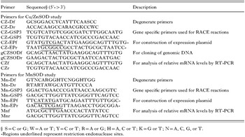
Table 1. Primers used in this study

Fig. 1. Nucleotide and translated amino acid sequences of Clonorchis sinensis Cu/ZnSOD and MnSOD cDNAs. (A) Cu/ZnSOD: the consensus poly(A) signal in the 3′ terminus is underlined; the degenerate primers employed to produce the first fragment of 260 base pairs are double underlined; 6 His and 1 Asp residues (boxed) form the copper/zinc binding sites; 3 Cys residues (bold) are believed to form intra-chain and/or inter-chain disulfide bonds; the arginine residue (italic) is necessary to guide the superoxide anion to the active site. The contagious nucleotide acid sequence from 54 to 527 was also found in the genomic DNA, indicting this is a one-exon gene. (B) MnSOD: the degenerate primers employed to produce the first fragment of 147 base pairs are double underlined; 3 His and 1 Asp residues (boxed) form a Mn-binding site.
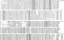
Fig. 2. Alignment of the deduced amino acid sequences of Clonorchis sinensis Cu/Zn-SOD and MnSOD with those of other organisms. Gaps are introduced to maximize alignment, (-) indicates a gap. The percentage identity of deduced amino acid sequences of C. sinensis SODs with those of other animals are presented at the end of each sequence alignments. (A) Cytosolic Cu/ZnSOD sequences, including C. sinensis (AY396147) in this study; P. westermani (AY396148); F. hepatica (AF071229); S. erinacei (AY396149); 5, T. solium (AF439353); S. mansoni (M97298); B. pahangi (X76284); B. malayi (AY428604); O. volvulus (X57150); A. viteae (Acanthocheilonema viteae, AJ240082); H. contortus (Haemonchus contortus, Z69621); D. immit (AF004949). The putative metal binding residues (boxed) are highly conserved. (B) Mitochondrial MnSOD sequences, their database Accession numbers are as follows: P. westermani (AY675508), S. erinacei (AY675509), C. elegans (Caenorhabditis elegans, NM_059889), O. voluulus (X82171 ), E. coli (NP_418344), Human (AAH12423), Cow (AAA30655), Chicken (AAG46055), Mouse (AAH18173 ), Zebrafish (AAH60895). The region boxed with a broken line includes putative mitochondrial targeting sequences. Amino acid residues in boxes are manganese-binding sites.
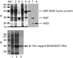
Fig. 3. Expression of Clonorchis sinensis Cu/ZnSOD and MnSOD in E. coli. (A) Cu/ZnSOD, showing Coomassie blue-stained SDS-polyacrylamide gel of proteins from lysates of E. coli without plasmid (lane1), uninduced pGEX-4T-2 (lane 2), pGEX-4T-2 induced with IPTG (lane 3), uninduced pGEX-Cu/ZnSOD (lane 4), pGEX-Cu/ZnSOD induced with IPTG (lane 5), purified GST-Cu/ZnSOD fusion protein (lane 6), purified GST-Cu/ZnSOD fusion protein cleaved with thrombin (lane 7), purified GST-free Cu/ZnSOD (lane 8). (B) MnSOD, showing Coomassie Blue-stained SDS-polyacrylamide gel of proteins from lysates of uninduced E. coli BL21 (DE3) harbouring plasmid pET-MnSOD (lane1), pET-MnSOD induced with IPTG (lane 2), Purified 6×His-tagged MnSOD protein (lane 3).
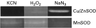
Fig. 4. SOD activity assay on a 12% SDS-polyacrylamide gel with NBT-riboflavin system. Cu/ZnSOD activity was significantly inhibited by KCN and H2O2, but not by NaN3; MnSOD activity was significantly inhibited by NaN3, but not by KCN or H2O2. NaN3, 10 mM NaN3; H2O2, 10 mM H2O2; KCN, 10 mM KCN.
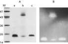
Fig. 5. Detection of disulfide bonds in the secondary structure of Cu/ZnSOD employing β-mercaptoethanol as reducing regent. Samples were separated on 12% SDS-PAGE and then were subjected to Coomassie Blue staining (A) or SOD activity staining (B). Purified SOD was treated with 5 mM (lane a), 50 mM (lane c) and or no (lane b) β-mercaptoethanol, and then heated for 5 min at 100 °C before loading the gels. The gel and sample treatment were identical for both panel A and panel B except the staining method. Two extra minor bands appearing on lanes a and b in panel A also showed SOD activity in panel B. M: molecular weight marker in kDa.
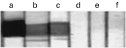
Fig. 6. Western blot analysis of pooled sera of clonorchiasis (a), paragonimiasis (b), fascioliasis (c), sparganosis (d), cysticercosis (e) patients and healthy human donors (f) versus recombinant Clonorchis sinensis Cu/Zn-SOD protein.
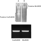
Fig. 7. The upper panel: activity detection and isoenzyme type identification of SOD in Clonorchis sinensis crude extract. (A) The soluble extract of adult C. sinensis (100 μg total protein) was separated on 12% non-denaturing polyacrylamide gels and stained immediately for SOD activity. (B) The sample and gel were identical to panel A except that the gel was treated with 10 mM KCN in the process of staining. The putative MnSOD and Cu/ZnSOD are indicted. The lower panel: reverse-transcriptase polymerase chain reaction (RT-PCR) of Cu/ZnSOD and MnSOD. PCR products were run on 1% agarose gels and stained with ethidium bromide.
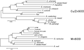
Fig. 8. Phylogenetic analysis of Clonorchis sinensis Cu/ZnSOD and MnSOD based on the amino acid sequences. The unrooted neighbour-joining method was used to construct the phylogenetic tree using the MEGA 2.1 program. The numbers at the nodes are the proportions of 1000 bootstrap re-samplings that support the topology shown. The scale bars indicate a distance of 0·05 or 0·1.
 You have
Access
- 12
- Cited by