Introduction
Chondrodermatitis nodularis chronica helicis is a common benign condition of the pinna. It presents as a painful, well demarcated nodule on the pinna that may be associated with surrounding erythema or an overlying crust. It usually develops on the apex of the helix but may also occur on the anti-helix. It typically affects men more than women, and usually appears on the side that the patient sleeps on.
The differential diagnosis of chondrodermatitis nodularis chronica helicis includes actin keratosis, squamous carcinoma and keratoacanthoma. Its exact aetiology is unknown, but trauma (including cold and pressure) and the anatomical characteristics of the pinna have been implicated. The apex of the pinna may be predisposed to this condition as the local blood supply to the epidermis, dermis and perichondrium is poor, with little supporting subcutaneous tissue. Upile et al. (2009) proposed a common endpoint of arteriolar narrowing in the perichondrium in areas furthest from the arterial blood supply. This leads to cartilage necrosis, extrusion and local inflammation, resulting in chondrodermatitis nodularis chronica helicis.Reference Upile, Patel, Jerjes, Singh, Sandison and Michaels 1
The patient may have already been treated by a dermatologist and had medical treatment (steroid injection),Reference Lawrence 2 or they may have had the cartilaginous lesion removed.Reference Lawrence 2 , Reference Hudson-Peacock, Cox and Lawrence 3 Other previously described surgical techniques include wedge excision, cartilage trimming and sutureless closure,Reference Hussain and Chalmer 4 curettage,Reference Jain and Jain 5 and the use of a CO2 laser.Reference Taylor 6 In addition, conservative management has been reported wherein pressure on the ear is relieved at night using a pillow with a hole for the ear or a custom-made prosthesis.Reference Sanu, Koppana and Snow 7 , Reference Singh, Wilson and Parkinson 8
Methods
We describe techniques for the excision of chondrodermatitis nodularis chronica helicis on both the helix and anti-helix, and reconstruction of the defect.
Anti-helical lesions
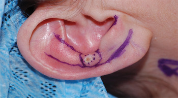
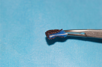
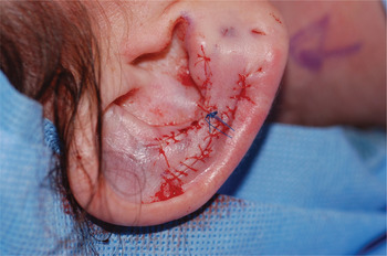
The procedure is performed under local anaesthetic. The lesion is demarcated and the superior and inferior advancement flaps are marked (Figure 1). The lesion is excised, including the overlying skin plus the affected cartilage. The remaining cartilage is smoothed down with an abrasive pad fashioned from a diathermy cleaning pad (Figure 2) in order to prevent recurrence. The superior and inferior advancement flaps are dissected off the underlying perichondrium and the surrounding skin is undermined. The edges of the superior and inferior flaps are sutured together and the sides are fastened in place (Figure 3) using a continuous absorbable suture. The wound is then dressed with adhesive strips and chloramphenicol ointment.
Anti-helical lesion and advancement flaps marked out.

Modified diathermy pad to smooth cartilage.

Anti-helical defect closed with advancement flaps.

Helical lesions
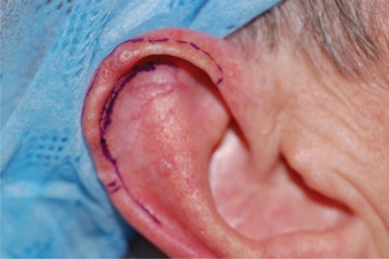
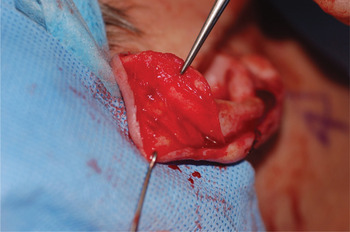
This method is suitable for multiple or single lesions. The procedure is performed under local anaesthetic. The lesion is demarcated with the incision planned around the helical lip (Figure 4). The lesion and overlying skin are excised and the underlying cartilage is smoothed down as above. The helical skin and skin on the posterior pinna are widely undermined and brought forward to cover the defect (Figure 5). The skin on the antihelix may also be undermined to facilitate closure. The incision is closed with a continuous absorbable suture and the wound is dressed with adhesive strips that are applied to refashion the helical rim. This is followed by the application of chloramphenicol ointment.
Multiple helical lesions marked out.

Posterior pinna skin undermined and sutured forward to cover defect.