Haematopoietic stem cell transplantation has been used with an increasing rate in treatment of many diseases occurring in the childhood including haematologic malignancy, immune deficiency, haemoglobinopathy, bone marrow failures, and congenital metabolism disorders. Reference Yeşilipek1,Reference Armenian, Sun and Kawashima2 Cardiovascular complications including heart failure, conduction disorders, valvular dysfunction, pericardial effusion, and intracardiac thrombosis may occur in patients after haematopoietic stem cell transplantation. Reference Armstrong, Chen and Yasui3 It has been reported that the 5-year cumulative incidence of cardiac impairment reached 26% in the patients who have been undergone haematopoietic stem cell transplantation during childhood. Reference Uderzo, Pillon and Corti4 It is also known that the development of cardiovascular complications may be related with the pre-transplantation therapies, especially anthracycline, as well as the preparation regimen which containing high dose cyclophosphamide and total body irradiation. Reference Yeşilipek1–Reference Armenian, Sun and Kawashima2,Reference Peres, Levine and Khaled5–Reference Karvandi, Piranfar and Yazdani9 Many studies evaluating haematopoietic stem cell transplantation patients have been focused on acute and late-onset cardiac side effects in adults and conducted by using conventional echocardiography. Reference Peres, Levine and Khaled5–Reference Murdych and Weisdorf8 The adverse effects of high-dose chemotherapy on left ventricular diastolic function after the bone marrow transplantation have been reported in an adult study by using tissue Doppler echocardiography. Reference Karvandi, Piranfar and Yazdani9 The diastolic dysfunction may be associated with the development of heart failure at follow-up. Reference Kane, Karon and Mahoney10 Evaluating of myocardial function by tissue Doppler echocardiography may provide early detection of patients at risk for cardiac impairment before heart failure symptoms develop and may be used as a guide in the treatment of haematopoietic stem cell transplantation patients. We hypothesised that children undergoing haematopoietic stem cell transplantation along with associated chemotherapy have myocardial injury before and after haematopoietic stem cell transplantation/chemotherapy and this impairment cannot be detected by conventional echocardiography. The effects of haematopoietic stem cell transplantation on ventricular diastolic functions have been evaluated in a few paediatric studies by using tissue Doppler echocardiography and conventional echocardiography Reference Daly, Colan and Blume11–Reference Kim, Moon and Yoon14 Some of these have focussed on only septal velocities Reference Yoon, Kim and Lee13–Reference Kim, Moon and Yoon14 or performed echocardiography in only one time point at follow-up. Reference Daly, Colan and Blume11–Reference Kim, Moon and Yoon14
In this study, we aimed to evaluate cardiac functions by serial echocardiography (at the five time points) in children who underwent haematopoietic stem cell transplantation. We also evaluated the effect of baseline echocardiographic parameters on mortality.
Materials and methods
This study was designed retrospectively and approved by the ethics committee of Akdeniz University. (KT/133/2015) Written informed consent was obtained from parents.
Study subjects
We evaluated 39 patients (under 18 years old) who underwent haematopoietic stem cell transplantation owing to non-malignant disease in our paediatric haematology and oncology clinics between January 2015 and May 2016. Patients with CHD, arrhythmia, or other systemic illness were excluded. The control group included 39 age-, gender-, and body surface area- matched healthy children who were evaluated for the aetiology of cardiac murmur and were determined to have normal cardiac findings. Our study was designed retrospectively and approved by the ethics committee of the hospital. Written informed consent was also obtained from parents.
We did not evaluate the myocardial effects of different types of conditioning regime and haematopoietic stem cell transplantation type on cardiac functions because this requires a lot more patients, time, and expenses.
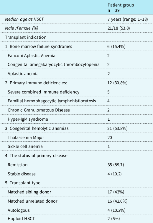
Transplant indications and haematopoietic stem cell transplantation types were reviewed and shown in Table 1. We have used various conditioning regimens according to underlying diseases and type of transplants. As myeloablative conditioning therapy, the following drugs were given; Busulfan (IV busulfan at myeloablative concentration according to plasma drug level) in combination with fludarabine (150 mg/m2), cyclophosphamide (160–200 mg/m2), melphalan (140 mg/m2), and thiotepa (10 mg/kg). Additionally, bendamustine, cytarabine, and etoposide were used in some protocols. As non-myeloablative (reduced intensity) conditioning therapy, the following drugs were given: Fludarabine (150 mg/m2) and cyclophosphamide 40–160 mg/m2. Cyclosporine was used as the back bone of GvHD prophylaxis, also anti-thymocyte globulin, methotrexate, and mycophenolate mofetil were used in combination. None of the patients had experienced cardiovascular symptoms/signs and haemodynamic disturbances during stem cell transfusion.
Baseline Clinical Features of HSCT Recipient

HSCT: Hematopoietic Stem Cell Transplantation.
Cardiovascular assessment
According to the standard protocol, cardiovascular status of the patient was determined by clinical and echocardiographic examination before haematopoietic stem cell transplantation (baseline) and after haematopoietic stem cell transplantation. Clinical manifestation of cardiotoxicity was defined as chest pain, palpitation, cardiac tamponade, and symptomatic heart failure. Basal echocardiographic examinations were performed within 1 month before haematopoietic stem cell transplantation, and repeated at the first, 3rd, 6th, and 12th months after haematopoietic stem cell transplantation.
Echocardiographic study
Echocardiographic studies were performed by using commercially available echocardiography machines (Vivid 7 pro, 3-MHz transducer; GE, Horten, Norway and IE33 Philips, Eindhoven, The Netherlands). One-lead electrocardiogram was recorded continuously during echocardiography examinations. We recorded all echocardiographic examinations in a computed recording system. All the echocardiographic measurements were averaged over three consecutive beats. Two-dimensional, M-mode, Doppler, conventional continuous wave, pulsed wave, and colour Doppler and tissue Doppler echocardiography measurements were obtained according to the paediatric guidelines of the American Society of Echocardiography. Reference Lopez, Colan and Frommelt15 The left ventricular ejection fraction, fractional shortening, the diastolic and systolic dimensions of the left ventricle, tricuspid valve annular plane excursion rate, and mitral valve annular plane excursion rate measurements were obtained by using M-mode echocardiography. The diastolic functions of both ventricles were measured (peak early [E] and late [A] wave velocities of the mitral and tricuspid flow and the E/A ratio).
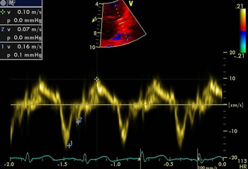
Myocardial tissue velocities (E’, A’, and S’ waves) obtained from mitral lateral annulus are shown in Figure 1.
Myocardial tissue velocities (E’,A’ and S’ waves) obtained from mitral lateral annulus were shown.

Measurement of the pulmonary artery systolic pressure by echocardiography
Assessment of pulmonary artery pressure can be challenging due to pyramidal shape and position of right ventricle in thorax. Measurement of pulmonary artery pressure by tricuspid regurgitation may be affected by preload and arrhythmia. The CW Doppler of tricuspid regurgitation and simplify Bernoulli equation were not used to calculate this pressure. Also, some patients had no tricuspid regurgitation to make this evaluation.
The pulmonary artery flow in the right ventricle outflow tract near the pulmonary valve was recorded by pulsed wave Doppler echocardiography in the parasternal short-axis view to measure the acceleration time of the pulmonary flow trace (AT), the right ventricle ejection time (RVET), and the AT/RVET ratio. The AT is the interval between the beginning of the flow and its peak velocity and RVET is defined as the interval from the onset of RV ejection to the point of systolic pulmonary arterial flow cessation. The mean pulmonary arterial pressure (mPAP) was calculated by the Mahan formula; mPAP (mmHg) = 90- (0.62 X AT). Reference Dabestani, Mahan and Gardin16
For Tissue Doppler imaging, transducer frequencies of 3 MHz were used with adjustment of the spectral pulsed Doppler signal filters until a Nyquist limit of 15–20 cm/s was reached and the use of minimal optimal gain. The monitor sweep speed was set at 50–100 mm/s to optimise the spectral display of myocardial velocities. The sampling window was positioned as parallel as possible with the myocardial segment of interest to ensure the optimal angle of imaging. Apical four-chamber views were obtained. The longitudinal peak annular velocities during systole (S’) as well as early (E’) and late (A’) diastole were measured at lateral and septal annuli of the mitral valve and the lateral annulus of the tricuspid valve.
Statistical analysis
Statistical analysis was performed using SPSS (Statistical Package for the Social Sciences, Chicago, IL, version 23). Numeric variables were expressed as mean + SD and categorical ones as percentages (%). The normality assumption of the variables was checked by the Shapiro–Wilks test. Independent T * test and Mann–Whitney U tests were used in the comparison of group averages according to the normal distribution. Statistical significance was assumed at p < 0.05. We calculated z scores of systolic and diastolic velocities in mitral lateral annulus, septum, and tricuspid lateral annulus in the control and the patient groups. Reference Eidem, McMahon and Cohen17 Mann–Whitney U test was used to compare z scores of each segment in the control and the patients group. Chi-square test was used to compare categorical variables and Spearman’s correlation coefficient was used to determine the correlation between continuous variables.
Results
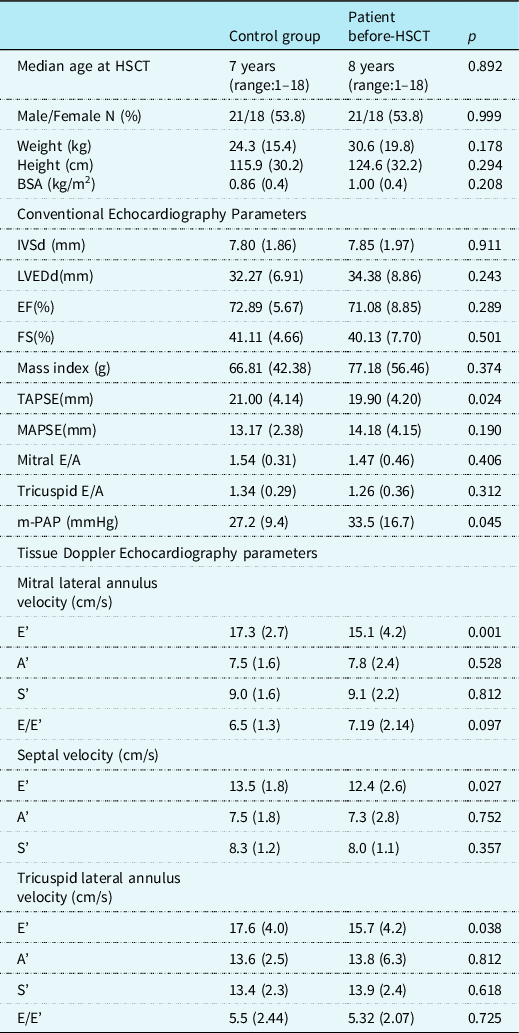
The patient and control groups had similar ages (mean + SD: 7.4 + 5.4 years) and gender distribution (Table 2). Except one case, all subjects had normal left ventricular ejection fraction before haematopoietic stem cell transplantation. However, the patient group had significantly greater m-PAP (p = 0.04) and lower tricuspid valve annular plane excursion rate value (p = 0.02) than the control group. The comparison of other conventional echocardiographic parameters showed no significant differences between patients and control groups. Tissue Doppler echocardiography showed lower E’ values for mitral lateral annulus, septum, and tricuspid lateral annulus (p = 0.02, p = 0.02 and p = 0.03) in the patient group before haematopoietic stem cell transplantation than the control group (Table 2). Consistent with the comparison of the mean values, baseline the mean z scores of E’ velocities in the three segments were lower in the patient group than those in the control group. The mean (SD) Z score of E’ velocities in mitral lateral annulus in the control group and the patient group were −0.04 (0.87) and −0.55 (1.31), respectively (p = 0.031). The mean (SD) Z score of E’ velocities in septum in the control group and the patient group were 0.28 (1.0) and −0.12 (1.3), respectively (p = 0.038). The mean (SD) Z score of E’ velocities in Tissue Doppler echocardiography in the control group and the patient group were 0.18 (1.2) and −0.27 (1.25), respectively (p = 0.038).
Comparison of the echocardiographic data between control (n = 39) and the patient group before HSCT (n = 39)

BSA: Body surface area. IVSd: Thickness of interventricular septum at end diastole. LVEDd: Left ventricular end diastolic diameter. EF: Ejection fraction. FS: Left ventricular fractional shortening. TAPSE: tricuspid valve annular plane excursion rate. MAPSE: mitral valve annular plane excursion rate. Mitral E/A: mitral E /A wave. Tricuspid E/A: tricuspid E/A wave. mPAP :The mean pulmonary arterial pressure. Mitral lateral annulus velocity E,A,S: Mitral valve lateral peak annular velocities during systole (S’) early (E’) and late (A’) diastole. Septal velocity E,A,S: Mitral valve septal annular velocities during systole (S’) early (E’) and late (A’) diastole. Tricuspid lateral annulus velocity E,A,S : Tricuspid valve lateral annular velocities during systole (S’) early (E’) and late (A’) diastole.
Comparison of the echocardiographic data in the patient group before and after haematopoietic stem cell transplantation
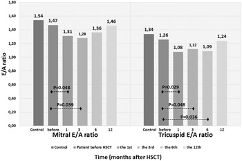
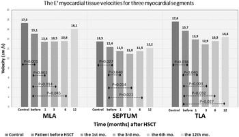
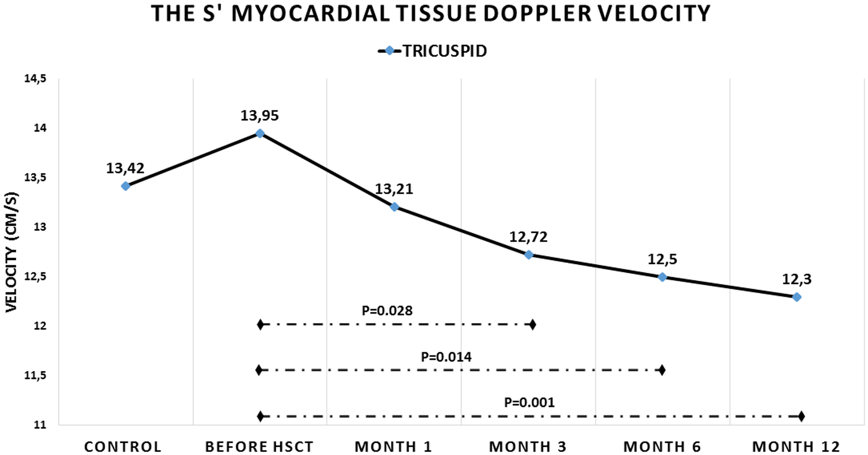
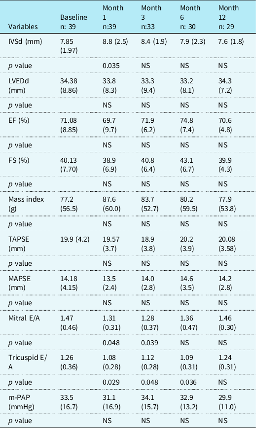
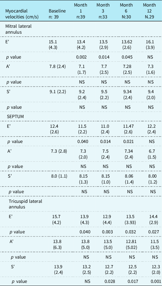
Seven patients (17.8%) had a new abnormality on echocardiogram during the first year of haematopoietic stem cell transplantation. The left ventricular systolic dysfunction was detected in one case, pericardial effusion occurred in six cases (15.4%), and right atrial huge thrombus developed in one case after haematopoietic stem cell transplantation. Showing not much difference than pre-haematopoietic stem cell transplantation, the lower values of the TAPSE persisted for the first year after haematopoietic stem cell transplantation (Table 3). The E/A ratio for the mitral valves was significantly lower for the 3 months after haematopoietic stem cell transplantation (p < 0.05, Table 3, Fig 2). Similarly, the E/A ratio for the tricuspid valves was significantly lower for the 6 months after haematopoietic stem cell transplantation (p < 0.05, Fig 2). Similar to pre-haematopoietic stem cell transplantation, the higher m-PAP values persisted for the 6 months after haematopoietic stem cell transplantation (Table 3). When compared to pre-haematopoietic stem cell transplantation, E’ velocities of all segments were lower in the patient group for 6 months after haematopoietic stem cell transplantation. E’ velocities for mitral lateral annulus and septum reached to the values of before haematopoietic stem cell transplantation at the first year after haematopoietic stem cell transplantation (Table 4, Fig 3). However, E’ velocity for tricuspid lateral annulus was still depressed in the first year after haematopoietic stem cell transplantation and did not reach the value of before haematopoietic stem cell transplantation (Table 4, Fig 3). Likewise, patient had lower S’ velocity for tricuspid lateral annulus at the third, sixth month of haematopoietic stem cell transplantation, (p = 0.028, p = 0.017) and it was still depressed at the first year after haematopoietic stem cell transplantation (p = 0.001) compared to basal levels (Table 4, Fig 4).
Changes in the mitral and tricuspid E/A ratio in the patient group at different time points for the first year of HSCT.

Changes in the E’ velocities for three myocardial segment of the patient group at different time points for the first year of HSCT.

Changes in the S’ velocities for tricuspid lateral annulus in the patient group at different time points for the first year of HSCT.

Conventional echocardiography data in patient group from baseline to the 12th month after HSCT

IVSd: Thickness of interventricular septum at end diastole. LVEDd: Left ventricular end diastolic diameter. EF: Ejection fraction. FS: Left ventricular fractional shortening. TAPSE: Tricuspid valve annular plane excursion rate. MAPSE: Mitral valve annular plane excursion rate. Mitral E/A: mitral E /A wave. Triküspit E/A: Tricuspid E/A wave. mPAP :The mean pulmonary arterial pressure. Mitral lateral annulus velocity E,A,S: Mitral valve lateral peak annular velocities during systole (S’) early (E’) and late (A’) diastole. Septal velocity E,A,S: Mitral valve septal annular velocities during systole (S’) early (E’) and late (A’) diastole. Tricuspid lateral annulus velocity E,A,S : Tricuspid valve lateral annular velocities during systole (S’) early (E’) and late (A’) diastole.
p values are the result of paired-samples t test comparing the baseline with each study point. NS: not significant (p > 0.1 for all of them).
Tissue Doppler Echocardiography Data in Patient Group from Baseline to the 12th Months after HSCT

Mitral lateral annulus velocity E,A,S: Mitral valve lateral peak annular velocities during systole (S’) early (E’) and late (A’) diastole. Septum velocity E,A,S: Mitral valve septal annular velocities during systole (S’) early (E’) and late (A’) diastole. Tricuspid lateral annulus velocity E,A,S : Tricuspid valve lateral annular velocities during systole (S’) early (E’) and late (A’) diastole.
p values are the result of paired-samples t test comparing the baseline with each study point. NS: not significant (p > 0.1 for all of them).
Relationship between mortality and baseline echocardiographic parameters
Ten patients died after haematopoietic stem cell transplantation and the mortality rate was 25.6 % in our cohort. The mortality was related to ventricular tachycardia in one case. The EF and SF value before haematopoietic stem cell transplantation were significantly lower in patients who died after haematopoietic stem cell transplantation than patients who survived (EF: 66.4 + 10.6 versus 72.7 + 7.6 p = 0.012 and SF: 36.8 + 9.9 versus 41.3 + 6.6 p = 0.014. However, EF and SF values were in normal range in both groups. In addition, the E’ velocity for septum before haematopoietic stem cell transplantation were significantly lower in patients who died after haematopoietic stem cell transplantation than patients who survived (10.5 + 2.4 versus 13.0 + 2.3 (cm/s) p = 0.009).
Discussion
This study showed subclinical impairment in myocardial functions before and after haematopoietic stem cell transplantation. The effects of haematopoietic stem cell transplantation on left ventricular diastolic functions persisted for 6 months, and then improved at the first year of haematopoietic stem cell transplantation. However, right ventricular systolic and diastolic velocities were found to be lower at the first year of haematopoietic stem cell transplantation. These findings mean that right ventricular functions have been affected for longer periods than left ventricle after haematopoietic stem cell transplantation.
The strategy of lowering cardiotoxic drug, as well as early detection and treatment of late effects have resulted in the extension of life spans for many survivors of childhood cancer. Reference Armstrong, Chen and Yasui3,Reference Uderzo, Pillon and Corti4 Subclinical left ventricular dysfunction has been recently reported in children undergoing haematopoietic stem cell transplantation for severe aplastic anaemia and acute leukemias. Reference Daly, Colan and Blume11–Reference Kim, Moon and Yoon14 Kim et al. Reference Kim, Moon and Yoon14 found no significant differences in tissue Doppler echocardiography or speckle tracking parameters between control and patient group before haematopoietic stem cell transplantation. They also did not observe any changes in echocardiographic parameters from pre-haematopoietic stem cell transplantation to post-haematopoietic stem cell period. Compared to control, the reduced EF, septal E' velocity, and longitudinal systolic strain rate were reported in their patient group at 6th months after haematopoietic stem cell transplantation. A study performed by Yong et al. Reference Yoon, Kim and Lee13 reported that septal E’ and A’ velocities in children planning for haematopoietic stem cell transplantation for acute leukaemia were lower than the healthy children group. However, they did not notice any significant alterations in these parameters after haematopoietic stem cell transplantation. Notwithstanding, these two authors did not examine mitral lateral annulus and tricuspid lateral annulus velocities in their studies.
ElMarsafawy et al. Reference El-Marsafawy, Matter and Sarhan12 examined myocardial velocities in children undergoing autologous haematopoietic stem cell transplantation for malignancy. Similar to our study, they showed significant reduction in all myocardial velocities before haematopoietic stem cell transplantation and myocardial velocities became more compromised for 3 months after haematopoietic stem cell transplantation. In our study, left ventricular systolic dysfunction was detected in one case, pericardial effusion occurred in six cases (15.4%), and atrial thrombus developed in one case after haematopoietic stem cell transplantation. Similar to previous studies Reference El-Marsafawy, Matter and Sarhan12–Reference Kim, Moon and Yoon14 , we found that E' velocities for all segments were reduced significantly after haematopoietic stem cell transplantation. The reduction of E’ and S’ velocities for tricuspid lateral annulus persisted at least the first year after haematopoietic stem cell transplantation. These findings can be explained by diastolic dysfunction usually preceding systolic disturbances. Reference Paulus, Tschöpe and Sanderson18 As seen in some cardiomyopathies, the right ventricular dysfunction may become evident prior to the left ventricular changes.
Daly et al. Reference Daly, Colan and Blume11 reported no changes in conventional echocardiographic parameters at 1-year post-haematopoietic stem cell transplantation. Interestingly, they observed a subclinical decline in the systolic and diastolic functions, even in the patient subgroup who did not receive anthracycline chemotherapy. They conclude that the decrease in LV contractility is likely to be secondary to other risk factors, such as haematopoietic stem cell transplantation conditioning therapy. Similar to our findings, they found that E’ velocities for mitral lateral annulus and tricuspid lateral annulus were reduced before haematopoietic stem cell transplantation and mitral and tricuspid E′ velocity was decreased in the first year of haematopoietic stem cell transplantation. They detected that baseline left ventricular ejection fraction and E' velocity for Mitral lateral annulus were slightly lower in their patients who died after haematopoietic stem cell transplantation when compared with all the study cohort. They concluded that these small changes might become clinically important over time. In our study, The EF and SF values before haematopoietic stem cell transplantation were significantly lower in patients who died after haematopoietic stem cell transplantation than patients who survived. However EF and SF values were in the normal range in the both patient groups. There was no left ventricular systolic dysfunction in our patient before haematopoietic stem cell transplantation, except one case.
Covi et al. Reference Covi, Ravindranath and Farooqi19 reported that haematopoietic stem cell transplantation was associated with initial worsening longitudinal strain values. They noticed that strain parameters returned to baseline levels in the first year after haematopoietic stem cell transplantation. Like our study, they showed pre-existing pulmonary hypertension in their patients. However, they reported that pulmonary hypertension persisted in their patients after haematopoietic stem cell transplantation.
In our study, most cases had a history of erythrocyte transfusions. A previously reported study showed a weak but significant correlation between serum ferritin and LV longitudinal strain parameters. Reference Kim, Moon and Yoon14 As implicated by Kim et al. Reference Kim, Moon and Yoon14 previous transfusion and iron load might be responsible for baseline cardiac findings in our cases with Thalassemia major or aplastic anaemia.
High dose radiotherapy and/or chemotherapy is usually used in preparation regimes and they may lead to early or late cardiac complications with varying severity. Since total body irradiation may lead to late endocrine complications in childhood and protocols containing only chemotherapy showed no difference in terms of prognosis, total body irradiation-based regimes usually were no't applied at all in children. Reference Yeşilipek1 The most commonly used regimes in children are the regimes in which cyclophosphamide and busulfan are used together. Reference Yeşilipek1
Our study showed that the baseline septal E' velocity was found lower in patients who died after haematopoietic stem cell transplantation than survived, we thought that the cardiac reserve should be taken into consideration for children planned to have haematopoietic stem cell transplantation. All our patients never received radiotherapy, so cardiotoxic effects of radiation were eliminated in our cases. However, cytokine release, sepsis, and acute graft versus host disease were likely to have a negative effect on myocardial function in our patient after haematopoietic stem cell transplantation.
Conclusion
This study showed deterioration in myocardial diastolic functions and increase in the mean pulmonary artery pressure in children before and after haematopoietic stem cell transplantation. After haematopoietic stem cell transplantation, the right ventricular function adversely affected for a longer time than the left ventricle. Tissue Doppler echocardiography can be used as a routine imaging technique to detect early myocardial dysfunction in haematopoietic stem cell transplantation recipients. The myocardial functions should be monitored after the first year of haematopoietic stem cell transplantation.
Limitations of the present study
This study was designed retrospectively, our cohort had heterogeneous primary disease, haematopoietic stem cell transplantation type and conditioning regimen. Since a larger number of patients was required, we did not evaluate the myocardial effects of different types of conditioning regime and haematopoietic stem cell transplantation type on cardiac functions. Myocardial abnormalities detected in our study population may be related to cytokine release sepsis and acute graft versus host disease. We could not examine the relationship between changes in echocardiographic parameters and complications such as sepsis and graft versus host disease, due to insufficient data with regard to complications (such as sepsis and acute graft versus host disease) in patient echocardiography reports. Lastly, it is not known that the early subclinical changes in myocardial function in post-haematopoietic stem cell transplantation period predict long-term cardiovascular impairment.
Acknowledgements
None.
Financial support
This research received no specific grant from any funding agency, commercial, or not-for-profit sectors.
Conflict of Interest
None.