Aging of the brain and molecular theories of aging
The brain houses about 600 km of blood vessels, and any problems with circulation due to aging of the brain can result in insufficient supply of oxygen and nutrients and, consequently, may induce various problems caused by hypoperfusion. In fact, hypoperfusion may arise from cholesterol deposition, diabetic changes, and atherosclerosis in general, and may result in tremor, minor memory impairment, stroke, and vascular dementia. Many researchers now believe that vascular dementia and dementia of the Alzheimer's disease-type (DAT) have common pathogenetic pathways.
Data in the literature comparing old and young brains show that learning is harder and short-term memory capabilities reduced in older age but that previously acquired memory remains to a great extent. Indeed, multiple memory functions decrease in the aged brain, including fluid intelligence, explicit memory (episodic prior to semantic), alertness (mainly vigilance and flexibility), and problem solving. These alterations in memory function result from changes in the brain at the biochemical and physiological levels, neurotransmitter and synaptic changes, and nerve cell degeneration and death.
Imaging studies that have investigated factors contributing to memory loss reveal altered functional activation of the prefrontal cortex and hippocampus, but gaps in our understanding remain. It is necessary, therefore, to consider the basic mechanisms of neuronal and cell aging. Neurons are long-lived, post-mitotic cells, and they can be affected by many different changes at the biochemical and molecular level. Of the numerous theories about the causes of cell aging, five are particularly prominent:
• Inflammation is believed to be a major factor in aging of the nervous system due to dysfunction of the immune system, inflammatory processes and toxic activity of immune modulators (Cheney and Walford, Reference Cheney and Walford1974).
• In a clearly defined senescence program, activity of defined gene products is a cause of progression of aging leading to a limited replicative life span of somatic cells (replicative senescence; Hayflick and Moorhead, Reference Hayflick and Moorhead1961).
• The telomer theory is still attractive and prominent, as continuous shortening of chromosomes indicates the lifespan of the cell and replicative senescence (Allsopp and Harley, Reference Allsopp and Harley1995).
• Free radicals and oxidative stress, which result in oxidation of biomolecules, have been proposed as drivers of aging, particularly neuronal aging, and have an impact on age-associated disorders (Harman, Reference Harman1965).
• More recent evidence indicates that caloric restriction slows down the rate of aging and increases lifespan (Witte et al,. Reference Witte, Fobker, Gellner, Knecht and Flöel2009).
Two of these theories – oxidative stress theory and the effects of caloric restriction – merit further consideration here.
Oxidative stress theory
The oxidative stress theory was first proposed in the 1960s but has seen a recent revival. Many different types of reactive oxygen and nitrogen radical species are continuously produced in the mitochondria and throughout the cell. These species are important triggers of physiological function and transcription, but a misbalance between production and detoxification of free radicals results in an accumulation of active species, which is known as oxidative stress. The resultant radicals can cause oxidation of different types of molecules; lipids were a focus of research for some time, but the spotlight has moved towards the oxidation of proteins, as oxidized proteins are found in aged brains, as well as the brains of people with Alzheimer's disease (AD). Extensive research in this area has led to theories about pathways resulting in aging and DAT (Behl et al., Reference Behl, Davis, Lesley and Schubert1994; Moosmann and Behl, Reference Moosmann and Behl1999; Behl, Reference Behl2002; Mocko et al., Reference Mocko, Kern, Moosmann, Behl and Hajieva2010), as well as some hypotheses about mechanisms that counteract oxidative stress (Sagara et al., Reference Sagara, Dargusch, Klier, Schubert and Behl1996; Lezoualc'h et al., Reference Lezoualc'h, Sagara, Holsboer and Behl1998; Greeve et al., Reference Greeve2000; Clement et al., Reference Clement, Gimpl and Behl2010). Oxidation of biomolecules has long-term detrimental effects: oxidized nucleic acids result in the production of abnormal proteins due to transcriptional and translational errors, oxidized proteins tend to build up into aggregates and cross-link with other materials, and oxidized lipids form end products that alter the cellular membrane and its function, with potentially catastrophic consequences for cells (Stadtman, Reference Stadtman2006). On the other hand, long-term strengthening of neurons and an induced neuron resistance against oxidative insults might explain why some cells in the brains of patients with AD survive, while other cells die (Sagara et al., Reference Sagara, Dargusch, Klier, Schubert and Behl1996; Lezoualc'h et al., Reference Lezoualc'h, Sagara, Holsboer and Behl1998; Greeve et al., Reference Greeve2000; Clement et al., Reference Clement, Gimpl and Behl2010). Such theories hint at future targets for intervention.
Caloric restriction
The effects of caloric restriction have been examined in animals including nematodes, mice, and primates. Results from a study in rhesus monkeys found that those with a reduced calorie diet (445 kcal) were slim, appeared younger and fitter, and were in general good health, whereas monkeys with a full calorie diet (885 kcal) appeared senile, lost hair and had aged skin, evidence of arthritis and poor blood parameters for their age (Colman et al., Reference Colman2009). Furthermore, in an interesting study of caloric restriction in humans, 50 healthy subjects aged 50–80 years were split into three dietary groups: calorie restriction (30% less calorie intake), increased unsaturated fatty acids, and controls (Witte et al., Reference Witte, Fobker, Gellner, Knecht and Flöel2009). Differences were noted after just three months, with significant improvements in verbal memory (word recall) and reduced levels of insulin and inflammatory markers in the calorie-restricted group. The brain thus responded to caloric restriction in a beneficial manner. The longer-term results of this study are eagerly awaited, as they might provide a new or additional strategy for maintaining cognitive function during aging.
Molecular targets and experimental therapies in AD: the view of a biochemist
Despite the many breakthroughs made during the past 100 years of research into AD, no effective causal therapy relevant to patients is available for use in the clinic and there is still no clear picture of the cause or causes of late-onset AD. This lack of progress may be attributed to a number of reasons. One of these is that much of the basic research into cellular systems has been undertaken in mice, but this species simply does not live long enough to develop AD. Research has also concentrated perhaps too much on extracellular amyloid beta (Aβ), aggregates, protofibrils, and senile plaque protein deposits at the expense of more detailed work on intracellular Aβ, which would be logical given that amyloid's production involves a recycling pathway that starts inside the cells themselves. Widespread extrapolation from work in genetic AD to sporadic AD may also be an important barrier to progress.
Amyloid cascade hypothesis
The almost exclusive focus on amyloid and tau hyperphosphorylation has resulted in detailed molecular insights into Aβ and tau biochemistry, supported by evidence in transgenic mice that amyloid is deposited in the brain and that oligomers of Aβ may change synaptic function, induce intracellular tau hyperphosphorylation, and induce inflammatory responses. Consequently, the amyloid cascade hypothesis and the tau hypothesis of AD were formulated. Aβ and tau have long been considered separate for the purposes of research; however, recent data suggest convergence of Aβ and tau in the mitochondria (Müller et al., Reference Müller, Eckert, Kurz, Eckert and Leuner2010; Eckert et al., Reference Eckert, Schmitt and Götz2011). Indeed, abnormal accumulation of Aβ and tau seems to result in neuritic plaques and neurofibrillar tangles, disturbed neuronal function, activation of astrocytes and microglia, and oxidative and inflammatory stress.
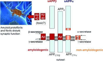
The amyloid precursor protein (APP) is not specific to the brain but is present in every cell of the body. This protein is cleaved and biochemically processed by different secretases: β-secretase, γ-secretase, and a combination of α- and γ-secretases. Cleavage of APP by γ-secretase followed by α- and γ-secretase results in a small peptide; this is harmless, as it is non-toxic, and is non-aggregating. However, cleavage achieved through a combination of β-secretase and γ-secretase results in the production of Aβ peptides comprising 40 or 42 amino acids, which are believed to have toxic effects (Figure 1).
Processing of the amyloid precursor protein APP.

The past five to ten years have seen widespread promotion of a theory in which fibrils (protofibrils) that form slowly outside of cells interact with synapses and disturb synaptic function. However, to date, there is no real correlation between the extent of memory loss and dementia and Aβ deposition, and although tau changes do correlate with dementia, tau is not specific for AD and is also involved in other neurodegenerative disorders.
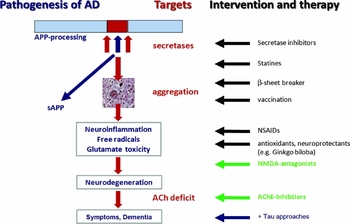
The anti-amyloid approach attempts to block secretases that result in forms of amyloid that could aggregate (Citron et al., Reference Citron2010). Blockade of β-secretase is not possible, because this enzyme is involved in cleavage of other substrates and is important for processes such as myelin sheath formation. γ-secretase is another critical enzyme, because it is involved in the notch signaling cascade that is essential to cells. Indeed, trials in AD with γ-secretase inhibitors (such as non-steroidal anti-inflammatory drugs and statins) either failed or were stopped after they showed no benefit or serious side-effects. Trials of Aβ vaccination, which clears Aβ aggregates outside of cells, are ongoing, although current clinical data do not indicate that immunization improves cognition in humans (Holmes et al., Reference Holmes2008).
Tau
Every protein that is synthesized in the neuronal cell body must be transported along axons before it can enter synapses. This axonal transport system runs through microtubuli, which are stabilized through an association with tau protein. In patients with AD, hyperphosphorylation of certain amino acids in the tau protein means that it dissociates from the microtubule, disturbing the transport structure and resulting in starvation of neurons and ultimately cell death. Hyperphosphorylated tau thus has an important role in the formation of neurofibrillary tangles and the pathogenesis of AD and related tauopathies (Noble et al., Reference Noble, Pooler and Hanger2011).
Diverse kinases have been implicated in tau phosphorylation, but protein phosphatase 2A (PP2A) seems to be most relevant. Recently, Kickstein et al. (Reference Kickstein2010) proposed that metformin, a drug long used in the treatment of diabetes, could be useful as a tau-targeting compound, as it changes the activity of PP2A so that it removes phosphorylated parts of tau protein. In murine primary neurons from wild-type and human tau transgenic mice, metformin induced PP2A activity and reduced tau phosphorylation in vitro and in vivo.
The anti-infection drug methylene blue is another “old” substance that may be of interest in the context of tau hyperphosphorylation. Methylene blue is a derivative of phenothiazine – one of the oldest pharmacological target compounds – and has been used as a stain in histology and molecular biology technology, as an antidote for intoxication, an antimalarial drug, and an antiseptic. Before tau phosphorylation had been identified as a potential pathogenetic mechanism, phenothiazine was identified as a powerful antioxidant. Recent work has shown that methylene blue has many activities, including antioxidant and tau dephosphorylation activity (Schirmer et al., Reference Schirmer, Adler, Pickhardt and Mandelkow2011), even though its penetration of the blood–brain barrier is low. No clinical data have yet been published, and the effect of the compound on the color of liquid excretions is a problem for clinical trials, but when given in a clinical setting, methylene blue has resulted in improvements in memory compared with other AD therapies, such as treatment with acetylcholinesterase inhibitors (reported at ICAD Meeting, 2008, by Dr. Wischik)
Figure 2 summarizes intervention and prevention approaches.
Summary of intervention and prevention approaches.

Novel hot topics in the search for AD therapy and prevention
A variety of novel targets for AD therapy and prevention have been developed recently and are proposed in addition to those described above.
Epigenetics
Genetic observations point towards mutations and genes that result in increased Aβ deposition, and specially designed secretase inhibitors may be used to treat early-onset familial AD, but clinicians are more likely to see sporadic cases than patients with hereditary diseases that are very rare. Epigenetics summarizes biochemical modifications that result in heritable changes in gene expression independent of changes in genetic sequences, which have long-lasting effects. To date, more than 50 histone modifications have been identified at the biochemical level. Differences in the methylation status of the APP gene have been identified between patients with AD or Pick's disease and healthy control individuals (West et al., Reference West, Lee and Maroun1995). Decreased amounts of methylcytosine within the APP promoter in the DNA from the parietal cortex of individuals without dementia have been shown with increasing age (Tohgi et al., Reference Tohgi, Utsugisawa, Nagane, Yoshimura, Genda and Ukitsu1999). Elevated levels of homocysteine have been observed in patients with AD and are associated with deficiency of folate and vitamin B12 (Seshadri et al., Reference Seshadri2002; Zhuo et al., Reference Zhuo, Wang and Praticò2011), which are essential cofactors required to regenerate methionine (the precursor of the methyl donor for DNA methylation) from homocysteine. Increased histone acetylation has been found in the brains of patients with AD, so the use of histone acetyltransferase (HAT) inhibitors may hold some potential for treatment of AD (Narayan and Dragunow, Reference Narayan and Dragunow2010). Use of biomarkers to define an individual's epigenome may allow for tailoring of drugs to specific epigenomes so that epigenetic switches could be turned back pharmacologically.
Protein homeostasis
Protein homeostasis deteriorates during aging, which results in protein dysfunction and aggregation. Simple signs of this are the so-called age spots, clusters of glycated proteins and lipids, observed not only in skin but also in the liver and neurons. In addition to changes in Aβ and tau in the hippocampi of patients with AD, disturbances of protein homeostasis resulting in protein aggregation also occurs in Parkinson's disease (for instance, with disturbance of α-synuclein creating Lewy bodies), in Huntington's disease (with disturbance of huntingtin in the striatum) and in amyotrophic lateral sclerosis (ALS) (Soto, Reference Soto2003). Age-associated disturbances of protein homeostasis thus seem to be causally related to the onset and progression of neurodegenerative disorders. A recent mathematical computer simulation of the appearance of the cytoplasm with respect to protein accumulation highlights the complex interplay between proteins, with some needing to interact and others needing to avoid any interaction (McGuffee and Elcock, Reference McGuffee and Elcock2010), which makes fine-tuned protein control and protein homeostasis vital. Correct folding of proteins results in proper function, while changes in and misfolding of proteins results in dysfunction, aggregation and, in some disorders, detrimental progress and changes.
Proteins formed during translation must be folded correctly if they are to become the native conformation proteins that act as enzymes or other key mediators within the body. Some proteins do misfold for a variety of reasons, and the heat shock protein 70 (HSP70) machinery is a key first line of defense. It identifies protein changes in cells and acts to maintain proteostasis, in combination with other partners, by refolding faulty proteins where possible or directing them to degradation if refolding is unachievable. Quality control factors that are also called chaperones (and co-chaperones) including HSP70, as well as HSP40, ubiquitin and carboxyl terminus of Hsp70-interacting protein (CHIP), are found in a variety of disease tissues, including AD, Parkinson's disease, Huntington's disease and spinocerebellar ataxia. Their presence indicates disturbed protein quality control, particularly a deficiency in protein degradation, which results in accumulation of proteins inside cells and consequent disturbance of physiology.
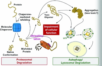
Cells have two ways to degrade proteins that are not folded correctly: the proteasomal system and the autophagy pathway (Figure 3). Proteasomal activity decreases in aged cells, particularly aged neurons, which means that autophagy becomes increasingly important. Recent work shows that when aged neurons or cells with an impaired proteasome are challenged with many proteins, the latter are not distributed widely throughout the cells but accumulate at a perinuclear site called the aggresome (Gamerdinger et al., Reference Garmerdinger, Kaya, Wofrum, Clement and Behl2011). This aggresome contains a variety of substances involved in autophagy, including poly-ubiquitin proteins, BAG3, p62, LC3 and protein aggregates. BAG3 is a co-chaperone of HSP70 and plays a role in the age-associated autophagy pathway, which takes place at an intracellular level (Behl, Reference Behl2011). It is therefore important in the future to focus some research on the intracellular activities around proteins rather than focusing solely on extracellular deposition. Indeed, when considering alternative explanations of AD at the basic science level, many observations suggest that failure of lysosomal proteolysis and autophagy driven by relevant genes and other risk factors in people with AD leads to complete blockade of lysosomal proteolytic pathways, with proteins that should be degraded becoming trapped in cells, leading to synaptic dysfunction and neurodegeneration (Pimplikar et al., Reference Pimplikar, Nixon, Robakis, Shen and Tsai2010; Shen and Ganetzky, Reference Shen, Ganetzky and 2009; Caijgas et al., Reference Caijgas, Will and Schuman2010; Willeumier et al., Reference Willeumier, Pulst and Schweizer2006; Thinakaran and Koo, Reference Thinakaran and Koo2008). This may explain at least in part the lack of an apparent relationship or association between extracellular amyloid load and dementia. It could be worthwhile, therefore, to focus on proteostasis and potential drugs to stabilize the intracellular protein homeostasis in the context of AD.
Principal pathways of protein degradation.

Summary
Research into AD has still to have a major impact on the day-to-day lives of patients, with the exact causes of the disease still not known and effective treatments out of reach. Research has, with good reason, focused on certain key pathways and potential mechanisms, but sometimes this has been at the expense of work on other theories and views, which may be slowing down progress in this field. With wider research, a variety of interesting theories are emerging, including different perspectives on oxidative stress and the Aβ cascade and tau hyperphosphorylation, as well as caloric restriction. Continued work in epigenetics and protein homeostasis may reveal suitable targets for intervention. Taken together, analysis of the biochemistry of aged neurons and the interplay with pathways of neurodegeneration may lead to a better understanding of AD and how to treat and prevent this condition.
Conflict of interest
None.
Acknowledgments
The author wishes to thank Dr. Christine Ziegler for fruitful comments and Michael Plenikowski for artwork.