Introduction
Tympanoplasty represents the most common otologic surgical procedure conducted in adults since the first description by Wullstein in 1950s to this day.Reference Telian, Schmalbach, Snow and Ballenger1 The main purpose of tympanoplasty type I (myringoplasty) is to restore the integrity of a perforated tympanic membrane, aiming at the sealing of the middle ear from the external auditory canal, maintaining the cleanliness of the area and protecting from infection. The improvement in hearing is a desirable but mostly secondary goal, as outcomes cannot be easily predicted. A tympanoplasty can be combined with concomitant ossiculoplasty (types II–IV) in an attempt to replace the eroded or malfunctioning ossicular chain.Reference Telian, Schmalbach, Snow and Ballenger1 Different techniques have been described during the history of otologic surgery and, in the past few decades, new methods seem to mark the progress. Post-auricular or endaural and the most recent endoscopic approach, in combination with the choice of overlay or underlay techniques, are some of the surgical options.Reference Adunka, Buchman, Adunka and Buchman2
Reported success rates of myringoplasties vary in literature, from 75–98 per cent, with an estimated average of 90 per cent. The success of a tympanoplasty is defined principally by the outcome of an intact neo-tympanum. Several factors have been correlated to the risk of failure, such as the technique, type of graft used, size and location of the perforation, function of the Eustachian tube, and other patient-related factors.Reference Salviz, Bayram, Bayram, Balikci, Chatzi and Paltura3 However, the main debate in literature concerns the type of graft used to reconstruct the perforated tympanic membrane.
Graft material usually consists of autologous tissue, such as fascia, fat, vein and cartilage, whereas alternative choices include implantable biomaterial composed of collagen. The temporal muscularis fascia remains the most commonly used type of graft, however cartilage often is used because of the stability and durability it provides to the tympanic membrane.Reference Jalali, Motasaddi, Kouhi, Dabiri and Soleimani4 Several studies support the advantages of cartilage use in reconstruction of the tympanic membrane and its superiority to fascia.Reference Bayram, Bayar Muluk, Cingi and Bafaqeeh5 Cartilage can be harvested from the tragus, the concha or even the septum, and several modifications in the technique have been described (palisades, butterfly grafts, etc.). The use of composite materials, such as the combination of cartilage with fascia or perichondrium, has also gained popularity because combining the strength of cartilage with the elasticity of a fascia can contribute to optimal results.Reference Tos6
The split two-layer tragal cartilage–perichondrium graft represents a composite type of graft, consisting of two separate layers: cartilage with the overlaying perichondrium, harvested from the tragus. The aim of this study is to determine the surgical outcome of tympanoplasties conducted with the use of this grafting technique, as defined by the post-operative tympanic membrane integrity, the need for further surgery and the improvement in hearing thresholds.
Materials and methods
Basic settings and patient selection
We carried out a retrospective observational study in a tertiary academic centre. Medical records of patients who underwent tympanoplasty, for chronic otitis media with tympanic membrane perforation, between January 2016 and June 2022, were reviewed.
The study was conducted in accordance with the Declaration of Helsinki principles for medical research involving human subjects. The Strengthening the Reporting of Observational Studies in Epidemiology (‘STROBE’) checklist for cross-sectional studies were followed.
Only adult patients who underwent a tympanoplasty with the use of split two-layer cartilage–perichondrium graft were included in the study. No limitations regarding the history of previous surgery, the presence of cholesteatoma or the ossicular chain integrity were applied. Both primary and revision tympanoplasty cases were included.
Demographic data, the presence of ossicular chain discontinuity, size and location of the perforation and audiology data, pre- and post-operatively were recorded.
Surgical Technique
All tympanoplasties were performed or directly supervised by the same surgeon. The surgical technique involved a unilateral tympanoplasty under general anaesthesia, of types I–IV, including cases with concomitant ossiculoplasty and the use of prosthesis, when necessary. Grafts were harvested from the ipsilateral tragus, consisting of tragal cartilage with perichondrium. The tragal cartilage and adjoining perichondrium were removed, separated, and the perichondrium flattened to maximise surface area for the best possible coverage of the perforation (Figure 1). The cartilage was remodelled, in thickness and size, according to the needs of each perforation, and the graft was placed with the underlay technique, in two layers (Figure 2). The cartilage was placed medially in underlay technique and the perichondrium was positioned more laterally, on top of the cartilage but under and in direct contact with the residual tympanic membrane (between the residual tympanic membrane and the cartilage). This technique was used in all cases to allow best possible structural support in the repair; the rims of the perforation were trimmed at an earlier stage to promote healing. All patients were treated with the same absorbable packing.
(a) Tragal cartilage with perichondrium being peeled off. (b) The prepared perichondrium, which will be used as a separate layer.

Left tympanic membrane perforation (a) with trimmed edges (b) reconstructed with a layer of cartilage (c, arrow) and an additional split layer of perichondrium (d, arrow); both (c) and (d) are shown with the tympanomeatal flap placed back.

Post-operative assessment and documented factors-analysis
Follow-up visits took place in the third and sixth week and six months after surgery. The principal outcome was measured by the anatomical integrity of the neotympanum, as observed with a microscope, six months postoperatively. Assessment of the hearing outcome was based on the measurement of the average air–bone gap (ABG) in pure tone audiogram thresholds (frequencies 0.5, 1, 2, 4 kHz) pre-operatively and six months postoperatively. We used ABG closures and hearing gain (hearing improvement following intervention) as hearing outcome markers.
We considered the outcome to be successful if there was complete healing of the graft at six months post-operatively without any defect, while failure was defined as post-operative residual defect and/or need for revision myringoplasty. We also recorded separately cases with near closure with a minimum residual defect, which did not affect either the hearing or contributed to infections. Nonetheless, these near-closure cases were recorded under failures, although they did not require further surgical or medical attention.
We also looked separately into the patients with total and/or sub-total or very anterior large perforations, given the known technical challenge of these cases. Absorbable and/or dissolvable packing in the middle-ear cavity to support the grafts was not used. Data were extracted and reviewed in a database. Statistical analysis was performed using a Microsoft Excel spreadsheet (t-test and Pearson's chi-square tests were used for statistical analysis). The level of significance was set at 0.05.
Results
A total of 108 consecutive patients underwent tympanoplasty with split two-layer tragal cartilage-perichondrium graft in this period via an endaural approach; there was a significant drop in the number of cases for a period of approximately 15 months due to the coronavirus disease 2019 (Covid-19) pandemic; 64 females (59.2 per cent) and 44 males (40.7 per cent) were included (female-to-male ratio = 1.45:1). The average age was 46.3 years (range 17–84 years); surgical procedures were conducted on 55 left ears and 53 right ears. Primary procedures were conducted in 79 (73.1 per cent) cases. Revision procedures were conducted in 29 (26.9 per cent) cases. A type I tympanoplasty was conducted in most patients (N = 79, 73.1 per cent), whereas in 29 cases (26.9 per cent) the procedure was combined with an ossiculoplasty, with the use of a prosthesis, either total or partial ossicular replacement prosthesis (TORP or PORP). The type of implant and related factors were outside the scope of this work. The presence of a sub-total or anterior perforation, considered as high risk, was observed in 44 cases, whereas in a group of 64 patients, a smaller and/or central or posterior pre-operative defect of the tympanic was recorded.
Overall, a successful outcome, as defined by graft integration, six months post-operatively, was measured in 97 (89.8 per cent) cases. In seven cases (6.4 per cent) the graft was rejected, and a perforation was present six months after surgery. In four cases (3.7 per cent) a marginal residual defect that did not require any further treatment was observed.
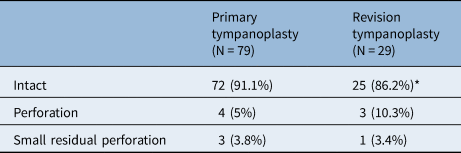
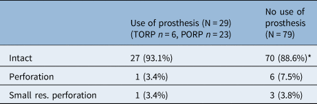
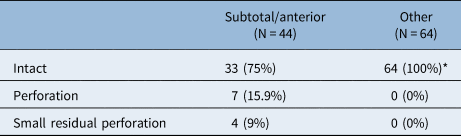
The success rates of the two groups (primary and revision tympanoplasty) are 91.13 per cent and 86.2 per cent, respectively. No statistically significant difference in success was observed (p = 0.621) (Table 1) between primary and revision tympanoplasties. Likewise, no correlation was present between the graft integration rate and the use of prosthesis (p = 0.686) (Table 2). All failed cases involved sub-total perforations with anterior extension, an association that seems to be statistically significant (p = 0.0004) (Table 3). No noticeable complications were recorded in any of the cases.
Graft integration rates between primary and revision tympanoplasty groups

Pearson's chi-square test *p = 0.621
Correlation between graft integration and the use of prosthesis

TORP = total ossicular replacement prosthesis; PORP = partial ossicular replacement prosthesis; Pearson's chi-square test: *p = 0.686
Graft integration and presence of a sub-total and/or anterior perforation

Pearson's chi-square test: *p = 0.0004
Regarding the hearing outcome, only patients with accessible audiometry records before surgery and six months post-operatively were recruited. Suitable records were available in 71/108 patients. Pre-operatively, all patients had conductive hearing loss of different severity.
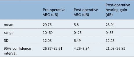
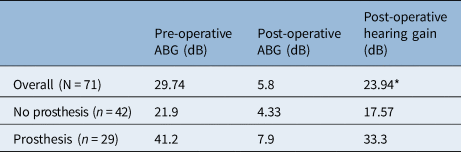
The average pre-operative ABG in the involved ear was 29.75 dB, with a minimum of 10 dB and a maximum of 60 dB. The average post-operative improvement of conductive hearing loss was 23.95 dB, with a post-operative average ABG of 5.8 dB (Tables 4 and 5). Overall, 94.3 per cent of the cases with audiological assessment were marked by a successful surgical outcome.
Hearing data, pre-operatively and six months after tympanoplasty (N = 71); ABG = air–bone gap; SD = standard deviation

Comparison of hearing data in relation to the use of prosthesis; ABG = air–bone gap

*Two paired t-test, t = 6.39, df = 49 p < 0.0001
Among the cases with a post-operative perforation, audiometry records were found in one patient with a large residual defect, and in three patients with small residual defects, two of whom appeared to have significant improvement in hearing, most possibly related to the concurrent use of a prosthesis. Twenty-nine of the included 71 cases (40.8 per cent) involved a simultaneous ossiculoplasty with prosthesis placement. A statistically significant difference was noted between the average hearing gain in two groups (p < 0.0001) (Table 5).
Discussion
Herein we assessed the surgical outcomes of tympanoplasty with the use of spilt two-layered, underlay cartilage–perichondrium technique. The overall success rate in our series (89.8 per cent) is considered amongst the higher ones reported in the literature in comparison to rates recorded for fascia grafts and compared with success rates reported in similar cartilage studies.Reference Jalali, Motasaddi, Kouhi, Dabiri and Soleimani4,Reference Yung, Vivekanandan and Smith7–Reference Bedri, Korra, Redleaf and Worku9 Given the 100 per cent success rates in non-total and/or large anterior perforations, it is sensible to recommend this technique for such perforations. With respect to total and/or anterior perforations, the reported success rates are lower, but still in agreement with those reported in the literature for such perforations.Reference Jung and Park10 Consequently, one could argue additional support of the grafts or even more robust reconstruction for such cases.
Although still controversial, the superiority of cartilage tympanoplasty has been widely supported in recent literature. In a metanalysis of 3606 patients, Jalali et al. observed success rates of 92 per cent for cartilage, compared to 82 per cent success in the fascia group.Reference Jalali, Motasaddi, Kouhi, Dabiri and Soleimani4 Yung et al. reported a success rate of 84.2 per cent and 80 per cent in primary myringoplasties using single-layer cartilage and fascia, respectively.Reference Yung, Vivekanandan and Smith7 However, based on the literature, the differences among different techniques of cartilage and composite grafts have not been thoroughly assessed. In a retrospective study of 120 patients, Demirpehlivan et al. highlighted the advantage of island cartilage–perichondrium graft in comparison with the temporalis fascia and the palisade cartilage grafts, with a difference of 97.7 per cent to 79 per cent and 80 per cent, respectively.Reference Demirpehlivan, Onal, Arslanoglu, Songu, Ciger and Can8 The double-layer graft technique, among others, was mentioned by Bedri et al. in a series of 622 patients, that concluded that there was a significantly higher success rate (90.1 per cent) in type I tympanoplasties compared with the single-layer fascia or cartilage technique (76 per cent and78 per cent, respectively).Reference Bedri, Korra, Redleaf and Worku9 A comparative study of 48 patients by Ismi et al. reported success rates with a difference of 96.1 per cent versus 68.2 per cent between the groups of single- and double-layered cartilage–perichondrium grafts.Reference Ismi, Gorur, Gur, Ozcan and Vayisoglu11 However, most of these studies did not thoroughly analyse the locations of the perforations or even included cases of nearly total closure, which are of clinical relevance.
The inclusion of revision surgery and cases with concomitant ossiculoplasty can be controversial, because in similar studies, the sample typically includes type I tympanoplasties. The reason we did not exclude revision cases was to focus on the effects of the graft material, independent of other factors. Overall, revision cases are associated with poorer results.Reference Andersen, Aabenhus, Glad and Sørensen12 However, the difference in our series was not statistically significant between the groups of revision and primary surgery. Likewise, the use of a prosthesis did not seem to affect the anatomical surgical outcome of the procedure.
There is debate in the literature about the effects of size and site of the perforation in the surgical outcome of a tympanoplasty, however central perforations appear to have better surgical outcomes.Reference Bayram, Bayar Muluk, Cingi and Bafaqeeh5 Large, sub-total perforations can be challenging for surgeons, as the poor remnants of the tympanic membrane result in lack of stability and poor vascular supply to the graft. Literature supports the use of cartilage as a graft material of choice in large perforations.Reference Lou, Lou, Wang, Zhang, Hu and Chen13 Anterior perforations are also considered to be associated with higher rates of failure because of the combination of a reduced anterior border vascularisation along with poor visualisation during surgery. Both factors can result in post-operative graft necrosis and rejection.Reference Castelhano, Correia, Colaço, Reis and Escada14 In our study, this hypothesis was confirmed, as the total of cases with graft failure had a perforation of this category, whereas posterior, central and/or smaller defects had a graft success rate of 100 per cent. Additional support of the grafts anteriorly with the use of dissolvable material or with little pieces of cartilage could be considered, depending on the technique used.
Overall, we found the technique beneficial as the two-layer design with the cartilage lying under the perichondrium offered additional support to the reconstruction. The surgeon has found this useful in cases with suboptimal middle-ear ventilation. While such factors were not formally assessed in our study, the authors have found the presented technique useful in challenging middle-ear situations.
• The split two-layer cartilage–perichondrium technique represents an alternative method of utilizing cartilage and perichondrium for tympanoplasties for perforations of any size
• In a series of more than 100 patients, the technique had 90 per cent success rates for total closure and approximately 94 per cent for no need of further intervention, while it also combined favourable audiological outcomes
• The success rates are similar for both primary and revision cases
• All failures were seen in sub-total tympanic membrane perforations with anterior extension, which appears to be a relative limitation of the technique, considering that such perforations are linked to lower success rates, regardless of the technique
• Where indicated, an ossicular prosthesis can be used simultaneously for optimizing audiological outcomes
Overall, the improvement in conductive hearing loss, as reflected by the ABG closure rates, is considered to be significant. However, we noticed that the biggest improvement was achieved in patients who had a prosthesis, a result mostly expected. It is well known that disruptions in the ossicular chain can affect hearing to a larger degree than defects of the tympanic membrane.Reference Lou, Lou, Wang, Zhang, Hu and Chen13,Reference Vaidya, Sharma and Singh15 Therefore, the optimal ABG closure is expected to be achieved in cases with a prosthesis placement, along with the restoration of the integrity of the tympanic membrane. Nonetheless, the hearing gain in type I tympanoplasty was also significant, with an average gap closure of 17.57 dB. In 20 cases (47.6 per cent) a full gap closure was achieved, with the postoperative gap minimised to zero.
The main limitation of our study is that it is not a case-control study, so no comparison to other techniques was conducted. Moreover, various potential risk factors affecting the outcome were not possible to be analysed further. However, these limitations, which are linked to the retrospective nature of our study and to the number of factors that could affect the outcome of a tympanoplasty, are difficult to quantify. We did include a large number of consecutive cases, performed and assessed in a standardised manner, in an attempt to minimise such limitations.
Conclusions
The use of the spilt two-layer cartilage–perichondrium underlay technique in tympanoplasty appears to be reliable and effective, with high success rates in both surgical and hearing outcomes for primary and revision cases. Total perforations remain a challenge. The efficacy of the method can prove a useful tool, particularly when a robust reconstruction is required.
Funding and financial disclosures
This research received no specific grant from any funding agency, commercial or not-for-profit sectors. No financial disclosures are declared.
Competing interests
None declared.
Ethical standards
The study was approved by the Research Ethical Committee as a retrospective study.
Author contributions
CM: data collection, manuscript preparation revision and editing; AM: data collection, statistical analysis, manuscript preparation; GK: conceptualisation, data analysis, manuscript revision and editing, supervision; all authors approved the final version.