Aortopulmonary window is a rare anomaly accounting for 0.2–0.6% of all congenital heart malformations. Reference Kutsche and Van Mierop1,Reference Richardson, Doty, Rossi and Ehrenhaft2 It refers to an intrapericardial communication between the ascending aorta and the pulmonary trunk before its bifurcation. This defect is caused by abnormal development of the spiral septum or conotruncal ridges that normally grow to divide the ascending aorta from the pulmonary trunk. Reference Kutsche and Van Mierop1 Aortopulmonary window may be an isolated anomaly or can be associated with additional cardiac defects such as ventricular septal defect, aortic arch anomalies, and tetralogy of Fallot in about half of the patients. Reference Li, Bin, Jiang and Shuang3
Classification of aortopulmonary window is descriptive according to the location of communication within the main pulmonary artery and extension towards the semilunar valves and the pulmonary bifurcation.
Here, we describe a very rare case of aortopulmonary communication that cannot be placed in a category within the typical classification system. An unusual type of aortopulmonary communication between the main pulmonary artery and the proximal ascending aorta was identified by fetal echocardiography in association with a right aortic arch and crossed pulmonary arteries at 23 gestational weeks.
Case
Prenatal findings
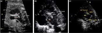
A 28-year-old woman, with no remarkable past medical history, was referred for fetal echocardiography at 22 gestational weeks due to identification of a right aortic arch during a routine anomaly scan. Fetal echocardiography at 23 gestational weeks revealed a normal fetal heart size in the four chambers view. Both atrioventricular and ventriculoarterial connections were concordant. The great arteries were normally related. There was no structural intracardiac abnormality. The three-vessels and three-vessel and trachea views revealed a large communication between the ascending aorta and the main pulmonary artery before its bifurcation (Fig 1a). The communication was above the semilunar valves and formed a tunnel-like connection rather than a window.
Fetal echocardiography ( a ) and postnatal echocardiography ( b and c ) showed a tubular AP window (APW) communicating the main pulmonary artery (MPA) and ascending aorta (Ao).

Although it was originally considered as the classical Type-I aortopulmonary window, it was accepted that the aorta and the pulmonary artery were related to each other through a tunnel-like tubular structure in a detailed ultrasound interrogation. Hence, it was considered aortopulmonary tunnel instead of window. In addition to this unusual communication between the two vessels, a right aortic arch and the absence of the ductus arteriosus were also noted. The origin of left pulmonary artery was on the right side of the pulmonary trunk and superior to the right pulmonary artery, with the pulmonary artery branches crossing one another. No extracardiac abnormality was identified. Karyotype and fluorescence in situ hybridisation analysis for 22Qdel were normal.
Postnatal follow-up
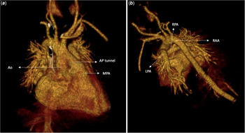
A male neonate weighting 3200 g was delivered by a caesarean section with Apgar scores of 8 and 9 at 1 and 5 minutes, respectively. Intracardiac anatomy, the heart size, and function were all normal on transthoracic echocardiography performed within 3 hours of birth. A tunnel-like aortopulmonary communication, a right aortic arch, crossed pulmonary arteries, and an absent ductus arteriosus were confirmed, consistent with the prenatal diagnosis (Fig 1b and c). A CT scan verified all echocardiographic findings and mirror-image branching pattern of the aortic arch (Fig 2). Follow-up physical examination demonstrated tachypnoea, abdominal breathing, and intercostal retractions. By auscultation, a machinery-type murmur was appreciated at the left upper sternal border, similar in quality to that found with patent ductus arteriosus. Treatment with diuretics and angiotensin-converting enzyme inhibitor was initiated, and a surgical repair was performed on the 12th day of life. The previously identified cardiac anomalies were verified by direct visual inspection and dissection during the operation. The intraoperative macroscopic inspection of the aortopulmonary tunnel bore a resemblance to the wall of an elastic artery, like the main pulmonary artery or the aorta. In fact, histopathological assessment of the AP tunnel specimen revealed the same structural elements as seen in the wall of the aorta. The patient underwent division and suturing using side-biting clamps, without cardiopulmonary bypass. After the successful operation, the infant was discharged home on the 8th postoperative day. The infant remained clinically well at 1-year follow-up without the need for any further intervention.
3D reconstruction of CT shows tubular aortopulmonary communication like a tunnel ( a ) and crossed pulmonary arteries ( b ).

Discussion
Any form of aortopulmonary window is a challenge for fetal diagnosis unless the specific views are obtained. This case describes a variation in the anatomy and hence the interest. Our case highlights the importance of identifying such a rare anomaly before surgery in order to decide on the best operative technique for repairing the communication.
Aortopulmonary window is characterised by a connection between the ascending aorta and the pulmonary trunk prior to its bifurcation. Fetal echocardiographic study helps to identify the morphological features of the defect, as well as its type, location, and size, along with other associated cardiac anomalies. Visualisation of a communication between the ascending aorta and the main pulmonary artery, in the short-axis view of aortopulmonary septum, high short-axis view of the great arteries, three vessel and three vessel trachea views, led us to the prenatal diagnosis by fetal echocardiography.
Richardson et al classified aortopulmonary window into three types. Reference Richardson, Doty, Rossi and Ehrenhaft2 Type I have only a minor rim towards the semilunar valves. Type II is located halfway between the pulmonary valve and its bifurcation. Type III extends up to the origin of the right pulmonary artery from the bifurcation. The most common type of aortopulmonary window is the Type 1. Reference Richardson, Doty, Rossi and Ehrenhaft2,Reference Tkebuchava, von Segesser and Vogt4 The aortopulmonary window can range from small, circular communication to total absence of the septum between the aorta and pulmonary artery. Rarely, some window defects have a tubular communication. Defects with tubular communication were rarely reported in the literature; the first reported case was described by necropsy and the second by echocardiography. The first case with tunnel like aortopulmonary window was described by Ho and colleagues in their series of 25 specimens. They noted a tunnel-like defect opening just above the commissural ridge of the right coronary sinus. Chidambarathanu et al reported the second case diagnosed with a tubular structure, connecting the aorta and MPA, measuring about 7 mm in length. Reference Ho, Gerlis, Anderson, Devine and Smith6 They noted that the intrapericardial location of the channel is sufficient to distinguish it from a persistent arterial duct. In our case, the tubular connection was in the intrapericardial location between the ascending aorta and the main pulmonary artery before its bifurcation. In light of the previous description by Ho and colleagues Reference Chidambarathanu, Agarwal, Hussain, Brown and Anderson5 and the other findings in the case described by Chidambarathanu, Reference Ho, Gerlis, Anderson, Devine and Smith6 this anomaly may be described as an aortopulmonary tunnel instead of an atypical aortopulmonary window, or a tubular window. Gowda et al reported the surgical results of 55 patients in which they noted that there was a medium-sized tubular window in three cases. Reference Gowda, Gajjar and Rao7 However, specific anatomical detail of these cases were not mentioned.
Although our case is the unusual type of aortopulmonary communication, its combination with the right aortic arch, a crossed pulmonary artery, and an absent ductus arteriosus have not been previously described. The association of an aortopulmonary window and a right aortic arch is rarely reported. Yen ho et al found right aortic arch in 1 specimen in 25 necropsy cases with aortopulmonary window and Gowda et al in 2 of 55 cases. Reference Chidambarathanu, Agarwal, Hussain, Brown and Anderson5,Reference Gowda, Gajjar and Rao7 However, no data were found on the association with the crossed pulmonary artery.
The prognosis of an uncorrected window is poor with 40% mortality rate within the first year of life. Various surgical techniques can be applied depending on the size of the communication, with or without cardiopulmonary bypass, by using transaortic or transpulmonary patches. We propose that in cases with tubular structure or tunnel-like aortopulmonary window, simple division technique should be preferred. Awareness of this unusual window type can guide surgeons to choose the best technique while repairing the defect.
Conclusion
It should be noted that unlike the usual aortopulmonary window cases, there may be rare and atypical aortopulmonary connections. The presence of a connection via a tubular structure between the ascending aorta and the main pulmonary artery before bifurcation can be described as an aortopulmonary communication, aortopulmonary tunnel, or tubular aortopulmonary window. This case illustrated that aortopulmonary window cases need to be redefined and reclassified according to the shape as well as localisation of the connection between the two great arteries. Identification of the anatomy of an aortopulmonary communication prior to any intervention is essential to determine the type of most effective surgical repair.
Supplementary material
To view supplementary material for this article, please visit https://doi.org/10.1017/S1047951121002225
Acknowledgements
The authors would like to thank Prof. Dr. Orhan Uzun for constructive criticism and proofreading of the manuscript.
Financial support
This research received no specific grant from any funding agency, commercial, or not-for-profit sectors.
Conflicts of interest
None.
Ethical standards
The authors assert that all procedures contributing to this work comply with the ethical standards of the relevant national guidelines and Helsinki Declaration of 1975, as revised in 2008.