Absence of the connections between the coronary arteries and the aorta are known to exist in the setting of pulmonary atresia with intact ventricular septum, but are very rare. We recently encountered a case that also demonstrated persistence of the left superior caval vein, which drained to an unroofed coronary sinus with integrity of the atrial septum. The left coronary artery arose anomalously from the right pulmonary artery, while a fistula from the right ventricle supplied the right coronary artery.
Case report
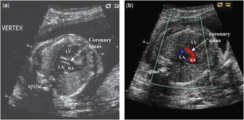
The stillborn male weighing 1510 grams was delivered at 30 weeks gestation. An ultrasonic examination at 20 weeks gestation had revealed what was thought to be hypoplasia of the left heart, along with a “strawberry-shaped” head, and ascities. Follow-up echocardiography 3 days later was limited due to technical issues, but revealed what was thought to be tricuspid atresia with discordant ventriculo-arterial connections and aortic atresia. The second echo, at 25 weeks, showed a large unroofed coronary sinus with reversal of flow from right to left, the atrial septum being intact (Fig. 1). The right atrium was seen to be dilated, and again tricuspid atresia was reported, with normal left ventricular function. Chromosomal analysis of the amniotic fluid showed a normal male karyotype.
The apical 4-chamber view (a) shows a dilated coronary sinus (arrow). The same image with color mapping (b) shows right to left flow (red) in the coronary sinus, which is unroofed, permitting drainage into the left atrium (blue).

Autopsy revealed anomalies of the head and neck, including a high forehead, hypertelorism, severe hypoplasia of the tip of the nose, redundant nuchal skin, and flattened ears. Anomalies of the limbs included slightly shortened fingers, bilateral crowding of the toes, and longitudinal folding of the plantar surface of the left foot. The features were deemed most consistent with Zellweger syndrome.1 External measurements were consistent with the estimated gestational age. There was desquamation over half of the body, and advanced autolysis of internal organs.
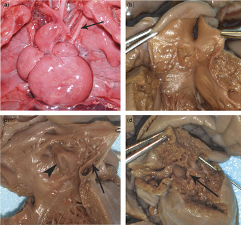
The heart and lungs had a combined weight of 38.1 grams. The atrial arrangement was normal. The superior and inferior caval veins were patent, and entered the right atrium in the normal fashion along with a patent left superior caval vein (Fig. 2a), which drained to the dilated coronary sinus. An adherent oval membrane with 2 fenestrations essentially occluded the atrial septum. The fenestrations, both less than 0.1 centimetres in diameter (Fig. 2c), were probe patent, being located at the superior border of the oval fossa, guarded by the infolding of the superior atrial septum. The fenestrations were visible only from the left side. The floor of the oval fossa itself bulged significantly from right to left. The right atrium was dilated, and its appendage extended along the entire right lateral heart border. There was a prominent terminal crest band and the pectinate muscles were thickened. Just left of the atrial septum, the coronary sinus was unroofed (Fig. 2c). The tricuspid valve was severely stenotic, inferiorly displaced, and had a circumference of 0.4 centimetres (Fig. 2d). The right ventricle was hypoplastic, with a widest diameter of 0.4 centimetres. The only component identified within the right ventricle was its inflow portion, with a single muscular band separating the outflow, which gave rise to a coronary arterial fistula (Fig. 2d). The ventricular septum was intact, and the pulmonic valve was atretic. No remnant of a pulmonary trunk was seen. A persistently patent arterial duct entered the aorta at an acute angle distal to the left subclavian artery. The pulmonary veins arose normally, and entered the left atrium in the usual fashion. The mitral valve had a circumference of 2.1 centimetres, possessing 2 leaflets which were normally formed and anchored. The mural thickness of the left ventricle measured 0.5 centimetres, and the ventricle was 2.1 centimetres long, being normally formed. The circumference of the aortic valve was 1.1 centimetres, with the valve possessing 3 normally formed leaflets. No coronary arterial connections to the aortic valvar sinuses were seen (Fig. 2b). An anomalous branch of the right pulmonary supplied the left coronary artery, which then pursued a normal course, branching to give rise to the circumflex and anterior interventricular arteries. A venous connection to the coronary sinus was seen on the diaphragmatic aspect of the heart in the same area as the unroofed portion of the coronary sinus, which perfused the ventricular wall in retrograde fashion. The ascending aorta bulged to the right, and was tethered by the arterial duct and the anomalous connection of the left coronary artery to the right pulmonary artery. Advanced autolysis of all internal organs precluded adequate histologic evaluation.
Pictures of the autopsied heart show (a) the left superior caval vein (arrow), (b) the aortic root with absence of coronary arterial orifices, (c) the left atrium, showing the unroofed coronary sinus (arrow) and the bulging intact atrial septum (arrow head), and (d) the hypoplastic right ventricle, showing the inferiorly displaced tricuspid valve (arrow), and a probe in the fistula to the right coronary artery.

Discussion
Pulmonary atresia with intact ventricular septum is a rare congenital cardiac anomaly, reported as occurring in 4.5 of every 100,000 live births.Reference Daubeney, Delany and Anderson2 The incidence of a persistent left superior caval vein in pulmonary atresia with intact ventricular septum is 2.7%.Reference Daubeney, Delany and Anderson2 The major morphological variation is the marked variation in size of the right ventricle, which is hypoplastic in nine-tenths of cases, being severely hypoplastic in just over half.Reference Daubeney, Delany and Anderson2 Fistulous communication with the coronary arteries are reported in three-quarters of these cases with severe hypoplasia, being found most commonly feeding the coronary arteries adjacent to the right ventricle.Reference Calder, Peebles and Occleshaw3 In our case, such a fistula was seen along the edge of the atretic outflow tract. The complete course of the right coronary and the fistula could not be traced at autopsy, and its demonstration would have required an angiocardiographic study. It has been postulated that the fistulous communications are a result of persistent fetal trabecular myocardial communications caused by high pressure generated in the ventricles.Reference Gerlis, Ho and Milo4, Reference Gittenberger-de Groot, Sauer, Bindl, Babic, Essed and Buhlmeyer5 The bulging right-to-left atrial septum, thickened and elongated right atrium, and dilated coronary sinus in our case attested to the increased pressure of the right heart.
The anterior and lateral components of the left ventricle were perfused by the anterior interventricular coronary artery, which was supplied by an anomalous connection from the right pulmonary artery. There were no direct connections from the aortic root to the coronary arteries. Such bilateral absence of proximal coronary arterial connections are an even rarer lesion, being reported in only 0.3% of patients with pulmonary atresia with intact ventricular septum.Reference Daubeney, Delany and Anderson2, Reference Calder, Peebles and Occleshaw3 The first case was reported in 1972,Reference Lenox and Briner6 and to the best of our knowledge, only 13 subsequent cases have been described.Reference Gerlis, Ho and Milo4, Reference Wald, Juraszek, Pigula and Geva7 Only the most recent infant, reported in 2006, has lived beyond infancy, and in this instance only because of successful cardiac transplantation.Reference Wald, Juraszek, Pigula and Geva7 To the best of our knowledge, this is the first reported case of pulmonary atresia with intact ventricular septum, absent aortocoronary arterial connections, and an intact atrial septum with an unroofed coronary sinus.
Intactness of the atrial and ventricular septums should be lethal in fetal development. In our case, it was the unroofing of the coronary sinus that allowed oxygenated blood to reach the left heart, with reversed flow through the arterial duct and anomalous connection with the right pulmonary artery providing coronary arterial perfusion that supported fetal life until the 30th week of gestation.