Background
Regional left ventricular systolic strain and 3-dimensional (3-D) global strain analysis have been reported to detect myocardial injury in patients with chronically increased left ventricular afterload before conventional 2-D echocardiographic parameters.Reference Di Salvo, Pacileo and Verrengia 1 – Reference Kowalski, Herbots and Weidemann 5 Selective reduction of the left ventricular long-axis function has been reported in patients with aortic stenosis.Reference Lam, Kaya, Li, Gatzoulis and Henein 6 , Reference Van Pelt, Stewart and Legget 7 Asymptomatic and mildly symptomatic aortic stenosis patients have a lower rest and exercise velocity–time integral and peak S’ velocity as determined by tissue Doppler techniques compared with normal controls.Reference Van Pelt, Stewart and Legget 7 One interpretation is that left ventricular longitudinal dysfunction might indicate pre-symptomatic myocardial abnormalities. The sub-endocardial fibres are predominantly longitudinal and are at greater risk from ischaemia compared with circumferential fibres in the mid-epicardial and sub-epicardial layers of the myocardium, especially in the presence of left ventricular dilation.
As with isolated analysis of the circumferential strain, longitudinal strain may fail to assess adequately the complex phenomenon of myocardial motion; prior studies using magnetic resonance imaging tissue tagging have attempted to overcome this problem by evaluating the left ventricular strain in three dimensions.Reference Stuber, Scheidegger and Fischer 8 – Reference Sandstede, Johnson and Harre 10 However, magnetic resonance imaging tissue tagging is technically demanding and expensive and has rather poor temporal resolution. Thus, Notomi et alReference Notomi, Setser and Shiota 11 compared quantification of the left ventricular rotation and torsion using tissue Doppler techniques to magnetic resonance imaging tissue tagging in patients with a variety of cardiac diseases and showed a close correlation with acceptable variability. Novel echocardiographic techniques such as 2-D speckle tracking echocardiography have recently been shown to provide reliable data on cardiac strain. However, this technique is not yet widely available and still has the limitation of being highly vendor dependent.
Accordingly, we sought to improve the understanding of myocardial mechanics – global longitudinal and circumferential strain and left ventricular rotation and twist – in children and adolescents with aortic valve disease using tissue Doppler techniques.
Methods
Patient selection
A total of 57 patients with aortic valve disease aged 19 ± 11 years examined in the echocardiography laboratory at the Children's Hospital Boston over a 6-month period were eligible for the study. Exclusion criteria included any additional cardiac abnormalities, left ventricular ejection fraction <55%, and segmental wall motion abnormalities. A total of 27 healthy subjects of similar age – 14 ± 5 years; p not significant – were also evaluated. The criteria used to classify the subjects as normal have been previously published.Reference Sluysmans and Colan 12 The height and weight were obtained at the time of echocardiography, and body surface area was calculated according to the formula of Haycock.Reference Haycock, Schwartz and Wisotsky 13
Classification of aortic valve disease
As all patients had a normal ejection fraction, aortic stenosis could be classified using continuous wave Doppler. Aortic stenosis was defined as mild when the peak instantaneous gradient was <40 mmHg or more than mild when the peak instantaneous gradient was ≥40 mmHg; aortic regurgitation was classified as mild when the vena contracta of the aortic regurgitation jet, adjusted for the square root of body surface area, was <3 mm/m2, left ventricular diameter and volume z-score were <2, and there was absence of pan-diastolic flow reversal in the descending aorta, or more than mild if any of these three criteria were not met. Patients with similar severity of aortic stenosis and aortic regurgitation in combination were classified as having mild or more than mild mixed aortic valve disease using the above criteria. Patients in whom one lesion predominated – more than mild – and the other was mild were classified as having the predominant lesion.
Data acquisition
All patients underwent a standard comprehensive 2-D echocardiogram, Doppler, and tissue Doppler examination. A series of short-axis images from base to apex, an apical four-chamber, and an apical long-axis image were recorded in 2-D tissue Doppler mode with a velocity equal to 20 cm/second. Two short-axis levels were defined: basal – mitral valve leaflets – and apical – distal to papillary muscles. The transducer was adjusted to achieve a circular left ventricular cross-section to ensure that the planes were normal to the major axis of the ventricle.
Image analysis
Image analysis was performed off-line using the Q-Lab software (Philips, Best, the Netherlands) and software developed at the Children's Hospital Boston (Marcus Laboratories). The endocardial and epicardial borders of the short-axis 2-D images were digitised frame by frame for calculation of the time-varying midwall radius and endocardial and midwall fibre circumferential strain. Endocardial and epicardial borders of the apical four-chamber and long-axis images were digitised at end-diastole and end-systole to derive the longitudinal strain. Left ventricular endocardial and epicardial volumes were calculated using a modified Simpson's rule algorithm. Left ventricular shape at end-diastole was quantified using: (1) the major-to-minor axis ratio, that is, sphericity, (2) the mass/volume ratio, and (3) the wall thickness/chamber dimension ratio. Meridional end-systolic stress was calculated as previously described.Reference Colan, Borow and Neumann 14 Basilar and apical short-axis tissue Doppler images were used to quantify the left ventricular torsion as described by Notomi et al.Reference Notomi, Setser and Shiota 11
Data analysis
The left ventricular volume and mass are reported as raw values and as z-scores relative to previously published normal data.Reference Sluysmans and Colan 12 The groups were stratified only according to disease category – controls, aortic stenosis, aortic regurgitation, and mixed aortic valve disease – and subgrouped according to disease severity – mild versus more than mild. Ventricular morphology and function were compared between the groups using analysis of variance. Interrelationships between ventricular torsion and the individual strain components – circumferential, longitudinal, and midwall circumferential – and end-systolic wall stress were analysed using linear regression analysis. We then conducted exploratory regression analyses of the interrelationships between these differences and global ventricular ejection performance and ventricular configuration.
Results
Patients
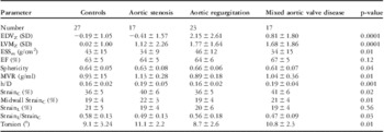
All patients who met the inclusion criteria and met no exclusion criteria were included for analysis on the basis of their diagnosis. No patients were excluded on the basis of image quality. The study included 57 patients with aortic valve disease and 27 controls. Patients were subdivided into six groups according to the type and severity of the valve disease as described above: nine patients with mild aortic stenosis, eight with more than mild aortic stenosis, 13 with mild aortic regurgitation, 10 with more than mild aortic regurgitation, 13 with mild mixed aortic valve disease, and four with more than mild mixed aortic valve disease (Tables 1 and 2).
Between-group comparisons stratified by severity of disease.

BSA = body surface area; EDV = end-diastolic volume; EF = ejection fraction; ESS = end systolic stress; LVM = left ventricular mass; MVR = mass-volume ratio
Between-group comparisons for left ventricular mechanical parameters.

EDV = end-diastolic volume; EF = ejection fraction; ESS = end systolic stress; LVM = left ventricular mass; MVR = mitral valve repair
Ventricular morphology and wall stress
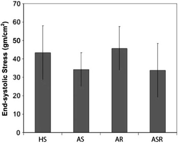
The most significant differences between the controls and the disease groups were the increased left ventricular end-diastolic volume and left ventricular end-diastolic volume z-score in the aortic regurgitation and the mixed aortic valve disease groups, and the increased left ventricular mass and left ventricular mass z-score in all three disease groups. Ventricular morphology was characterised by an increased mass/volume ratio and wall thickness/chamber dimension ratio in aortic stenosis and normal mass/volume ratio and wall thickness/chamber dimension ratio in aortic regurgitation, accompanied by normal sphericity in both. The magnitude of the observed differences varied as a function of the severity of the disease. As a result of the abnormalities in cardiac geometry, patients with aortic stenosis had significantly lower meridional end-systolic stress (Fig 1) and higher circumferential strain, as previously described.Reference Borow, Colan and Neumann 15 Mixed aortic valve disease was similar to aortic stenosis in ventricular morphology, wall stress, and function, but left ventricular dilation was significantly higher in the more than mild mixed aortic valve disease group.
Comparison of LV ESSm in patients with AS, AR, ASR, and HS. Myocardial afterload (ESSm) was significantly lower (p = 0.01) in both the AS and ASR groups. LV = left ventricle; ESS = end-systolic wall stress; AS = aortic stenosis; AR = aortic regurgitation; ASR = aortic stenosis/regurgitation or mixed aortic valve disease; HS = healthy subjects.

Patterns of strain and torsion
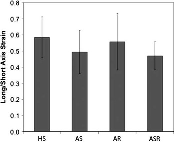
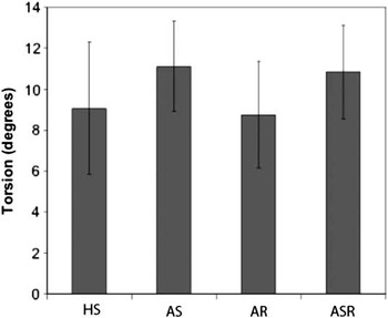
Circumferential strain and left ventricular rotation and twist (Fig 2) were significantly elevated in patients with aortic stenosis, whereas longitudinal strain tended to be reduced – p-value not significant. The increase in circumferential strain and left ventricular rotation and twist was proportional to the severity of stenosis. The ratio of longitudinal strain/circumferential strain was significantly lower in aortic stenosis (Fig 3). Circumferential strain, longitudinal strain, left ventricular rotation, and twist were similar in the aortic regurgitation patients and controls. Patients with mixed aortic valve disease exhibited a pattern of strain and left ventricular rotation and twist similar to those with aortic stenosis.
Comparison of the ratio of strainL with strainC and torsion in patients with AS, AR, ASR, and HS. The strain ratio was significantly lower (p = 0.03) in both the AS and ASR groups. AS = aortic stenosis; AR = aortic regurgitation.

Comparison of LV torsion in patients with AS, AR, ASR, and HS. Torsion was significantly elevated (p = 0.01) in both the AS and ASR groups. LV = left ventricle; AS = aortic stenosis; AR = aortic regurgitation.

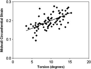
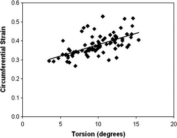
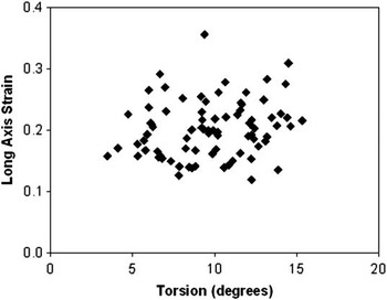
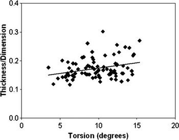
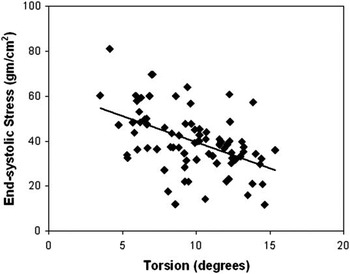
The left ventricular rotation and twist correlated strongly with midwall (r = 0.62, p < 0.0001; Fig 4) and endocardial (r = 0.61, p < 0.0001; Fig 5) circumferential strain, but not significantly with longitudinal strain (r = 0.18, p = 0.09; Fig 6). Therefore, left ventricular rotation and twist appear to contribute significantly to circumferential shortening, but negligibly to longitudinal shortening. Left ventricular rotation and twist are positively correlated with left ventricular wall thickness/chamber dimension ratio (r = 0.29, p = 0.007; Fig 7) and negatively with meridional end-systolic stress (r = −0.48, p < 0.0001; Fig 8).
There was a significant (p < 0.0001) correlation between LV midwall strainC and torsion in the total patient population. There was a significant (p < 0.0001) correlation between LV strainC and torsion (b) in the total patient population (all four groups combined). LV = left ventricle.

There was a significant (p < 0.0001) correlation between LV midwall strainC and torsion in the total patient population (all four groups combined). LV = left ventricle.

There was no significant correlation between LV strainL and torsion in the total patient population (all four groups combined). LV = left ventricle.

LV h/D ratio with positively correlated with torsion (r = 0.29, p = 0.007) in the total patient population (all four groups combined). LV = left ventricle.

LV rotation and twist are negatively correlated with ESSm (r = −0.48, p < 0.0001) in the total patient population (all four groups combined). ESS = end-systolic wall stress; LV = left ventricle.

We did not find evidence of non-normal distribution of the primary outcome variables, but the sample size was too small to permit definitive testing.
Discussion
In our young patients with aortic valve disease, multi-dimensional strain analysis has provided new and useful information concerning ventricular remodelling and patterns of ventricular mechanics that are characteristic of aortic stenosis and mixed aortic valve disease. In addition, because of its high temporal resolution, tissue Doppler has an advantage over magnetic resonance imaging and speckle tracking in the assessment of left ventricular multi-dimensional strain and torsion in children whose heart rate is higher than adult patients.
Strain analysis in patients with aortic valve disease
The disease-specific differential behaviour of circumferential strain versus longitudinal strain is of interest. Several authors have noted diminished longitudinal strain in aortic stenosis, thus suggesting that this may represent an early sign of myocardial deterioration.Reference Kowalski, Herbots and Weidemann 5 – Reference Van Pelt, Stewart and Legget 7 Carasso et alReference Carasso, Cohen and Mutlak 16 showed that aortic stenosis is associated with differential changes in left ventricular longitudinal and circumferential mechanics. Our findings confirm that reduced longitudinal strain in aortic stenosis represents an alteration in the pattern of ventricular mechanics with a shift in strain from longitudinal to circumferential, rather than a net diminution of fibre shortening. Indeed, in aortic stenosis patients, owing to chronic pressure overload, the ventricle undergoes concentric remodelling, resulting in reduced end-systolic wall stress. Therefore, our findings indicate that the increase in circumferential strain and torsion, together with the decrease in longitudinal deformation in our aortic stenosis patients, represents an adaptive response to the chronic pressure overload of aortic stenosis.
Our study suggests the importance of multi-dimensional strain analysis to identify early alterations of left ventricular function in aortic stenosis, before changes in ejection fraction occur.Reference Kowalski, Herbots and Weidemann 5 , Reference Lam, Kaya, Li, Gatzoulis and Henein 6 , Reference Carasso, Cohen and Mutlak 16 , Reference Carasso, Cohen and Mutlak 17 In addition, it confirms that individual strain measurements, reflective of ventricular wall motion in a single plane, are not sufficient to provide a complete representation of the complex motion of the ventricle. It is also worth noting that our patients with compensated aortic regurgitation did not exhibit alterations in circumferential strain, longitudinal strain, or torsion, even in the presence of left ventricular dilation. These results are consistent with previous studies using magnetic resonance imaging tissue tagging before aortic valve replacement in asymptomatic patients with chronic aortic regurgitation and normal left ventricular ejection fraction.Reference Pomerantz, Wollmuth and Krock 2 , Reference Ungacta, Dávila-Román and Moulton 18 On the other hand, abnormal myocardial structure can be seen microscopically even in patients with preserved indexes of systolic function.Reference Taniguchi, Kawamaoto and Kuki 19 – Reference Krayenbuehl, Hess, Monrad, Schneider, Mall and Turina 21 Moreover, abnormal accumulation of connective tissue – interstitial fibrosis – can affect the long-term outcome after mixed aortic valve disease despite improvement in left ventricular ejection fraction and a decrease in left ventricular size.Reference Pomerantz, Wollmuth and Krock 2 , Reference Krayenbuehl, Hess, Monrad, Schneider, Mall and Turina 21 Secondary eccentric hypertrophy, as seen in chronic aortic regurgitation, is invariably accompanied by an increase in interstitial connective tissue.Reference Krayenbuehl, Hess, Monrad, Schneider, Mall and Turina 21 , Reference Hein, Arnon and Kostin 22 Further studies are needed to define the relationship between such myocardial changes and echocardiographic parameters or symptoms.
Left ventricular torsion and ventricular load
Our data show that left ventricular torsion is correlated with meridional end-systolic stress, supporting the hypothesis that afterload has a significant effect on cardiac systolic rotation. There has been disagreement about the load dependency of left ventricular torsion,Reference MacGowan, Shapiro and Salvador 23 – Reference Moon, Ingels, Daughters, Stinson, Hansen and Miller 26 but our data confirm that fibre shortening is dependent on ventricular afterload.
Left ventricular torsion in patients with aortic valve disease
The cause of increased torsion in patients with aortic stenosis is likely to be multifactorial. Theoretical models predict greater torsion in conjunction with higher wall thickness/chamber dimension ratio and circumferential strain.Reference Taber, Yang and Podszus 27 In the aortic valve disease patients, the dominant influence appears to be myocardial afterload (Fig 8). Our findings support the observation that left ventricular pressure-overload hypertrophy is associated with increased ventricular torsion, and thus a reduction in basal and an increase in apical rotation.Reference Stuber, Scheidegger and Fischer 8 , Reference Carasso, Cohen and Mutlak 17 The physiological explanation for reduced rotation of the left ventricular base might be the increased stiffness of the atrioventricular valve plane in the presence of eccentric hypertrophy, with a compensatory increase of the apical rotation and torsion. In addition, it has been recently demonstrated that in patients with hypertension and diastolic dysfunction, cardiac torsion is significantly increased.Reference Park, Miyazaki, Bruce, Ommen, Miller and Oh 28 Owing to the change in the angle of the myofibres, sub-endocardial fibres exert a clockwise force, whereas sub-epicardial fibres exert a counterclockwise force. The larger moment arm for the epicardial fibres leads to a net counterclockwise torsion, although attenuated by the sub-endocardial fibres.Reference Partho, Jamil, Krishnaswamy, Bijoy and Khandheria 29 The increase in cardiac torsion in hypertension might be the result of sub-endocardial damage, reducing the normal forces opposing epicardial longitudinal fibre shortening, with a consequent increase in apical rotation.
Interestingly, left ventricular torsion did not differ between our patients with chronic aortic regurgitation and the controls. To the best of our knowledge, this is the first study that has assessed left ventricular torsion in young patients with chronic aortic regurgitation. These findings are consistent with the known adaptive response of the left ventricle to chronic volume overload, eccentric hypertrophy with normalisation of afterload, and maintenance of a normal left ventricular ejection fraction. Despite the fact that severity of aortic stenosis and aortic regurgitation was equivalent in our patients with mixed aortic valve disease, the ventricular mechanical behaviour was similar to aortic stenosis. Therefore, it is likely that aortic stenosis dominated the ventricular remodelling response and the functional characteristics of these ventricles.
Limitations
An inevitable drawback of tissue Doppler is the angle dependency of the acquired myocardial velocity data. We sought to minimise this issue by extracting tangential velocity from two sampling points – lateral and septal regions – of the left ventricle. Prior detailed magnetic resonance imaging has demonstrated in humans that peak strain and torsional components are typically greatest in the anterolateral wall and minimum in the septum. Owing to this regional inhomogeneity, tissue Doppler velocity data in the two regions observed could represent left ventricular rotational velocity while simultaneously correcting for cardiac translation. Speckle tracking technology overcomes the angle dependency of tissue Doppler by estimating rotation directly from tissue motion rather than from Doppler velocity estimates. However, speckle tracking techniques are derived from vendor-specific algorithms, and data obtained show poor agreement when analysis is performed on different echocardiographic equipment, with the relevant drawback of reducing the clinical applicability of the results in other centres. In addition, Doppler-based techniques have the advantage of having a high temporal resolution, which improves the assessment of left ventricular multi-dimensional strain and torsion in children whose heart rate is high.
Through-plane motion of the myocardium due to longitudinal shortening results in sampling from different myocardial segments throughout systole.Reference Gentles, Cowan, Occleshaw, Colan and Young 30 Tissue Doppler underestimated basal slice rotation more than apical rotation compared with magnetic resonance imaging and speckle tracking measurements, probably because longitudinal motion is greatest at the base and least at the apex. The close agreement between magnetic resonance imaging and echocardiography in Notomi's studyReference Notomi, Setser and Shiota 11 suggests that this effect is not large enough to cause clinical concern. The relative importance of total torsion versus torsion per unit length is controversial.Reference Henson, Song, Pastorek, Ackerman and Lorenz 31 We elected to report only total torsion because of significant technical obstacles to measuring the exact distance between the basilar and apical sample sites and the impact of through-plane motion on this measurement.
Conclusions
We have provided new insights into the mechanical adaptation of the left ventricle to chronic pressure and volume loading in young patients with aortic valve disease. In our study, increase in left ventricular torsion in patients with congenital aortic stenosis was associated with increased circumferential strain and reduced meridional end-systolic stress. Further longitudinal studies on systolic cardiac mechanics are needed to understand its relationship to the clinical status of the patient and the response to aortic valve surgery.
Acknowledgement
The authors sincerely thank Dr Andreina Santoro for her kind revision of the manuscript.