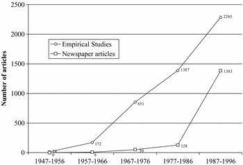


Autism is a neurodevelopmental disorder characterized by significant deficits in social reciprocity, language and communicative skills, and the presence of repetitive, formulaic behaviors and interests. There has been a striking increase in interest in this disorder, as documented by the exponential rise in both scientific and lay publications; see Figure 1. The causes of autism; mechanisms underlying the disorder; biological, cognitive, and behavioral features; and treatment efficacy have all provided a focus for investigation.
Figure 1.

Autism is diagnosed via behavioral rather than medical criteria, and involves multiple domains of functioning; the latter underlies its status as a pervasive developmental disorder. Neurodevelopment is one of the more promising areas of research in autism; studies have consistently shown differences in brain structure and function (although the specific findings have often varied across these studies). Although a number of labs have been working to develop animal models of autism spectrum disorder (ASD), to date, our knowledge is based on investigations with human subjects. The most important challenge for the future is to link differences in the developing brain to cognitive and behavioral features of the disorder.
Autism Spectrum Disorders (ASD)
The autism spectrum comprises a set of disorders (including autism, Asperger's disorder, Rett syndrome, childhood disintegrative disorder, and pervasive developmental disorder not otherwise specified) that span a wide spectrum of cognitive functioning (Folstein & Rosen-Scheidley, Reference Folstein and Rosen-Scheidley2001). While 50%–75% of individuals with autism experience comorbid intellectual disability, the remainder have intellectual abilities that range from low average to above average, and some 16% are able to function independently (Howlin, Goode, Hutton, & Rutter, Reference Howlin, Goode, Hutton and Rutter2004). Individuals with ASD have an uneven cognitive profile, performing well on most visuospatial (Happé & Frith, Reference Happé and Frith1996) and rote memory tasks, but poorly on tasks that demand planning, interpretation, and organizational aspects of memory (Bennetto, Pennington, & Rogers, Reference Bennetto, Pennington and Rogers1996; Ozonoff et al., Reference Ozonoff, Cook, Coon, Dawson, Joseph and Klin2004). Due to significant changes in diagnostic criteria from the 1980 to 1994 versions of the Diagnostic and Statistical Manual of Mental Disorders, broader public awareness of the disorder, and, of course, the possibility of actual increases in prevalence, the reported prevalence of the five diagnoses on the autism spectrum has increased dramatically (from 5 in10,000 in the 1960s to as many as 1 in 150 today).
Although the presentation of language skills in ASD is reviewed elsewhere in this volume, we must mention that the diagnosis of ASD involves marked abnormalities in acquisition and pragmatic language skills in as many as 25%–50% of individuals with the diagnosis (Eigsti, Bennetto, & Dadlani, Reference Eigsti, Bennetto and Dadlani2007; Rapin & Dunn, Reference Rapin and Dunn2003; Tager-Flusberg & Joseph, Reference Tager-Flusberg and Joseph2003). The remaining 50%–75% have a failure to develop language skills at any level. Just as the severity of the disorder is variable, language abilities in children with ASD range anywhere from nearly normal to profoundly impaired. Nonverbal individuals with ASD are a distinct minority in research reports, in part because an understanding of communicative impairments is hoped to lead to an understanding of failed communicative skills. In addition, verbal individuals are overrepresented in the imaging literature, largely because experimental demands (e.g., exposure to loud noise, the need to be physically still, the need to focus on sometimes tedious tasks) present a significant challenge. Thus, there are limitations on the utility of the work reviewed here for illuminating the mechanism underlying the failure to develop verbal skills in some individuals with ASD.
The ultimate goal of this review, and this area of research, is to increase our understanding of language skills in autism, and specifically, the neural underpinnings of the communicative impairments that characterize this disorder. We provide an overview of electrophysiological and brain imaging studies of language in autism, and we argue that a growing body of literature suggests that information integration is impaired in autism, as exhibited in reduced connectivity between neural networks and abnormally increased connectivity within the isolated individual neural assemblies (e.g., Rippon, Brock, Brown, & Boucher, Reference Rippon, Brock, Brown and Boucher2007).
We turn now to a brief technical review of the principal methodologies that are used in elucidating the neural bases of language skills in autism, encompassing both electrophysiological (electroencephalography [EEG] and magnetoencephalography [MEG]) and brain imaging (magnetic resonance imaging [MRI], functional MRI [fMRI], diffusion-tensor imaging [DTI]) techniques, and a review of structural brain differences in ASD that have been replicated across multiple samples and studies, followed by an overview of electrophysiological and brain imaging research related to language processes.
Electrophysiological Approaches
Electroencephalography (EEG)
The most common method for measuring the detection and encoding of auditory stimuli is electroencephalography (EEG), a noninvasive procedure that records electrical activity in the brain through electrodes placed on the scalp. Event-related potentials (ERPs) are electrophysiological brain responses that are time-locked to a stimulus event. In auditory tasks, ERPs represent the activation of neural structures in primary and association areas of the auditory cortex (Bomba & Pang, Reference Bomba and Pang2004). Typically, stimuli are repeated over multiple (often, thousands of) trials, to increase the signal-to-noise ratio so that a specific ERP can be detected (Friederici, Reference Friederici2004). Although not particularly sensitive to spatial localization (a limitation that may decrease as source localization techniques improve), ERPs are exquisitely sensitive to temporal changes. EEG is particularly valuable in studies of ASD because behavioral responses are not required, and because ERPs can detect differences in cortical activation when behavioral data shows no differences (Dunn, Gomes, & Gravel, Reference Dunn, Gomes and Gravel2007). This review will focus on auditory ERP studies because of their centrality to language processing in ASD.
When a sound is repeatedly presented at a fast rate, EEG records a waveform consisting of multiple high and low peaks including P1, N1, P2, N2, and P3 (Jansson-Verkasalo et al., Reference Jansson-Verkasalo, Ceponiene, Kielinen, Suominen, Jantti and Linna2003; Lepisto et al., Reference Lepisto, Kujala, Vanhala, Alku, Huotilainen and Näätänen2005). The names reflect the timing and polarity of these peaks; thus, P1 refers to a positive electrical signal, or waveform, occurring 100 ms poststimulus; N2 refers to a negative waveform occurring 200 ms poststimulus. These peaks, or components, are thought to reflect distinctive neural processes in response to a given stimulus. The shape, amplitude, and intensity of these components has a typical developmental time course; for example, P1 and N1 latency decreases and amplitude increases with age, reaching a stable shape by the end of adolescence (Cunningham, Nicol, Zecker, & Kraus, Reference Cunningham, Nicol, Zecker and Kraus2000). Decreases in latency (i.e., faster processing times) indicate increased efficiency and seem to be related to changes in the strength and efficiency of synaptic connections across neural networks. The P1, N1, P2, N2, and P3 components have been explored in ASD, in a variety of age ranges.
Magnetoencephalography (MEG)
Magnetoencephalography (MEG) is a method for quantifying the time course of neural activity based on the magnetic field elicited from activation in neural structures, much in the same fashion that EEG records electrical output from neural activation. MEG differs from EEG in several ways; first, the signal is not distorted by the bones of the skull, and the scalp; second, MEG is more sensitive to intracellular currents, whereas EEG is more sensitive to extracellular currents (a difference of time scale); and third, MEG does not require a physiological reference point. MEG's components map onto the ERPs found in EEG; for example, the mismatch negativity component (described later) has been studied using MEG to quantify low-level auditory processes, as well as stimulus anticipation, and even higher-order cognitive processes such as grammatical and semantic comprehension (Näätänen, Paavilainen, Rinne, & Alho, Reference Näätänen, Paavilainen, Rinne and Alho2007).
Brain Imaging Methods
For several decades, it has been possible to experimentally investigate neuroanatomical structure and function in typical and atypical development. Early techniques including computerized tomography (CT), positron emission tomography (PET), and single photon emission computerized tomography (SPECT) all entailed exposing subjects to ionizing radiation (Casey & Cohen, Reference Casey and Cohen1996). Although these methods have been employed in autism, MRI allows us to safely explore the developing brain, and to examine links between brain development and behavioral change.
Magnetic Resonance Imaging (MRI)
Magnetic resonance technologies have introduced a new set of tools for capturing features of brain development in living, developing humans. MRI provides exquisitely accurate anatomical images (Kennedy, Haselgrove, & McInerney, Reference Kennedy, Haselgrove and McInerney2003). With MRI, it is possible to scan children of all ages, though children younger than 5 or 6 are less able to stay sufficiently still without sedation. It is also safe to scan the same individual repeatedly, providing more precise measurement of neuroanatomical and functional changes due to developmental processes or clinical interventions.
Functional Magnetic Resonance Imaging (fMRI)
Whereas structural MRI is used to produce images of the brain that are useful for anatomical and morphometric studies, functional MRI allows in vivo measurement of brain activity by quantifying changes in blood oxygenation in the brain that are assumed to reflect changes in neural activity (Logothetis, Pauls, Augath, Trinath, & Oeltermann, Reference Logothetis, Pauls, Augath, Trinath and Oeltermann2001).
Diffusion tensor imaging (DTI)
DTI is an imaging process that uses the MRI modality in providing information about structural brain connectivity. The DTI signal arises from the relative diffusion of water molecules, which tend to align in a direction parallel to axon bundles (reflecting anisotropy, defined as the diffusion of water molecules in a direction parallel to nerve bundles). DTI is sufficiently sensitive to detect myelination (the white sheath around axons that increases the efficiency of interneuron signaling) and changes in white-matter microstructure (Watts, Liston, Niogi, & Ulug, Reference Watts, Liston, Niogi and Ulug2003). This technique has been used to assess the connectivity between structures and facilitates a focus on distributed neural networks rather than a simplistic modular set of isolated brain regions. DTI is an important new addition to investigations of language in ASD because language acquisition and processing demand significant coordination of information, and thus require the integration of information across brain regions. If there are structural impediments to connectivity, one would expect to find deficits in complex information processing (Minshew, Goldstein, & Siegel, Reference Minshew, Goldstein and Siegel1997). Such deficits are central to ASD.
Although MRI, fMRI, and DTI are perhaps the most influential tools in human developmental neuroscience today, it is important to note that they provide an indirect measure of brain structure and function. MRI studies of changes in the volume of a structure or amount of activity lack the resolution to definitively characterize the mechanism of change (e.g., dendritic arborization, cell death, synaptic pruning, myelination). As such, the imaging work reviewed here suggests biological substrates that must be tested with postmortem and animal models.
Brain Development and ASD
While a thorough description of the neuroanatomical findings in ASD is beyond the scope of this article (see, e.g., Bauman & Kemper, Reference Bauman and Kemper2005; Eigsti & Shapiro, Reference Eigsti and Shapiro2003), we will briefly summarize this literature as far as it is relevant for language skills. Imaging and pathological (e.g., autopsy) studies of brain development have indicated an absence of gross structural brain lesions. However, studies have consistently found an increase in cerebral volume in ASD, with some reports documenting increases in white matter (the myelinated axons of neurons that are critical in communication between brain cells; see, e.g., Hendry et al., Reference Hendry, Devito, Gelman, Densmore, Rajakumar and Pavlosky2006) and other studies finding increases in gray matter (the bodies of neurons and glial cells; see, e.g., Waiter et al., Reference Waiter, Williams, Murray, Gilchrist, Perrett and Whiten2004), with magnitude associated with symptom severity. Volumetric studies of lobes and regions are highly inconsistent.
One well-replicated finding is macrocephaly (increased volume of brain), as measured by head circumference (HC; Lainhart et al., Reference Lainhart, Piven, Wzorek, Landa, Santangelo and Coon1997; Piven et al., Reference Piven, Arndt, Bailey, Havercamp, Andreasen and Palmer1995). Children with autism have smaller HC at birth, but grow more rapidly and outstrip their typical peers in HC at age 14 months (Courchesne, Carper, & Akshoomoff, Reference Courchesne, Carper and Akshoomoff2003). Between 20% and 50% of individuals with ASD have a head circumference above the 97th percentile at some point in development (Courchesne et al., Reference Courchesne, Carper and Akshoomoff2003; Dementieva et al., Reference Dementieva, Vance, Donnelly, Elston, Wolpert and Ravan2005), and the degree of difference appears to predict cognitive impairment later in life (Akshoomoff et al., Reference Akshoomoff, Lord, Lincoln, Courchesne, Carper and Townsend2004).
In addition to differences in brain volume, studies have consistently documented abnormalities in the amygdala and related structures, areas thought to be central to emotional processing (Sparks et al., Reference Sparks, Friedman, Shaw, Aylward, Echelard and Artru2002). Surprisingly, MRI studies have documented both increased and decreased volume of this structure. In both children and adults, anatomical (postmortem) studies have indicated that brains in autism are characterized by small, densely packed cells in the limbic system, specifically the hippocampus, amygdala, subiculum, entorhinal cortex, septal nuclei, anterior cingulate gyrus, septum, and mamillary body (reviewed in Bauman & Kemper, Reference Bauman and Kemper2005). Another finding is a decreased number of Purkinje cells, which are large, densely branching neurons found in the cerebellum and brain stem (Bauman & Kemper). MRI studies have documented both increases (Sparks et al., Reference Sparks, Friedman, Shaw, Aylward, Echelard and Artru2002) and decreases (Hashimoto et al., Reference Hashimoto, Tayama, Murakawa, Yoshimoto, Miyazaki and Harada1995) in the cerebellum and parietal cortex (Courchesne et al., Reference Sparks, Friedman, Shaw, Aylward, Echelard and Artru2001). Decreased gray matter in the right paracingulate gyrus, left occipitotemporal cortex, and left inferior frontal sulcus, and increased gray matter in the left amygdala, right inferior temporal gyrus, left middle temporal gyrus, and cerebellum may be another correlate of autism (Abell et al., Reference Abell, Krams, Ashburner, Passingham, Friston and Frackowiak1999). With the exception of the cerebellum, these brain structures project to or from the amygdala, which may impact the regulation of emotional states.
In sum, structural neuroanatomical studies show that specific areas of the brain are consistently implicated in autism; however, the nature of these abnormalities varies. The most consistent finding thus far appears to be increased brain size in children with autism, which suggests a more global abnormality in brain development (Dementieva et al., Reference Dementieva, Vance, Donnelly, Elston, Wolpert and Ravan2005).
Functional Connectivity
In the course of development, neural systems typically become more stable, at a cost of increased rigidity. The brain goes through both a brain-building phase including cell migration and myelination, and a “pruning” phase, during which synaptic projections to inappropriate targets are removed to refine cell connectivity. If the process of synaptic pruning has gone awry in autism, one may expect excessive connections—that is, neurons that are connected to more other neurons than is typical. If cellular networks are pruned too early in development, it may prohibit the typical refining of connections and lead to impaired learning. Computational models of neural networks have demonstrated that these overly connected systems present a liability, not an advantage, as they would learn less efficiently (McClelland, Reference McClelland2000).
Several studies using DTI to assess connectivity in ASD have found low anisotropy (i.e., a more disorganized alignment of water molecules along axons, reflecting less efficient transmission along nerve bundles) of the corpus callosum (Alexander et al., Reference Alexander, Lee, Lazar, Boudos, DuBray and Oakes2007), a structure critical for interhemispheric communication and brain lateralization; of the superior temporal gyrus and temporal stem (Lee et al., Reference Lee, Bigler, Alexander, Lazar, DuBray and Chung2007), structures that are important in language and sound processing; and of the ventromedial prefrontal cortices, the anterior cingulate gyri, and the temporoparietal junction, all structures that are critical for social cognition (Barnea-Goraly et al., Reference Barnea-Goraly, Kwon, Menon, Eliez, Lotspeich and Reiss2004). In contrast to these findings of reduced connectivity in adolescents and adults, a recent study in children with ASD found increased anisotropy, reflecting an early, accelerated maturation of white matter, particularly in the left hemisphere and in the frontal lobes (Ben Bashat et al., Reference Ben Bashat, Kronfeld-Duenias, Zachor, Ekstein, Hendler and Tarrasch2007). Taken together, these preliminary findings suggest that the microstructure of white matter (and thus, brain connectivity) in autism is atypical, possibly reflecting a developmentally early overgrowth followed by a significant reduction in connectivity.
Consistent with this physiological finding, several studies have documented differences in the distribution of white matter, including decreases in volume, supporting findings of underconnectivity between or within cognitive, perceptual, and motor circuits in ASD (Herbert et al., Reference Herbert, Ziegler, Deutsch, O'Brien, Kennedy and Filipek2005; Waiter et al., Reference Waiter, Williams, Murray, Gilchrist, Perrett and Whiten2005). Recent work examining the water content of neural tissues has shown increases in overall cerebral white matter for school-age children with autism, as well as increases in gray matter and associated white matter of parietal lobes and in white matter of frontal lobes (Hendry et al., Reference Hendry, Devito, Gelman, Densmore, Rajakumar and Pavlosky2006). White matter abnormalities might result from irregular development or maintenance of unnecessary connections (Just, Cherkassky, Keller, & Minshew, Reference Just, Cherkassky, Keller and Minshew2004), or atypical neuronal migration (Bailey et al., Reference Bailey, Luthert, Dean, Harding, Janota and Montgomery1998). These white matter changes appear to be primarily postnatal (Herbert et al., Reference Herbert, Ziegler, Makris, Filipek, Kemper and Normandin2004) and may provide a cellular (cytoarchitectural) basis for the differences in functional connectivity reviewed here.
As one of the largest and most prominent axonal pathways in the brain, the corpus callosum is responsible for interhemispheric transfer of information. Structural MRI studies have indicated that the body and posterior regions of the callosum are significantly decreased in autism (Chung, Dalton, Alexander, & Davidson, Reference Chung, Dalton, Alexander and Davidson2004; Waiter et al., Reference Waiter, Williams, Murray, Gilchrist, Perrett and Whiten2005) and that callosal changes may play a significant role (Steiner, Guerreiro, & Marques-de-Faria, Reference Steiner, Guerreiro and Marques-de-Faria2004). Differences in the corpus callosum, along with reduced connectivity in tracts adjoining ventromedial prefrontal cortex, anterior cingulate, superior temporal sulcus, and occipitotemporal tracts, have been documented in ASD using DTI (Barnea-Goraly et al., Reference Barnea-Goraly, Kwon, Menon, Eliez, Lotspeich and Reiss2004).
A series of recent studies has demonstrated atypical functional connectivity in autism using fMRI (Just et al., Reference Just, Cherkassky, Keller and Minshew2004; McAlonan et al., Reference McAlonan, Cheung, Cheung, Suckling, Lam and Tai2005; Villalobos, Mizuno, Dahl, Kemmotsu, & Muller, Reference Villalobos, Mizuno, Dahl, Kemmotsu and Muller2005). For example, Just and colleagues asked high-functioning adults with autism to identify the actor in a series of passive and active sentences. Compared to typically developing adults matched on verbal IQ, the autism group produced more activation in Wernicke's area (left laterosuperior temporal gyrus) and less activation in Broca's area (left inferior frontal gyrus). In addition, the authors assessed the relative correlations in brain activity across a set of prespecified neural areas and found that there was less correlation in activity for the autism group, supporting the notion that language-related processes are poorly integrated in autism.
Data from several methods (DTI, fMRI, anatomy) support the hypothesis that brain function in ASD is constrained by less efficient communication between right and left hemispheres. Courchesne, Redcay, Morgan, and Kennedy (Reference Courchesne, Redcay, Morgan and Kennedy2005) have suggested that frontal, cerebellar, and temporal pathologies occur before higher order neural systems first begin to form their circuitry. These pathologies may lead to impairments in the ability of the frontal cortex to integrate information from diverse functional systems (emotional, sensory, autonomic, memory, etc.) and provide context-based and goal-directed feedback to lower level systems.
Neurobiological Studies of Language in ASD
Electrophysiological Findings
A variety of studies suggest that ASD is characterized by atypical processing of both speech and nonspeech auditory information. Behavioral observations strongly support this hypothesis; findings include hyposensitivity and hypersensitivity (Hoshino, Reference Hoshino1980) and savantlike skills in music perception (Heaton, Reference Heaton2003; Mottron & Burack, Reference Mottron, Burack, Burack, Charman, Yirmiya and Zelazo2001; O'Riordan & Passetti, Reference O'Riordan and Passetti2006).
Early processing of auditory information seems to comprise at least three stages: sound encoding (detecting a sound and encoding its acoustic properties), discrimination (comparing and distinguishing novel sounds), and pre-attentional orienting (shifts of attention toward an auditory stimulus outside the current focus of attention; Lepisto et al., Reference Lepisto, Kujala, Vanhala, Alku, Huotilainen and Näätänen2005). The three stages, which seem to require no conscious effort, are typically quantified with an oddball paradigm. In the ERP “oddball” paradigm, subjects repeatedly hear a “standard” sound, and infrequently hear a “deviant” sound qualitatively differing (e.g., in frequency) from the standard sound.
Sound Encoding
Sound detection and encoding of acoustic properties such as pitch, intensity, and duration form the necessary basis of complex auditory processing and, thus, language processing. This initial stage of auditory processing is reflected in a set of short latency (within 250 ms of the stimulus onset) components, the P1, N1, P2, and N2.
Relative to typically developing controls, children and adolescents (ages 6–13 across studies) and adults have shown no differences in P1 latency (i.e., time to onset) for speech sounds, suggesting that sensory sound processing of speech stimuli is intact in ASD. In contrast, nonspeech stimuli including clicks (Buchwald et al., Reference Buchwald, Erwin, Van Lancker, Guthrie, Schwafel and Tanguay1992) and tones (Ceponiene et al., Reference Ceponiene, Lepisto, Shestakova, Vanhala, Alku and Näätänen2003; Gage, Siegel, Callen, & Roberts, Reference Gage, Siegel, Callen and Roberts2003; Jansson-Verkasalo et al., Reference Jansson-Verkasalo, Ceponiene, Kielinen, Suominen, Jantti and Linna2003; Lepisto et al., Reference Lepisto, Kujala, Vanhala, Alku, Huotilainen and Näätänen2005) elicit diminished P1 amplitudes, suggesting some abnormality in the processing of basic auditory information. This finding of decreased P1 amplitude has not, however, been replicated in MEG studies (Gage et al., Reference Gage, Siegel, Callen and Roberts2003; Oram Cardy, Flagg, Roberts, & Roberts, Reference Oram Cardy, Flagg, Roberts and Roberts2005; Tecchio et al., Reference Tecchio, Benassi, Zappasodi, Gialloreti, Palermo and Seri2003), which may be a reflection of MEG's temporal resolution or of the fragility of the effect.
A second ERP component, N1, is thought to reflect early encoding of physical stimulus features and to arise from processing in the supratemporal plane of the auditory cortex, auditory association areas in the temporal lobe, and frontal and parietal networks (Giard et al., Reference Giard, Perrin, Echallier, Thevenet, Froment and Pernier1994). The activity of frontal and parietal networks suggests that N1 is an index of early attention-shifting mechanisms.
Although studies of the N1 component have been limited by sampling and methodological concerns, findings suggest that N1 amplitude is diminished in ASD for nonspeech sounds, with no difference in latency (Lincoln, Courchesne, Harms, & Allen, Reference Lincoln, Courchesne, Harms and Allen1995). One study comparing ASD to typical controls found significantly longer latency in response to tones in the ASD group, but only in a limited portion of the N1, called N1c, which the authors interpreted as indicating a slowed transmission of information within secondary auditory cortex (Bruneau, Roux, Adrien, & Barthelemy, Reference Bruneau, Roux, Adrien and Barthelemy1999). Consistent with the P1 results, the N1 results suggest an impairment in low-level auditory encoding.
Developmental changes may have also played a role in the variability and null findings that have characterized N1 results as a whole. The “generator” (the population of neurons that appears to contribute the electrical signal) of the N1 shifts in location over development, with children having more frontal generators until around age 16, when the location shifts more centrally (Bruneau, Roux, Guerin, Barthelemy, & Lelord, Reference Bruneau, Roux, Guerin, Barthelemy and Lelord1997).
The N2 and P2 components are also thought to reflect processing of specific features of auditory information. Although several studies have examined these waveforms in ASD (e.g., Ceponiene et al., Reference Ceponiene, Lepisto, Shestakova, Vanhala, Alku and Näätänen2003; e.g., Lepisto et al., Reference Lepisto, Kujala, Vanhala, Alku, Huotilainen and Näätänen2005), results have been conflicting, and no conclusions can be drawn at this point.
Impairments in sound encoding could clearly contribute to language impairments in ASD. For example, findings of greater amplitude and decreased latency of N1 during active stimulus paradigms could reflect greater attention to potentially irrelevant stimulus distinctions.
Sound Discrimination
After the acoustic properties of sounds have been encoded, they must be differentiated from one another. The second stage of auditory processing involves sound discrimination, and it is typically studied using mismatch negativity (MMN, e.g., Näätänen & Winkler, Reference Näätänen and Winkler1999). The repetition of a stimulus leaves a sensory memory trace that represents the properties of the standard stimulus. MMN reflects the detection of the difference between the memory trace and a novel deviant stimulus. Thus, whereas sound encoding ERPs are evoked by both standard and deviant sounds, sound discrimination-related MMN is a response to novel sensory input and is calculated as the difference in processing of standard and deviant stimuli.
MMN peaks between 150 and 250ms after stimulus onset (Seri, Pisani, Thai, & Cerquiglini, Reference Seri, Pisani, Thai and Cerquiglini2007) and is thought to have two main generators. The first, in the supratemporal area of auditory cortex, represents discrimination of acoustic features, and the second, located in frontal or prefrontal cortical areas, reflects involuntary switching of attention to a change in stimulus (Gomot et al., Reference Gomot, Bernard, Davis, Belmonte, Ashwin and Bullmore2006). Both the temporal and frontal components of MMN are evident by 5 years of age. The temporal component has greater amplitude in school-age children relative to adults, whereas the frontal component seems to mature in early childhood. By age 10, MMN is similar in both amplitude and latency to adults (Gomot, Giard, Roux, Barthelemy, & Bruneau, Reference Gomot, Giard, Roux, Barthelemy and Bruneau2000).
A number of studies have presented children with ASD aged 5–13 (and in one study, Kasai et al., Reference Kasai, Hashimoto, Kawakubo, Yumoto, Kamio and Itoh2005, adults with ASD,) with vowels, syllables, and simple or complex tones. When control groups are matched for chronological and mental age, findings have demonstrated an enhanced MMN amplitude (Ferri et al., Reference Ferri, Elia, Agarwal, Lanuzza, Musumeci and Pennisi2003; Lepisto et al., Reference Lepisto, Kujala, Vanhala, Alku, Huotilainen and Näätänen2005) and, generally, longer latency of the MMN component (Ferri et al.; Jansson-Verkasalo et al., Reference Jansson-Verkasalo, Ceponiene, Kielinen, Suominen, Jantti and Linna2003; Oram Cardy et al., Reference Oram Cardy, Flagg, Roberts and Roberts2005). These studies differ in whether the tasks involve active listening (in which a response is required) or passive exposure. Based on their finding that individuals with ASD made discriminations in active but not passive conditions, contrary to findings in controls, Dunn et al. (Reference Dunn, Gomes and Gravel2007) suggested that detecting auditory changes requires explicit attention in ASD.
In general, these data suggest delays in ASD in the discrimination of speech, particularly vowel sounds. Importantly, several studies have found that the degree of atypicality in these early auditory processes correlated with ASD symptom severity in both children ages 1–4 (Kuhl, Coffey-Corina, Padden, & Dawson, Reference Kuhl, Coffey-Corina, Padden and Dawson2005) and adults (Kasai et al., Reference Kasai, Hashimoto, Kawakubo, Yumoto, Kamio and Itoh2005). In the Kuhl study, the ASD group also had a preference for nonspeech sounds, unlike typically developing controls.
Pre-Attentional Orienting
A third stage of auditory processing is characterized as involuntary pre-attentional orienting and involves a shift of attention toward a new stimulus occurring outside of the current focus of attention. This attentional shift is reflected in a long-latency ERP component (P3) and is linked to higher-level, modality-general cognitive processing (Gomot et al., Reference Gomot, Bernard, Davis, Belmonte, Ashwin and Bullmore2006). Researchers in the field of autism have often posited that children with ASD consciously prefer the sounds of nonspeech over speech (Klin, Reference Klin1992). However, abnormalities in this pre-attentional orienting would suggest that this preference is due to basic impairments in auditory processing rather than a conscious aversion or dispreference for speech. A lack of responsiveness to auditory stimuli, specifically speech, could decrease the quantity and salience of language experiences, with likely detrimental effects on language development.
In experimental paradigms, the P3 waveform (associated with pre-attentional or involuntary orienting) is elicited by a deviant stimulus that is perceptually salient (Ceponiene et al., Reference Ceponiene, Lepisto, Shestakova, Vanhala, Alku and Näätänen2003). P3 amplitude is thought to reflect both memory and the degree to which attentional resources are engaged; latency reflects stimulus processing and classification (Kutas, McCarthy, & Donchin, Reference Kutas, McCarthy and Donchin1977). Because shorter latencies are associated with better cognitive performance, it is critical to control for cognitive ability to clarify whether differences in latency reflect task difficulty or general intellectual ability (Polich & Martin, Reference Polich and Martin1992).
The P3 generators are located in dorsolateral prefrontal cortex, the temporoparietal junction, the posterior hippocampal region, and auditory cortex. The P3 is typically divided into the P3a (frontal lobe, hippocampus) and P3b subcomponents. P3a is highly responsive to novel information and is more prominent in tasks that use novel rather than deviant stimuli. The P3b is elicited during difficult discrimination tasks and is localized to the temporoparietal pathway (Polich, Reference Polich2004). Latency of P3b is highly variable during childhood, decreasing steadily until 15 years of age and reaching a plateau in early adulthood (Martin, Barajas, & Fernandez, Reference Martin, Barajas and Fernandez1988).
No differences in P3a amplitude have been found in response to simple or complex tones (Lepisto et al., Reference Lepisto, Silokallio, Nieminen-von Wendt, Alku, Näätänen and Kujala2006), though diminished P3a amplitudes have been found for changes in prosody (pitch, Lepisto et al., Reference Lepisto, Kujala, Vanhala, Alku, Huotilainen and Näätänen2005, Reference Lepisto, Silokallio, Nieminen-von Wendt, Alku, Näätänen and Kujala2006) and vowel categories (Lepisto et al., Reference Lepisto, Kujala, Vanhala, Alku, Huotilainen and Näätänen2005, Reference Lepisto, Silokallio, Nieminen-von Wendt, Alku, Näätänen and Kujala2006). Interestingly, in one study, children with ASD remembered more of the nonspeech than speech stimuli in a later recall task (Ceponiene et al., Reference Ceponiene, Lepisto, Shestakova, Vanhala, Alku and Näätänen2003).
No abnormalities in P3b latency have been found in ASD (Courchesne, Lincoln, Yeung-Courchesne, Elmasian, & Grillon, Reference Courchesne, Lincoln, Yeung-Courchesne, Elmasian and Grillon1989; Lincoln et al., Reference Lincoln, Courchesne, Harms and Allen1995), suggesting that the time required to process auditory stimuli is normal. In contrast, the amplitude of P3b was diminished across numerous studies (in both children ages 7–13 and adolescents ages 15–24), using tones (Lincoln et al.), novel sounds (Ciesielski, Ciesielski, Courchesne, & Elmasian, Reference Ciesielski, Ciesielski, Courchesne and Elmasian1990), clicks (Novick, Vaughan, Kurtzberg, & Simson, Reference Novick, Vaughan, Kurtzberg and Simson1980), phonemes (Dawson, Finley, Phillips, Galpert, & Lewy, Reference Dawson, Finley, Phillips, Galpert and Lewy1988), and words (Courchesne, Courchesne, Hicks, & Lincoln, Reference Courchesne, Courchesne, Hicks and Lincoln1985).
There are few EEG studies of adults with ASD, but a recent study found that adults with ASD were less efficient in localizing auditory information in space (Teder-Salejarvi, Pierce, Courchesne, & Hillyard, Reference Teder-Salejarvi, Pierce, Courchesne and Hillyard2005), and exhibited poor spatial tuning of the N1 (reflecting the initial stage of attention filtering) to irrelevant sounds. Less effective tuning of attention was also demonstrated by later sensory processes including P3 amplitude. These findings suggest a fundamental deficit in the spatial focusing of auditory attention in autism, a deficit that could underlie communicative deficits.
In addition to the highly automatic and mostly unconscious processes described earlier, there are later ERP components that are sensitive to semantic aspects of linguistic input. The N4 component (elicited 400 ms after stimulus onset) is speech-specific and is elicited when there is a mismatch between the semantic classification of the current word and the preceding word or context. Studies of ASD have demonstrated a N4 diminished amplitude to speech (Lepisto et al., Reference Lepisto, Silokallio, Nieminen-von Wendt, Alku, Näätänen and Kujala2006), idioms (Strandburg, et al., Reference Strandburg, Marsh, Brown, Asarnow, Guthrie and Higa1993), and tasks of semantic processing (Dunn & Bates, Reference Dunn and Bates2005).
Brain Imaging Studies of Language in ASD
There have been a few studies using functional brain imaging to investigate language processing in ASD. One fMRI study of adults with ASD found abnormal language-related responses in Broca's area (left inferior prefrontal cortex, BA45) and left middle temporal gyrus (BA21) during “deep” (semantic) vs. “shallow” (perceptual) processing of visually presented words (Harris et al., Reference Harris, Chabris, Clark, Urban, Aharon and Steele2006). Subjects with ASD also showed diminished activation to concrete versus abstract words. Data were consistent with the possibility that participants with ASD were performing more semantic and phonological processing during perceptual tasks, and were thus less able to suppress this processing. In a related semantic category task, a small sample of adults with autism was compared to matched controls on the ability to categorize a set of words as tools, colors, or feelings (Gaffrey et al., Reference Gaffrey, Kleinhans, Haist, Akshoomoff, Campbell and Courchesne2007). The autism group had less activation of left inferior frontal gyrus (Broca's area) than controls, but more activation in visual areas (extrastriate visual cortex bilaterally), suggesting an important role of perceptual components (possibly visual imagery) during semantic decision.
Moving from semantic knowledge to the social aspects of language, an fMRI study of pragmatic skills in ASD indicated that high-functioning children with ASD were behaviorally less able to determine whether a speaker was sincere or ironic, given either prosodic or contextual cues. The imaging data indicated that ASD participants showed increased activity within the network of regions (right inferior frontal gyrus, bilateral temporal cortex) also recruited in the typical control sample. When explicitly directed to do so, children with ASD seem to be able to recruit the normative neural tissues during a social language task (Wang, Lee, Sigman, & Dapretto, Reference Wang, Lee, Sigman and Dapretto2006). In a follow-up study, again using an irony decision task, Wang and colleagues (Wang, Lee, Sigman, & Dapretto, Reference Wang, Lee, Sigman and Dapretto2007) found that brain activity in the ASD group, but not controls, was modulated by explicit instructions to attend to facial expression and tone of voice only, suggesting that individuals with ASD are more likely to look atypical in passive task conditions.
As described previously, Just and colleagues used correlational approaches to show a decrease in synchronization between brain areas as adults with ASD performed a sentence comprehension task (judging the performer of the action in passive and active sentences; Just et al., Reference Just, Cherkassky, Keller and Minshew2004). The same group followed up with an fMRI study contrasting participants’ responses while making true/false judgments about series of low- or high-imagery sentences, demonstrating decreased synchrony between language and spatial areas in the autism group. In addition, the autism group had similar activations of parietal and occipital brain regions associated with imagery for both the low and high-imagery sentences (rather than just for high-imagery items, in controls; Kana, Keller, Cherkassky, Minshew, & Just, Reference Kana, Keller, Cherkassky, Minshew and Just2006).
Lateralization of Language-Related Activity
The lateralization (e.g., which hemisphere of the brain) of language functioning in ASD has been shown to be aberrant in multiple studies. Typically, MMN will show a greater amplitude over left hemisphere, reflecting the typical organization of language as a primarily left-hemisphere process (Studdert-Kennedy & Shankweiler, Reference Studdert-Kennedy and Shankweiler1970). In participants with ASD, Gomot and colleagues found that MMN was more diffuse across the left hemisphere, rather than showing the typical front and central locations (Gomot, Giard, Adrien, Barthelemy, & Bruneau, Reference Gomot, Giard, Adrien, Barthelemy and Bruneau2002). Kuhl and colleagues (Reference Kuhl, Coffey-Corina, Padden and Dawson2005) found that ASD children had positive rather than negative increases in left hemisphere, in response to novel syllables. Taken together, findings of a larger MMN amplitude for syllable change over the right hemisphere (Lepisto et al., Reference Lepisto, Silokallio, Nieminen-von Wendt, Alku, Näätänen and Kujala2006), prolonged latency in left hemisphere phoneme change (Kasai et al., Reference Kasai, Hashimoto, Kawakubo, Yumoto, Kamio and Itoh2005), and shortened latency and lack of MMN centralization in left hemisphere to tones (Gomot et al., Reference Gomot, Giard, Adrien, Barthelemy and Bruneau2002) all indicate abnormal functioning of the left hemisphere (or atypical lateralization of speech processes).
Lateralization differences have emerged for other ERP components as well. Decreased P3b amplitudes are found in the left hemisphere, as described earlier. Attenuated P3 amplitudes in left hemisphere, which is important in decoding rapid temporal acoustic information (Zatorre, Belin, & Penhune, Reference Zatorre, Belin and Penhune2002), suggest impairments in the ability to orient to speech and may predict language abilities (Dawson, Reference Dawson, Molfese and Segalowitz1988). Larger P3b amplitudes in right hemisphere could indicate more effortful processing, greater attentional focus, or greater demand on memory resources. Atypical right hemisphere P3b waveforms have been found in response to pitch and duration differences in speechlike sounds (Lepisto et al., Reference Lepisto, Silokallio, Nieminen-von Wendt, Alku, Näätänen and Kujala2006). Greater difficulty with the discrimination of temporal information would likely impact the perception of phonologically meaningful contrasts in duration.
In short, findings of a larger MMN amplitude for syllable change over the right hemisphere (Lepisto et al., Reference Lepisto, Silokallio, Nieminen-von Wendt, Alku, Näätänen and Kujala2006), prolonged latency in left hemisphere phoneme change (Kasai et al., Reference Kasai, Hashimoto, Kawakubo, Yumoto, Kamio and Itoh2005), shortened latency and lack of MMN centralization in left hemisphere to tones (Gomot et al., Reference Gomot, Giard, Adrien, Barthelemy and Bruneau2002), and decreased P3b amplitude in the left hemisphere in combination with increased amplitude in the right hemisphere (Lepisto et al.) all indicate abnormal lateralization of speech processes.
Abnormal asymmetry in temporal and frontal cortices has been reported in several MRI studies using speech and complex tonal stimuli (Gomot et al., Reference Gomot, Bernard, Davis, Belmonte, Ashwin and Bullmore2006; Herbert et al., Reference Herbert, Harris, Adrien, Ziegler, Makris and Kennedy2002; Just et al., Reference Just, Cherkassky, Keller and Minshew2004; Takeuchi, Harada, Matsuzaki, Nishitani, & Mori, Reference Takeuchi, Harada, Matsuzaki, Nishitani and Mori2004). PET studies of adults with ASD have shown greater activation in auditory temporal cortex of the right hemisphere for sentences and speechlike stimuli (Boddaert et al., Reference Boddaert, Belin, Chabane, Poline, Barthelemy and Mouren-Simeoni2003). Both children and adults with ASD show increased activation in right middle frontal gyrus and left anterior cingulate gyrus, areas commonly activated during auditory attention tasks. Reduced activation has been found in the left anterior cingulate gyrus, left medial orbitofrontal region, and left inferior frontal gyrus, areas crucial for acoustic processing and stimulus evaluation, and the left middle temporal gyrus, an area important for word processing (Gomot et al., Reference Gomot, Bernard, Davis, Belmonte, Ashwin and Bullmore2006).
Structural imaging studies have found decreases in the relative lateralization of language-related areas in ASD (Bigler et al., Reference Bigler, Mortensen, Neeley, Ozonoff, Krasny and Johnson2007). These imaging studies in combination with EEG evidence suggest a reduction in the differentiation of hemispheric functions in autism, consistent with a decrease in the efficiency or specialization of these tissues.
Conclusions
Recent electrophysiological and brain imaging studies indicate a neural basis for language impairments in autism spectrum disorders. Specifically, a host of findings has documented differences in ASD for the structures that are critical to the efficient transmission of information within and between brain structures (e.g., the corpus callosum), in the context of increased brain volume (at least at some developmental stages). Thus, anatomically, the brain in autism appears globally abnormal and less prepared or able to support the complex, information-integration functions that are likely critical for language acquisition and language processing. Analyses of functional connectivity from fMRI and anatomical findings from DTI suggest that brain connectivity in autism is consistent with a pattern of developmentally early overgrowth followed by a significant reduction in connectivity. Overall, it appears that communication between hemispheres is less efficient. Furthermore, the brain structures that are important to language functioning in typical adults seem less strongly lateralized in ASD.
Research on functional brain activity in ASD is also consistent with the presence of neural differences in language-related processes. Studies using EEG and MEG methodologies suggest that both early (sound encoding) and later (sound discrimination and pre-attentional orienting) stages of auditory processing may differ in ASD. Findings include decreases in the P1, P3a, and N1 amplitudes in response to non-speechlike sounds; decreased P3b amplitude to both speech and non-speechlike sounds; and an enhanced MMN amplitude to speech sounds. Some studies support the hypothesis that hypersensitive perceptual abilities may be critical in ASD (e.g., Mottron, Dawson, Soulieres, Hubert, & Burack, Reference Mottron, Dawson, Soulieres, Hubert and Burack2006). For example, findings of greater amplitudes and decreased latency of N1 during active stimulus paradigms could reflect greater responsiveness in ASD to (potentially irrelevant) stimulus distinctions. Subacute perceptual tuning could of course lead directly to an inability to distinguish speech from nonspeech sounds and thus to language delays. Furthermore, to learn native phonetic categories, the learner must discriminate differences in pitch, intonation, sound duration, and intensity to decipher different phonemic units of speech. Abnormalities in auditory processing could prevent a learner from efficiently forming phonetic categories. Similarly, a failure of orienting to sounds may result in missed opportunities for language interaction.
The studies reviewed here have some clinical implications. For example, children with ASD show difficulties with the perception of different stimulus durations. Explicit training on stimulus duration has been helpful in some cases for children with dyslexia, who may also have impairments in duration perception (Strehlow, et al., Reference Strehlow, Haffner, Bischof, Gratzka, Parzer and Resch2006), suggesting that this training might be helpful in ASD as well. Furthermore, given that a variety of studies have shown a difficulty in orienting to speech sounds, using visual cues to in some way explicitly mark speech events and direct attention to these events might be useful in compensating for perceptual limitations.
The consistently identified difficulties for individuals with ASD in the lateralization of language processes and differences in brain structure that may lead to inefficient coordination of activity (i.e., functional connectivity) are both likely to play a significant role in the search for the neural bases of language impairments in autism spectrum disorders.