Syndromes of lipodystrophy have been known for many years, and in the past decade several genes have been identified whose disruption causes partial or generalised loss of adipose tissue in humans (Refs Reference Garg and Agarwal1, Reference Hayashi2, Reference Hegele3, Reference Rubio-Cabezas4). These genes provide valuable lessons about the molecular process of human adipocyte and adipose tissue development because their importance in vivo is evident. While some of these genes have previously identified roles in the adipocyte, for others relatively little was known about their molecular function at the time of their identification. Several reviews have described the genetic disruptions causing congenital lipodystrophies and the clinical phenotypes of affected individuals (Refs Reference Garg and Agarwal1, Reference Hegele3, Reference Agarwal and Garg5). However, the aim of this review is to focus on the molecular mechanisms underlying these diseases. Various syndromes of acquired lipodystrophy also exist, perhaps most notably that caused by treatment of HIV patients with antiretroviral therapies (Refs Reference Hegele3, Reference Mallewa6, Reference Villarroya, Domingo and Giralt7). There is considerable interest in this area and in the molecular mechanisms via which adipose tissue is lost in these individuals. However, because these mechanisms do not stem from a single, identified molecular defect, they do not reveal the specific role of a single protein in the process of adipose tissue development in the same way as monogenic congenital lipodystrophies. For this reason they will not be discussed in detail here.
Importance of functional adipose tissue
The development of metabolic disease in individuals with lipodystrophy clearly demonstrates the importance of functional adipose tissue for human health. Those with the most severe generalised lipodystrophy typically suffer severe insulin resistance, hepatic steatosis, hypertriglyceridaemia and cardiovascular disease (Ref. Reference Agarwal and Garg5). In recent years the adipocyte has emerged as a multifunctional cell, and has a role in nutrient storage and also in the secretion of numerous factors affecting appetite, insulin sensitivity, inflammation and angiogenesis (Ref. Reference Waki and Tontonoz8). This is all the more remarkable for a cell in which over 95% of the volume is typically taken up with the lipid droplet, of which triglyceride (TG) constitutes the major lipid species. The secretory function of adipocytes has received much attention; however, interest has recently increased in the proteins controlling the formation, maintenance and utilisation of the lipid droplet (Refs Reference Guo9, Reference Brasaemle10, Reference Ducharme and Bickel11). It is now clear that several of the genes disrupted in lipodystrophies directly impact some aspect of lipid droplet biology. Concurrent with lipid biosynthesis, adipogenesis involves a complex and highly orchestrated programme of gene transcription, wherein waves of transcription factors and accessory molecules are induced and repressed (Refs Reference Lefterova and Lazar12, Reference Rosen and MacDougald13, Reference White and Stephens14). This has been a major focus of research and probably constitutes the best understood aspect of adipocyte development. Despite frequently being studied in isolation, the transcriptional and lipogenic programmes active in the developing adipocyte are inextricably linked. Thus, there is much to learn about the crosstalk between the enzymes and lipids of the nascent lipid droplet and the regulation of gene expression in the nucleus.
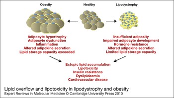
Although this review will focus on the molecular mechanisms underlying lipodystrophies, the information gleaned from the work in this field has importance beyond these rare diseases. The metabolic consequences of insufficient adipose tissue depots are similar to those of excess adiposity in obese individuals. Insulin resistance, type 2 diabetes, dyslipidaemia and cardiovascular disease are all associated with either pathologically increased or decreased adiposity (Refs Reference Garg and Agarwal1, Reference Savage15). Concepts unifying these apparently paradoxical observations include those of adipose tissue expandability and lipotoxicity (Fig. 1). Essentially, these propose that although individuals may have abundant adipose tissue, this is functioning poorly and so remains insufficient for the body's requirements in terms of nutrient intake, storage and metabolism. As a result, lipids that cannot be safely accommodated in adipose tissue accumulate ectopically in other tissues such as skeletal muscle, liver and the pancreas. Inappropriate lipid in these tissues then impairs insulin sensitivity or secretion, resulting in metabolic disease (Refs Reference Savage, Petersen and Shulman16, Reference Unger and Scherer17, Reference Virtue and Vidal-Puig18). Thus if obese individuals were able to generate new insulin-sensitive, metabolically active adipose tissue, they might become metabolically healthy, even in the face of further weight gain. Notably, recent studies have demonstrated that the turnover rate of adult human adipocytes might be around 10% a year, regardless of age or body mass index (Ref. Reference Spalding19). This indicates that interventions merely affecting the function of these newly forming adipocytes could be of significant therapeutic benefit.
Lipid overflow and lipotoxicity in lipodystrophy and obesity. The lipid overflow hypothesis proposes that in obesity, as a result of overnutrition, the lipid storage capacity of hypertrophic adipocytes has been exceeded. While the precise mechanisms are not clear, there is an inability to further expand adipose mass and impaired function of existing adipocytes. This may include altered secretion of adipokines, macrophage infiltration and secretion of inflammatory cytokines, exacerbating insulin resistance. Instead of being safely stored in adipose tissue, lipids spill over and accumulate ectopically in other tissues such as the liver, skeletal muscle and pancreas with damaging metabolic consequences. Lipodystrophic individuals may have similarly restricted lipid storage capacity owing to absent, poorly developed or dysfunctional adipose tissue. Again insulin resistance, inflammation and altered adipokine secretion may occur and lipids accumulate ectopically in other tissues. The failure to appropriately handle and store lipids in both conditions may therefore be a unifying theme, resulting in similar metabolic diseases.

Partial versus generalised lipodystrophies
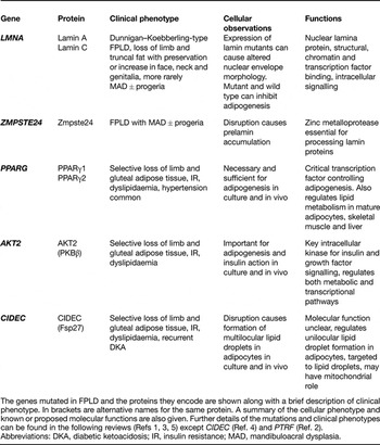
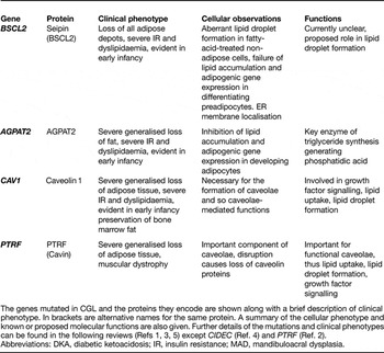
The lipodystrophies arising from genetic mutations are broadly divided into familial partial lipodystrophies (FPLD) and congenital generalised lipodystrophies (CGL) (Refs Reference Garg and Agarwal1, Reference Hegele3). A list of the genes mutated in FPLD and CGL, their protein products and a summary of the resultant phenotypes is provided in Tables 1 and 2.
Familial partial lipodystrophies (FPLD)

The genes mutated in FPLD and the proteins they encode are shown along with a brief description of clinical phenotype. In brackets are alternative names for the same protein. A summary of the cellular phenotype and known or proposed molecular functions are also given. Further details of the mutations and clinical phenotypes can be found in the following reviews (Refs Reference Garg and Agarwal1, Reference Hegele3, Reference Agarwal and Garg5) except CIDEC (Ref. Reference Rubio-Cabezas4) and PTRF (Ref. Reference Hayashi2).
Abbreviations: DKA, diabetic ketoacidosis; IR, insulin resistance; MAD, mandibuloacral dysplasia.
Congenital generalised lipodystrophies (CGL)

The genes mutated in CGL and the proteins they encode are shown along with a brief description of clinical phenotype. In brackets are alternative names for the same protein. A summary of the cellular phenotype and known or proposed molecular functions are also given. Further details of the mutations and clinical phenotypes can be found in the following reviews (Refs Reference Garg and Agarwal1, Reference Hegele3, Reference Agarwal and Garg5) except CIDEC (Ref. Reference Rubio-Cabezas4) and PTRF (Ref. Reference Hayashi2).
Abbreviations: DKA, diabetic ketoacidosis; IR, insulin resistance; MAD, mandibuloacral dysplasia.
As the name implies, FPLD patients suffer selective loss of adiposity from some, but not all, depots (Ref. Reference Hegele3). The reason for the selective sparing of some adipose tissue is not clear, but it is evident that the metabolic functions of different adipose depots, and thus the consequences of their loss, vary significantly. Broadly speaking, visceral adiposity is associated with metabolic disease whereas subcutaneous, and especially gluteal, adiposity is not (Refs Reference Kissebah and Krakower20, Reference Snijder21). Fat transplantation studies have shown that implantation of subcutaneous but not visceral fat from donor mice into the visceral location in recipient mice can improve overall adiposity, glucose tolerance and insulin sensitivity in recipient animals (Ref. Reference Tran22). This elegantly demonstrates that inherent differences exist between these adipose depots. Indeed, the adipocyte precursor cells in these depots also exhibit intrinsic differences, as do the resulting adipocytes when these cells are differentiated in culture (Ref. Reference Tchkonia23). The observed differences between the metabolic flexibility of subcutaneous and visceral adipose tissues are likely to involve several factors. These include adipocyte insulin sensitivity, rate of lipolysis, number of resident macrophages, level of inflammatory cytokines, the transcription factor profile (notably the expression of PPARγ) and differences in tissue vascularisation (Ref. Reference Tran and Kahn24). As several partial lipodystrophies involve the selective loss of gluteal and limb subcutaneous adipose tissue, this is likely to contribute to the apparently disproportionate metabolic disease associated with relatively small decreases in absolute fat mass.
In CGL, adipose tissue development is dramatically impaired in almost all depots throughout the body. Even here, subtle differences exist. For example, individuals with mutations in BSCL2 lack adipose tissue on the soles of the feet, palms and under the scalp, whereas these depots are preserved in other forms of CGL. In addition, bone marrow fat is retained in individuals with disruption of the CAV1 gene but not other forms of CGL (Refs Reference Garg and Agarwal1, Reference Hegele3, Reference Simha and Garg25, Reference Kim26).
The correlation between overall adiposity and metabolic disease is far from perfect in lipodystrophy, and probably reflects the relative importance of the adipose depot affected, the diversity of processes in which the affected proteins are involved and the tissues in which they play a role. For example, as described below, some molecules, such as PPARγ and AKT2, may have a particular importance for insulin sensitivity in skeletal muscle and/or liver as well as adipose tissue, in addition to a role in adipogenesis per se (Refs Reference Pearce, Komander and Alessi27, Reference Tontonoz and Spiegelman28).
Genes disrupted in familial partial lipodystrophies
LMNA and ZMPSTE24
Mutations in the LMNA gene, which encodes the nuclear lamina proteins lamin A and lamin C, were the first identified genetic causes of FPLD (Refs Reference Shackleton29, Reference Hegele30, Reference Hegele31, Reference Cao and Hegele32). In addition to causing lipodystrophy, mutations in LMNA cause several other syndromes collectively described as laminopathies (Ref. Reference Worman and Bonne33). These include muscular dystrophies, cardiomyopathies, neuropathies and progeroid syndromes. At least 11 different diseases have been described associated with over 300 different LMNA mutations. Some LMNA mutations cause complex phenotypes of lipodystrophy with additional progeroid features (mandibuloacral dysplasia-associated lipodystrophy, type 1), whereas others more selectively cause FPLD (Dunnigan variety). Lamins are highly post-translationally processed, including farnesylation and carboxymethylation of a CAAX motif on the C-terminus of the prelamin proteins (Ref. Reference Dechat34). In lamin A, this motif is subsequently cleaved by the protease Zmpste24. Interestingly, mutation of the ZMPSTE24 gene gives rise to another form of FPLD with progeria (Ref. Reference Agarwal35). Disruption of Zmpste24 in mice causes a similar phenotype and accumulation of prelamin A (Ref. Reference Pendas36).
Potential roles for lamin A/C in adipose tissue
Despite the many studies investigating lamin A/C function, the reason why mutations in the same gene cause such diverse phenotypes and how mutation of such a widely expressed nuclear lamina protein could affect specific tissues remains unclear. However, lamins are now appreciated to have diverse roles in the nucleus, including maintaining the structural integrity of the nuclear membrane and binding to chromatin and several transcription factors (Ref. Reference Dechat34). One mechanism via which lamin A/C mutation may impair adipose development involves the transcription factor SREBP1c, which was found to interact with lamin A in a yeast two-hybrid screen (Ref. Reference Lloyd, Trembath and Shackleton37). SREBP1c has an essential role in adipocyte differentiation, inducing the expression of numerous genes of the mature adipocyte (Ref. Reference Kim and Spiegelman38). In addition, SREBP1c is believed to be essential in the production of an endogenous activating ligand for PPARγ in developing adipocytes (Ref. Reference Kim39).
SREBP1c is produced as an integral endoplasmic reticulum (ER) membrane precursor protein that is activated upon regulated transport to the Golgi and proteolytic cleavage. This releases a transcriptionally active N-terminal fragment that translocates to the nucleus (Ref. Reference Bengoechea-Alonso and Ericsson40). Although several studies have suggested a link between lamin A/C and SREBP1c, there is little consensus about the precise mechanism involved. An initial study suggested that the R482W mutant of lamin A/C localised correctly when expressed in HeLa cells but showed impaired binding to SREBP1c in vitro (Ref. Reference Lloyd, Trembath and Shackleton37). However, subsequent studies in patient fibroblasts suggested that the R482L mutant, despite altering the same amino acid, can cause impaired processing, aggregation and targeting of lamin A/C, which sequesters SREBP1c and decreases the active pool of this transcription factor (Refs Reference Capanni41, Reference Capanni42). Further studies show that expression of the R482W and R482Q mutants, but also wild-type lamin A/C, is sufficient to inhibit adipogenesis in cultured 3T3-L1 preadipocytes. This causes impaired induction of both PPARγ and the insulin-sensitive glucose transporter Glut-4, without affecting the induction of SREBP1c mRNA (Ref. Reference Boguslavsky, Stewart and Worman43). This would be consistent with impaired SREBP1c activity in these cells.
Overall, these studies suggest that impaired SREBP1c function may contribute to the development of lipodystrophy in individuals with LMNA or ZMPSTE24 mutations but without clear, consistent mechanistic insight. It should also be noted that the mechanisms via which each of the lamin mutations cause lipodystrophy may differ, even when these affect the same amino acid.
Given the pleiotropic roles of lamin A/C, it seems unlikely that only SREBP1c-mediated mechanisms are involved. Several other transcription factors have been shown to interact with, or be regulated by, lamins, and some of these, such as pRb, have also been proposed to regulate adipogenesis (Ref. Reference Classon44). Moreover, lamins are involved in RNA-polymerase-II-driven transcription, chromatin organisation, nuclear membrane disassembly and reassembly during mitosis, appropriate formation of the nuclear pore complexes, maintaining the structural integrity of the nuclear envelope and communication with the cytoskeleton and cytoplasm (Ref. Reference Dechat34). The majority of these functions have yet to be investigated as potential mechanisms via which lamins might be important for adipose tissue development, but can be expected to yield further insight.
PPARG
PPARγ is a critical transcription factor that has a central role in the regulation of adipogenesis (Ref. Reference Tontonoz and Spiegelman28). Indeed, the finding that PPARγ is both necessary and sufficient for adipocyte differentiation has led to it being dubbed the master regulator of adipogenesis. It is therefore no surprise that impaired PPARγ function can lead to the development of lipodystrophy. Indeed one might wonder why PPARγ disruption does not cause a more severe lipodystrophic phenotype. The answer is likely to lie in the observation that Pparg-null mice die in utero as a result of defects in placental development (Ref. Reference Barak45). However, chimeric rescue experiments in mice to circumvent embryonic lethality have demonstrated that cells lacking PPARγ cannot develop into mature adipocytes in vivo (Refs Reference Barak45, Reference Rosen46). Thus a certain level of PPARγ activity appears to be essential for survival in utero, implying that the mutant forms observed in humans must retain some residual activity.
PPARG mutations in adipocyte development
To date, 14 different mutations in PPARG have been described to cause lipodystrophy. These are the subject of a recent review which describes the detailed molecular mechanisms for each (where known) and so will not be individually described here (Ref. Reference Jeninga, Gurnell and Kalkhoven47). The PPARG gene encodes two isoforms, PPARγ1 and PPARγ2, the latter containing an additional 28 amino acids at the N-terminus (Ref. Reference Tontonoz and Spiegelman28). Both PPARγ1 and PPARγ2 have high expression in adipose tissue, whereas PPARγ2 is almost exclusively expressed in adipocytes. Recent genome-wide analyses of direct PPARγ target promoters in adipocytes have revealed the extent of its involvement in regulating critical genes of the adipogenic and lipogenic programmes, and also demonstrated that it cooperates directly on many of these targets with the C/CAAT enhancer binding protein C/EBPα, another crucial transcription factor in adipogenesis (Refs Reference Lefterova48, Reference Nielsen49). While endogenous ligands for PPARγ are believed to exist in adipocytes, their identity has remained elusive, although several lipid metabolites have been proposed for this role (Ref. Reference Tontonoz and Spiegelman28). By contrast, the thiazolidinedione (TZD) class of antidiabetic drugs act as potent ligands for PPARγ and have been widely used clinically and extensively studied both in vitro and in vivo (Refs Reference Tontonoz and Spiegelman28, Reference Semple, Chatterjee and O'Rahilly50). The impaired effectiveness of TZDs in mice without fat or selectively lacking PPARγ in adipose tissue suggests that adipocyte PPARγ has a crucial role in insulin sensitivity and metabolic health (Refs Reference Chao51, Reference He52). However, these and other studies also indicate that PPARγ is important for lipid metabolism and insulin sensitivity in the liver and skeletal muscle (Refs Reference Chao51, Reference He52, Reference Hevener53).
PPARγ in metabolic regulation
The mechanism via which PPARγ activation improves metabolic health (and hence whereby impaired PPARγ function causes lipodystrophy and metabolic disease) is likely to principally involve effects on lipid storage and metabolism. Many of the genes regulated by PPARγ in adipocytes drive the uptake of lipid substrates [e.g. lipoprotein lipase (LPL), the fatty acid transporters FATP and CD36] or their storage and metabolism (lipin 1, perilipin) (Refs Reference Tontonoz and Spiegelman28, Reference Nielsen49). In addition, PPARγ activation appears to induce a futile cycle of hydrolysis and re-esterification of TG (Ref. Reference Guan54). Overall, the effect is to trap lipids in adipose tissue, decreasing circulating fatty acids and reducing lipid accumulation in non-adipose tissues. As ectopic lipid accumulation in skeletal muscle, liver and the pancreas (so-called lipotoxicity) has deleterious effects on insulin sensitivity and secretion, PPARγ activation can systemically improve insulin sensitivity by preventing this (Ref. Reference Medina-Gomez, Gray and Vidal-Puig55). PPARγ is also likely to directly improve insulin sensitivity within target cells by increasing the expression of proteins such as the insulin-sensitive glucose transporter Glut-4 (Ref. Reference Tontonoz and Spiegelman28). Moreover, PPARγ activation increases expression of the circulating adipokine adiponectin, which may improve skeletal muscle and liver insulin sensitivity (Refs Reference Yamauchi56, Reference Yamauchi57). TZDs also decrease adipocyte and macrophage expression of proinflammatory cytokines, such as TNFα and resistin, which may contribute to insulin resistance (Ref. Reference Tontonoz and Spiegelman28).
Thus, impaired PPARγ function is likely to cause partial lipodystrophy and insulin resistance through several overlapping mechanisms. The presence of significant remaining adipose depots in these individuals suggests only modest impairment of adipogenesis per se. However, pathogenic PPARγ mutations may significantly impair adipocyte function and lead to dysregulation of lipid storage and insulin sensitivity in multiple tissues, decreased adiponectin secretion and increased circulating fatty acids and inflammatory cytokines (Ref. Reference Tontonoz and Spiegelman28). In addition, TZD treatment appears to promote selective accumulation of subcutaneous adipose depots (Refs Reference Kawai58, Reference Kelly59, Reference Mori60). Therefore, PPARγ could be particularly important for developing or maintaining these more metabolically active and beneficial adipose tissues.
AKT2
A family with partial lipodystrophy due to a dominant negative mutation in the protein encoded by the AKT2 gene was identified in 2004 (Ref. Reference George61). AKT2 is a kinase with essential roles in growth factor and insulin signalling (Ref. Reference Pearce, Komander and Alessi27). Three isoforms of AKT have been identified, AKT1 (also called PKBα), AKT2 (PKBβ) and AKT3 (PKBγ). AKT is activated by phosphorylation of two residues, T308 and S473 in the case of AKT1, in response to insulin and growth factors via phosphoinositide 3-kinase (PI3K) (Ref. Reference Pearce, Komander and Alessi27). Active AKT is capable of phosphorylating a plethora of substrates with roles in cell fate, cell survival, proliferation, nutrient uptake or storage, and protein synthesis (Ref. Reference Pearce, Komander and Alessi27). The mutation of AKT2 identified in individuals with lipodystrophy causes an amino acid change of R274H (Ref. Reference George61). R274 is highly conserved in AKT isoforms across several species and lies in the catalytic loop of AKT2 (Ref. Reference Johnson, Noble and Owen62). This amino acid forms critical hydrogen bonds with phosphorylated T309 (equivalent to T308 in AKT1) and is key for the appropriate positioning of substrates for phosphorylation by the active AKT2 (Ref. Reference Yang63). Affected individuals are heterozygous for this mutation, which appears to act as a dominant-negative form of the enzyme, inhibiting both wild-type AKT2 and AKT1 (Ref. Reference George61). Thus the R274H mutation would be expected to inhibit both AKT1 and AKT2 in cell types where AKT2 is abundantly expressed, but not in cells selectively expressing AKT1.
Mice lacking AKT2 show impaired glucose metabolism and age-dependent decreases in adipose mass (Refs Reference Cho64, Reference Garofalo65), whereas loss of AKT1 does not impair glucose metabolism but leads to growth retardation. Knockout of both AKT1 and AKT2 leads to early postnatal death with a complex phenotype, including defects of skin, skeletal muscle and bone, but also significantly impaired adipose development (Ref. Reference Peng66). Studies in mouse embryo fibroblasts (MEFs) from AKT-knockout mice have suggested a greater role for AKT1 than AKT2 in adipogenesis (Refs Reference Peng66, Reference Yun67). However, this may be distorted by the fact that AKT1 is the major isoform in MEFs and this could be less relevant in vivo. Moreover, additional loss of AKT2 in AKt1-null MEFs further inhibits adipogenesis, suggesting that AKT2 is also involved in adipocyte development consistent with studies comparing adipose development in AKT1, AKT2 and double-knockout animals (Refs Reference Garofalo65, Reference Peng66).
Roles of AKT2 in insulin action
Lipodystrophy and insulin resistance in individuals carrying the AKT2 mutation are likely to involve modestly impaired adipogenesis compounded by insulin resistance and poor metabolic function in both adipose and non-adipose tissues. Impaired insulin action in skeletal muscle, where AKT is important for insulin-stimulated translocation of Glut-4, glucose transport and glycogen synthesis, almost certainly contributes significantly to insulin resistance in these individuals (Ref. Reference Bouzakri68). Likewise, the suppression of hepatic gluconeogenesis by insulin is also regulated via AKT, which acts as a key intermediate signalling for the inhibition of glucose-6-phosphatase (G6P) and phosphoenolpyruvate carboxykinase (PEPCK) expression (Refs Reference Pearce, Komander and Alessi27, Reference Barthel69, Reference Liao70). In adipocytes, AKT, particularly AKT2, appears to be important for insulin-stimulated glucose uptake via phosphorylation of AS160, a Rab GTPase activating protein (GAP) that promotes Glut-4 translocation to the plasma membrane (Refs Reference Bae71, Reference Jiang72, Reference Larance73, Reference Miinea74, Reference Ng75). AKT may also contribute to the inhibition of lipolysis by phosphorylating PDE3B, lowering cellular cAMP and hence PKA activity, thus suppressing its activation of hormone-sensitive lipase (HSL) (Ref. Reference Giorgino, Laviola and Eriksson76).
AKT2 in adipocyte differentiation
At the transcriptional level, AKT has been proposed to regulate adipogenesis through the inhibition of various forkhead transcription factors, including Foxo1, Foxc2 and Foxa2, each of which can negatively regulate adipocyte differentiation (Refs Reference Gerin77, Reference Nakae78, Reference Wolfrum79, Reference Davis, Moldes and Farmer80). AKT phosphorylation of forkhead factors causes translocation from the nucleus and hence loss of transcriptional activity. Constitutively active Foxo1 increases the expression of the inhibitory C/EBP isoform CHOP10 and inhibits the induction of PPARγ and several downstream adipocyte genes such as Glut-4 (Ref. Reference Nakae78). Active Foxa2 induces the expression of the inhibitor of adipogenesis Pref1, whereas Foxc2 activity inhibits the expression and activity of PPARγ and the induction of C/EBPα and various PPARγ targets (Refs Reference Wolfrum79, Reference Davis, Moldes and Farmer80). In addition to the regulation of forkhead factors, AKT has been suggested to regulate SREBP1c function at several levels, including expression, translocation, processing, activation and, via GSK3, degradation (Ref. Reference Krycer81). Although not determined in differentiating adipocytes expressing AKT2-R274H, it is possible that disturbance of all or any of these pathways by this mutant AKT impairs adipogenesis.
Finally, AKT has important proliferative/pro-survival effects and antagonises p53 tumour suppressor activity (Refs Reference Jiang82, Reference Levine83). Given the high levels of AKT2 expression in adipose tissue and the ability of AKT2-R274H to inhibit AKT1, it is conceivable that developing adipocytes in these patients are also prone to reduced proliferation or increased cell death.
Overall, the R274H mutation in AKT2 could therefore affect both insulin sensitivity and the transcriptional programme of adipogenesis, causing the phenotype of insulin resistance and partial lipodystrophy. However, this has yet to be experimentally tested in detail at the cellular level.
CIDEC
Mutation of CIDEC is the most recently described cause of partial lipodystrophy in humans, with the identification of an individual homozygous for a mutation causing a premature stop codon E186X in the CIDEC protein (Ref. Reference Rubio-Cabezas4). The cell-death-inducing DNA fragmentation factor-α-like effector (CIDE) family of proteins were initially characterised as potential mitochondrial inducers of apoptosis (Ref. Reference Inohara84). However, the true physiological roles of members of this family (CIDEA, CIDEB and CIDEC) are likely to be in metabolism. Mice lacking CIDEA or CIDEB are lean and obesity resistant, the former because of brown adipose tissue mitochondrial uncoupling and the latter as a result of increased fatty acid oxidation and decreased lipogenesis in the liver (Refs Reference Li85, Reference Zhou86). CIDEC is the third member of this family and is highly expressed in white adipose tissue, earning it the name Fsp27 (fat-specific protein of 27 kDa) in mice (Ref. Reference Danesch, Hoeck and Ringold87).
CIDEC disruption affects lipid droplet morphology
Disruption of Fsp27 in mice causes the development of multilocular adipocytes in adipose tissue (Refs Reference Nishino88, Reference Toh89) whereas Fsp27 knockdown in differentiating cultured adipocytes reduces lipid droplet size (Refs Reference Keller90, Reference Puri91). Similarly, the patient homozygous for the E138X mutation in CIDEC also has characteristic white adipocytes with multiple lipid droplets (Ref. Reference Rubio-Cabezas4). Fsp27 loss protects mice from diet-induced obesity and insulin resistance, promoting a more brown-fat-like morphology and increasing mitochondrial function (Refs Reference Nishino88, Reference Toh89). However, human CIDEC disruption leads to insulin-resistant diabetes with dyslipidaemia, highlighting the potential difficulties of extrapolating findings in mice to human metabolism (Ref. Reference Rubio-Cabezas4). An important caveat to this comparison, however, is that the reported mutation of human CIDEC results in a truncated protein, which may not be mechanistically equivalent to the gene knockout described in mice. The high fasting plasma fatty acid levels observed with CIDEC disruption in the lipodystrophic patient were not so evident in the Fsp27-null mice. In addition, unlike the mice, there was no evidence of increased mitochondrial number in the adipose tissue of the lipodystrophic patient (Refs Reference Rubio-Cabezas4, Reference Nishino88, Reference Toh89). Together, these factors may contribute to a discrepancy in the phenotypes observed with disruption of CIDEC in mice versus humans.
CIDEC is localised to lipid droplets and the ER in adipocytes, and appears to translocate to lipid droplets in response to lipolytic stimulation (Refs Reference Rubio-Cabezas4, Reference Puri91, Reference Nian92). Moreover, binding to the lipid droplet appears to stabilise CIDEC, delaying its ubiquitin-dependent degradation which otherwise occurs rapidly (Ref. Reference Nian92). Critically, the E138X form of CIDEC is truncated early in the functionally important and conserved CIDE-C domain and does not associate with lipid droplets (Ref. Reference Rubio-Cabezas4). Thus, CIDEC expression influences the pattern of lipid accumulation in cultured adipocytes and plays an important role in the formation of a single lipid droplet in mature adipocytes in vivo. As a lipid droplet protein, this could involve altering lipid droplet fusion, the synthesis of phospholipids coating the lipid droplet or the function or localisation of enzymes of lipogenesis or lipolysis. However, the precise molecular function of CIDEC in adipocytes remains to be determined.
Genes disrupted in congenital generalised lipodystrophies
BSCL2
In 2001, Magre and colleagues identified the first specific mutations in the gene BSCL2, which lies in the second locus known to cause CGL (Ref. Reference Magre93). The name BSCL2 arises because CGL is also referred to as Berardinelli–Seip Congenital Lipodystrophy (BSCL) after the physicians who first described the condition. The BSCL2 gene encodes the protein seipin. As BSCL2 patients almost completely lack adipose tissue, this makes seipin arguably the first protein unequivocally demonstrated to play an essential role in the development of human adipose tissue.
Given its importance, it is therefore remarkable that the first insights regarding the molecular function of this protein have only come recently. BSCL2 is highly expressed in adipose tissue and several regions of the brain with lower expression in skeletal muscle and little in the liver (Refs Reference Magre93, Reference Payne94). Two translations of the BSCL2 transcript have been proposed, the longer having an additional sequence of 64 amino acids at the N-terminus (Refs Reference Magre93, Reference Agarwal and Garg95). Seipin is inserted in the ER membrane via two transmembrane domains such that its N- and C-termini are exposed to the cytosol (Refs Reference Ito96, Reference Lundin97). However, seipin has no homology to known proteins that indicate a molecular function.
The majority of the mutations causing lipodystrophy involve deletions or insertions that cause dramatic disruption of seipin, indicating that loss of functional protein causes the phenotype (Refs Reference Magre93, Reference Miranda98, Reference Van Maldergem99). However, three point mutations causing single amino acid changes have now been described: T78A, L91P and A212P (numbered according to the −64 version of seipin) (Refs Reference Magre93, Reference Miranda98). Interestingly, all these occur in the region of the protein lying in the ER lumen. These mutants are of particular interest as they indicate an important functional role for this region of the protein. Indeed, preliminary studies suggest that A212P seipin may be, at least in part, mistargeted to the nuclear envelope, although it is not clear how or if this is involved in its dysfunction (Ref. Reference Payne94).
It should also be noted that two specific mutations affecting an N-glycosylation site in seipin (N88S and S90L) cause silver syndrome, an autosomal dominant form of spastic motorneuropathy (Ref. Reference Windpassinger100). The molecular pathology of this has been investigated in some detail and involves aggregation of mutant and wild-type seipin, causing ER stress and neuronal dysfunction (Ref. Reference Ito96). Affected individuals do not have a reported adipose phenotype, whereas lipodystrophic BSCL2 patients have little or no apparent neurological problems, suggesting that the molecular mechanisms at play are very different (Refs Reference Van Maldergem99, Reference Windpassinger100). However, studies of seipin in the silver syndrome have provided some of the clearest insights into the domain structure and localisation of seipin so far.
Phenotypically, BSCL2 patients are distinctive owing to the loss of retro-orbital adipose tissue and that under the scalp and on the soles of the feet and palms, which are all typically preserved in other forms of CGL (Refs Reference Garg and Agarwal1, Reference Hegele3, Reference Simha and Garg25). In addition, when compared with individuals with AGPAT2 disruption, BSCL2 patients have higher circulating adiponectin levels, albeit lower than in control individuals (Ref. Reference Antuna-Puente101). This latter observation is perhaps surprising, given the lack of adipose tissue that should represent the sole source of adiponectin. It has been proposed that this may reflect secretion from partially developed adipocytes, but this remains to be determined (Ref. Reference Antuna-Puente101).
Molecular function(s) of seipin
The first suggestion of a biological function for seipin came through a screen to identify factors influencing lipid droplet formation and morphology in yeast in which one of the genes identified, YLR404w, was found to be the orthologue of BSCL2 (Ref. Reference Szymanski102). Two such studies revealed that YLR404w is important for the characteristic accumulation of lipid droplets in yeast incubated with the fatty acid oleate (Refs Reference Szymanski102, Reference Fei103). In addition, the yeast seipin orthologue was found at the junctions of the ER and forming lipid droplets, suggesting a role in the generation of the latter (Refs Reference Szymanski102, Reference Fei103). Wild-type human seipin can rescue the yeast phenotype, suggesting functional conservation of the protein in evolution, although, interestingly, a yeast version of A212P seipin cannot. Moreover, fibroblasts from a patient with disrupted BSCL2 exhibited similarly disrupted lipid droplet formation when incubated with oleate in culture (Ref. Reference Szymanski102).
Work from our own lab was the first to examine the role of seipin in developing adipocytes, demonstrating that this protein has a crucial role in adipocyte formation in culture. Loss of seipin was shown to comprehensively inhibit both lipid accumulation and adipogenic gene expression in cultured C3H10T1/2 mesenchymal stem cells (Ref. Reference Payne94), a finding subsequently replicated in 3T3-L1 adipocytes (Ref. Reference Chen104). Both studies implicate PPARγ in this effect. We demonstrated that the initial induction of PPARγ is preserved in cells lacking seipin, but is not then sustained while the broader adipogenic gene expression programme is dramatically impaired (Ref. Reference Payne94). This suggests that preadipocytes lacking seipin undergo some initial stages of adipogenesis but subsequently fail. Given the implied role for seipin in lipid droplet development, it is tempting to speculate that the loss of some vital signals from the forming lipid droplet may stall the adipogenic programme in cells lacking seipin. One study suggested that PPARγ activation by TZD treatment can, at least partially, rescue adipogenesis in the face of seipin loss (Ref. Reference Chen104). Our own preliminary studies suggest that although PPARγ activation can improve differentiation, seipin-knockdown cells still have a marked defect of adipogenesis when compared with control cells similarly treated with TZDs. Therefore, a failure of PPARγ activation may alone not explain the lack of differentiation in cells lacking seipin.
Most recently, analysis of fatty acid composition of lipids in lymphoblastoid cells from BSCL2 patients demonstrated decreased Δ9-desaturation compared with cells from control individuals or AGPAT2 patients (Ref. Reference Boutet105). This indicates that the activity of the enzyme responsible for Δ9-desaturation, steroyl-CoA desaturase-1 (SCD1), is selectively impaired in cells lacking functional seipin. SCD1 has important roles in providing monounsaturated fatty acids for the synthesis of TGs as well as other lipid species; hence this may contribute mechanistically to defective adipose tissue development in these patients (Ref. Reference Paton and Ntambi106).
Overall, these studies indicate an important cell-autonomous role for seipin in lipid droplet formation and adipogenesis. However, these studies clearly provide only the first step in understanding the function of seipin in developing adipocytes, which remains a key unanswered question.
AGPAT2
AGPAT2, encoding 1-acylglycerol-3-phosphate-O-acyltransferase 2, was identified in 2002 as the gene disrupted in the first locus described to cause CGL (Ref. Reference Agarwal107). As AGPAT2 is a critical enzyme of TG synthesis in the developing adipocyte, it seems obvious that disruption of this gene would cause lipodystrophy. However, as described below, this apparently simple defect of lipogenesis affects numerous aspects of adipogenesis, demonstrating the crosstalk that exists between the different molecular pathways active in the developing adipocyte.
The AGPAT2 enzyme catalyses the addition of an acyl chain to lysophosphatidic acid (LPA) generating phosphatidic acid (PA), and is also referred to as lysophosphatidic acid acyltransferase β (LPAATβ) (Ref. Reference Shindou and Shimizu108). PA is subsequently dephosphorylated by lipin 1 to generate diaglyglycerol (DAG) before DAG acyltransferase (DGAT) adds a third acyl chain to generate TG (Ref. Reference Takeuchi and Reue109). Although several enzymes with proposed AGPAT activity exist, AGPAT1 and AGPAT2 are the best characterised, whereas other putative AGPATs have more limited or debatable AGPAT activities (Ref. Reference Shindou and Shimizu108). AGPAT2 deficiency dramatically and selectively impairs the development of adipose tissue, indicating a lack of functional redundancy among these proteins. AGPAT1 is widely expressed in different tissues whereas the highest expression of AGPAT2 is seen in adipose tissue, liver and heart. AGPAT2 is the predominant AGPAT isoform in human adipose tissue, which may for the most part explain the dramatic but selective dysfunction of this tissue with AGPAT2 disruption. Structurally, AGPAT2 has four putative transmembrane domains, is localised to the ER, the site of adipocyte TG synthesis, and has a preference for 18:1, 18:0 and 16:0 acyl-CoAs (Refs Reference Gale110, Reference Hollenback111).
AGPAT2 loss affects both TG synthesis and adipocyte gene expression
The simple model whereby loss of AGPAT2 disrupts TG synthesis, so preventing the development of a lipid droplet that defines the adipocyte, was challenged by a study in cultured preadipocytes. Knockdown of AGPAT2 expression in these cells not only inhibited TG accumulation but also prevented the normal induction of adipogenic gene expression, particularly inhibiting the expression of C/EBPβ and delaying PPARγ expression (Ref. Reference Gale110). While only a limited range of genes were analysed, this demonstrates that AGPAT2 has an important role in gene transcription as well as in lipogenesis in developing adipocytes. The mechanism via which this occurs is not clear, but could involve the generation of lipid species or derivatives that either directly or indirectly control transcription factors.
Because AGPAT2 is likely to have a significant role in the production of PA in adipocytes, it has the potential to influence several pathways. PA is an important intracellular-signalling phospholipid. While PA generated from phosphatidylcholine via the action of receptor activated phospholipase D enzymes has been more extensively studied in this regard, PA generated by AGPAT can also activate intracellular signalling. Pharmacological inhibition of AGPAT2 in vascular smooth muscle cells can suppress the activation of AKT and MAPK signalling pathways (Ref. Reference Coon112). Indeed, selective inhibitors of AGPAT2 have been investigated as potential anticancer agents given their antiproliferative effects (Refs Reference Coon112, Reference La Rosee113). In addition, via the intermediate cytidine diphosphate DAG (CDP-DAG), PA is essential for the synthesis of phosphatidylinositol, and via DAG is a precursor of phosphatidylethanolamine (PE) and phosphatidylcholine (PC), all key structural and/or signalling phospholipids (Ref. Reference Carrasco and Merida114). Given that the developing lipid droplet must be coated with phospholipids, their generation is also likely to be particularly important in developing adipocytes. Indeed PC appears to have a critical role in lipid droplet development and morphology (Ref. Reference Guo115). Few of these molecular pathways via which AGPAT2 disruption could influence adipocyte development have been examined so far in this cell type, and more detailed studies of the effects of AGPAT2 loss in developing adipocytes are clearly warranted.
While the precise molecular consequences of AGPAT2 loss in developing adipocytes remain poorly characterised, Agpat2-null mice have been generated to study the metabolic implications of losing AGPAT2 function. Consistent with the human phenotype, Agpat2-null mice are lipodystrophic with severe insulin resistance (Ref. Reference Cortes116). The livers of these mice display dramatically decreased AGPAT activity but significantly increased TG accumulation. This appears to be due to increases in other enzymes of de novo lipogenesis, particularly monoacylglycerol acyltransferase 1 (MGAT1). MGAT1 adds an acyl chain to the sn-2 position of monoacylglycerol to generate DAG, thereby feeding substrates into the TG-synthesis pathway below AGPAT2. Thus, while this model has not revealed significant detail about why individuals with AGPAT2 mutations fail to develop adipose tissue, it does provide valuable insight into the molecular mechanisms underlying resultant metabolic complications.
CAV1 and PTRF
Caveolin proteins are critical components of membrane invaginations called caveolae. Although caveolae are present on the cells of many tissues, they are particularly abundant on adipocytes, typically representing 20% of the plasma membrane surface (Refs Reference Cohen117, Reference Fan118). The CAV1 gene encodes the caveolin 1 protein. Two other caveolins have been described, although caveolin 3 is selectively expressed in muscle. Caveolin 1 and 2 have broader expression and although both are present in adipocytes, however, caveolin 1 is crucial for the development of caveolae (Refs Reference Cohen117).
As described below, caveolae have important functions in lipid uptake, lipogenesis and insulin signalling (Ref. Reference Le Lay119). Moreover, Cav1-null mice have progressive adipose tissue loss with age, reduced leptin and adiponectin levels and increased TGs (Ref. Reference Razani120). Magre and colleagues screened the CAV1 gene in individuals with CGL not accounted for by AGPAT2 or BSCL2 mutations. They identified an individual bearing a homozygous premature stop mutation (E38X) in caveolin 1, causing a severe truncation of the protein (Ref. Reference Kim26). Further analysis revealed that caveolin 1 was undetectable in fibroblasts from the patient, indicating that they are essentially rendered null for this protein. In addition, as observed in Cav1-null mice, caveolin 2 was also undetectable in patient fibroblasts lacking caveolin 1 (Refs Reference Kim26, Reference Drab121).
Caveolae and caveolin 1 have several crucial roles in signalling and lipid metabolism in the mature adipocyte (Ref. Reference Le Lay119). Loss of caveolin 1 impairs fatty acid uptake, probably as a result of loss of fatty acid translocase (FAT) localisation to caveolae (Ref. Reference Ring122). Caveolin 1 ablation in adipocytes leads to decreased cholesterol content, whereas cholesterol treatment induces translocation of caveolin 1 from the plasma membrane to the lipid droplet, consistent with a role in cholesterol uptake (Ref. Reference Le Lay123). In addition, the association of caveolin 1 with the lipid droplet appears to be highly regulated and to influence lipid droplet size (Ref. Reference Blouin124). This may at least in part reflect the abundance of phospholipids required to coat the lipid droplet, because caveolin 1 loss significantly reduces phospholipid but not neutral lipid content and results in smaller lipid droplets (Ref. Reference Blouin125). In addition, caveolin 1 has been proposed to have a role in lipolysis, translocating to the lipid droplet in lipolytically stimulated cells (Refs Reference Brasaemle126, Reference Cohen127). Loss of caveolin 1 decreases PKA-dependent phosphorylation of the lipid-droplet-coating protein perilipin, despite increased PKA activity (Ref. Reference Cohen127). It is proposed that caveolin 1 facilitates perilipin phosphorylation, which is critical for permitting access of lipases such as hormone-sensitive lipase (HSL) and adipose triglyceride lipase (ATGL) to the lipid droplet, so controlling lipolysis.
Aside from these roles in lipid transport and droplet formation or utilisation, caveolin has essential roles in insulin signalling in adipocytes (Ref. Reference Cohen128). Receptors for insulin and other growth factors are localised to caveolae, which act as critical signal-transduction nodes (Ref. Reference Fruhbeck, Lopez and Dieguez129). Loss of caveolin 1, and hence caveolae, dramatically impairs insulin signalling. This results in large part from loss of insulin receptor protein expression because caveolin 1 appears to be essential for stabilising the insulin receptor, thus preventing proteasomal degradation (Ref. Reference Cohen128). Thus, loss of caveolin 1 is likely to render developing adipocytes resistant to the actions of insulin and other plasma-membrane-receptor-mediated signalling molecules. This may impair the process of adipocyte development in addition to rendering any maturing adipocytes or other cells expressing caveolin 1 insulin resistant.
Most recently, mutations have been identified in the PTRF gene in individuals with a syndrome, including generalised lipodystrophy (Ref. Reference Hayashi2). PTRF encodes cavin, another key protein of caveolae that appears to play an important role in stabilising caveolins and hence caveolae (Refs Reference Liu130, Reference Hill131). Loss of cavin appears to cause a secondary deficiency of caveolin proteins, and in mice this results in a phenotype with features of lipodystrophy such as reduced adipose mass, hypertriglyceridaemia, hyperinsulinaemia and impaired glucose tolerance (Ref. Reference Liu130). As all three caveolin isoforms are affected, the disruption of PTRF causes a more complex syndrome than CAV1 disruption, with lipodystrophy accompanied by muscular dystrophy (Ref. Reference Hayashi2). As cavin has been found to be associated with caveolin 1 on the lipid droplet as well as in plasma membrane caveolae, it is likely that PTRF and CAV1 mutations cause lipodystrophy via many of the same mechanisms (Ref. Reference Blouin125).
Perspectives
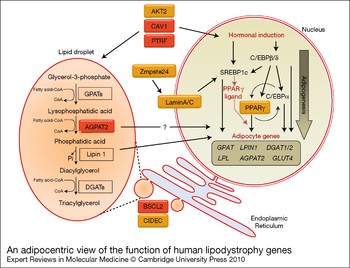
The products of genes disrupted in syndromes of lipodystrophy have been implicated in multiple metabolic or signalling pathways, suggesting a variety of potential mechanisms of action (summarised in Fig. 2). So far only some of these have been experimentally assessed in developing or mature adipocytes, and hence the precise molecular mechanisms via which disruption of these genes causes lipodystrophy remain incompletely understood. Ironically, the most recently identified lipodystrophy genes, CAV1 and PTRF, are perhaps best understood in terms of their molecular function, although it is not yet clear which of the many roles of caveolin 1 are most critical for the development of adipose tissue in vivo. By contrast, how the disruption of LMNA and BSCL2 cause lipodystrophy remains much less clear, even though they were identified almost a decade ago. Of perhaps most current interest is the finding that several genes, BSCL2, AGPAT2, CAV1, PTRF and CIDEC, have roles in the development or maintenance of the lipid droplet. Far from being an inert energy store, this is a fascinating organelle that clearly signals to other parts of the cell by mechanisms that we do not yet understand. It is to this area that the study of lipodystrophy genes may bring the greatest insight.
An adipocentric view of the function of human lipodystrophy genes. Although most genes disrupted in congenital lipodystrophies have roles in other tissues, each has a significant function in the developing adipocyte. Adipogenesis requires a complex programme of gene transcription that can be hormonally induced in cultured preadipocytes but is poorly defined in vivo. Key transcription factors are shown upstream of the genes that they regulate, including members of the C/CAAT enhancer binding protein (C/EBP) family, SREBP1c and PPARγ. These drive the expression of genes of the mature adipocyte such as the insulin-sensitive glucose transporter GLUT4, lipoprotein lipase (LPL) and enzymes of triglyceride synthesis. Lipogenesis occurs concurrently at the membrane of the endoplasmic reticulum (ER). Here, triacylglycerol is synthesised from glycerol-3-phosphate via the addition of fatty-acyl chains by glycerol-3-phosphate acyltransferase (GPAT) and acyl-glycerophosphate acyltransferase 2 (AGPAT2) and dephosphorylation of phosphatidic acid by lipin 1, which can also regulate nuclear gene expression (Ref. Reference Koh132). A final acyl chain is added to diaylglycerol by diacylglycerol acyltransferase (DGAT). Transcription and lipogenesis appear tightly linked in the developing adipocyte. Disruption of the nuclear lamina protein lamin A/C may inhibit the actions of the key adipogenic transcription factor SREBP1c whereas the metalloprotease Zmpste24 is required for the processing, and hence function, of lamin A. The PPARγ transcription factor is at the core of the adipogenic programme of gene expression and directly regulates the expression of several genes in the mature adipocyte that control lipid storage and metabolism. AKT2 is a key intracellular signalling kinase and regulates adipogenic gene transcription and also nutrient uptake and so may influence the supply of substrates for lipid droplet formation. Similarly, caveolin 1 and PTRF/cavin are essential for caveolae formation at the plasma membrane and so may influence hormonal signalling to both adipogenesis in the nucleus and lipogenesis. In addition, caveolin 1 and PTRF/cavin have important roles in the uptake of substrates for the lipid droplet and its maintenance. AGPAT2 is an essential enzyme in the biogenesis of triglyceride where the lipid droplet forms on the ER, but also influences adipogenic transcription in the nucleus by an unknown mechanism. Seipin is found in the ER membrane and is involved in lipid droplet formation although its molecular function is unclear. CIDEC/Fsp27 also regulates lipid droplet development by localising to the lipid droplet surface and facilitating the formation of a single large lipid droplet in developing adipocytes.

Another key process that has not been discussed in detail here is the development of adipocytes from stem cell precursors and the precise identity of these cells. Given the potential therapeutic benefits of using these to expand poorly functioning adipose tissue and improve metabolic disease, there is much interest in this area (Ref. Reference Tran and Kahn24). While a role has been demonstrated for PPARγ in the commitment of pluripotent stem cells to adipocytes, the involvement of other lipodystrophy genes in this process remains undefined (Ref. Reference Rosen46). The disruption of precursor commitment, the potential of these precursors, their proliferation and/or their maintenance could clearly also underlie the development of lipodystrophy. This is clearly an area worthy of future investigation with regard to many of the genes discussed above.
Overall, ongoing studies of lipodystrophy genes are likely to reveal important new details about the development and function of metabolically flexible adipocytes. As several of these affected genes exert effects in non-adipose tissues, it will be challenging to unravel all the mechanisms underlying these conditions. The use of drugs that target PPARγ demonstrates that activation of proteins whose disruption causes lipodystrophy can improve metabolic disease. Thus, a better understanding of the molecular functions of lipodystrophy genes, and the pathways they affect, is likely to suggest novel targets and therapeutic strategies for treating all disorders involving adipocyte dysfunction, including obesity.
Acknowledgements
The author is supported through an MRC New Investigator Research Grant. The reviewers are thanked for their many helpful suggestions.