Introduction
Necrotising fasciitis is a fulminant soft-tissue infection which occurs most frequently in the extremities, abdomen and perineum.Reference Gallia and Johnson1, Reference Kantu and Har-El2 When it occurs in the head and neck, it is very aggressive and often life-threatening. In this region, instead of ‘walling off’ to form an abscess, the (often polymicrobial) infection spreads along the superficial fascial planes of the neck and can easily track down into the mediastinum. It usually spares the overlying skin initially, but eventually the skin becomes erythematous and necrotic when feeding microvessels become thrombosed.Reference Bahu, Shibuya, Meleca, Mathog, Yoo and Stachler3 Historically, necrotising fasciitis has occurred following surgery or minor trauma,Reference Gallia and Johnson1 but most current cases of cervical necrotising fasciitis are odontogenic in origin.Reference Maisel and Karlen4 Predisposing factors include immunosuppression, peripheral vascular disease, cirrhosis and alcoholism.Reference Kantu and Har-El2, Reference Spankus, Flint, Smith and Miller5–Reference Reed and Anand7
Previously reported mortality rates for cervical necrotising fasciitis have ranged from 50 to 73 per cent,Reference Kantu and Har-El2 but mortality has decreased in more recent studies to 0 to 10 per cent.Reference Bahu, Shibuya, Meleca, Mathog, Yoo and Stachler3, Reference Spankus, Flint, Smith and Miller5, Reference Tovi, Fliss and Zirkin6
The complications of cervical necrotising fasciitis include airway obstruction, vascular occlusion or thrombosis, and mediastinal extension. The infection spreads to the mediastinum via two routes: by fascial spread along the carotid sheath inferiorly into the mediastinum, or by spread through the retropharyngeal space into the prevertebral space (the so-called ‘danger space’).Reference Spankus, Flint, Smith and Miller5 Once infection reaches the mediastinum, it can result in mediastinitis, pericarditis, pleural or pericardial effusion, empyema, pneumonitis, cardiac tamponade, and oesophageal bleeding.Reference Bahu, Shibuya, Meleca, Mathog, Yoo and Stachler3, Reference Reed and Anand7
Early identification of the disease process, broad-spectrum intravenous antibiotics, aggressive surgical debridement and wound care, and supportive measures such as hyperbaric oxygen are essential to a successful outcome.
In this article, we describe a case of cervical necrotising fasciitis arising from poorly managed acute tonsillitis.
Case report
A 23-year-old Nigerian woman presented to our emergency department with a two-week history of fever and an eight-day history of painful neck swelling. Nine days before presentation, she had undergone digital manipulation of her throat by her (medically untrained) neighbour, due to throat symptoms suggestive of acute tonsillitis. Oral bleeding had immediately resulted, and the next day a rapidly progressive, generalised, painful neck swelling had developed. Other major complaints included dysphagia and odynophagia. The patient had received intravenous ceftriaxone for two days at a teaching hospital, but was subsequently referred to our institution due to her worsening clinical condition.
On presentation, the patient was noted to have marked trismus and tender, diffuse swelling of the whole neck. A test aspiration of the neck swelling was initially negative.
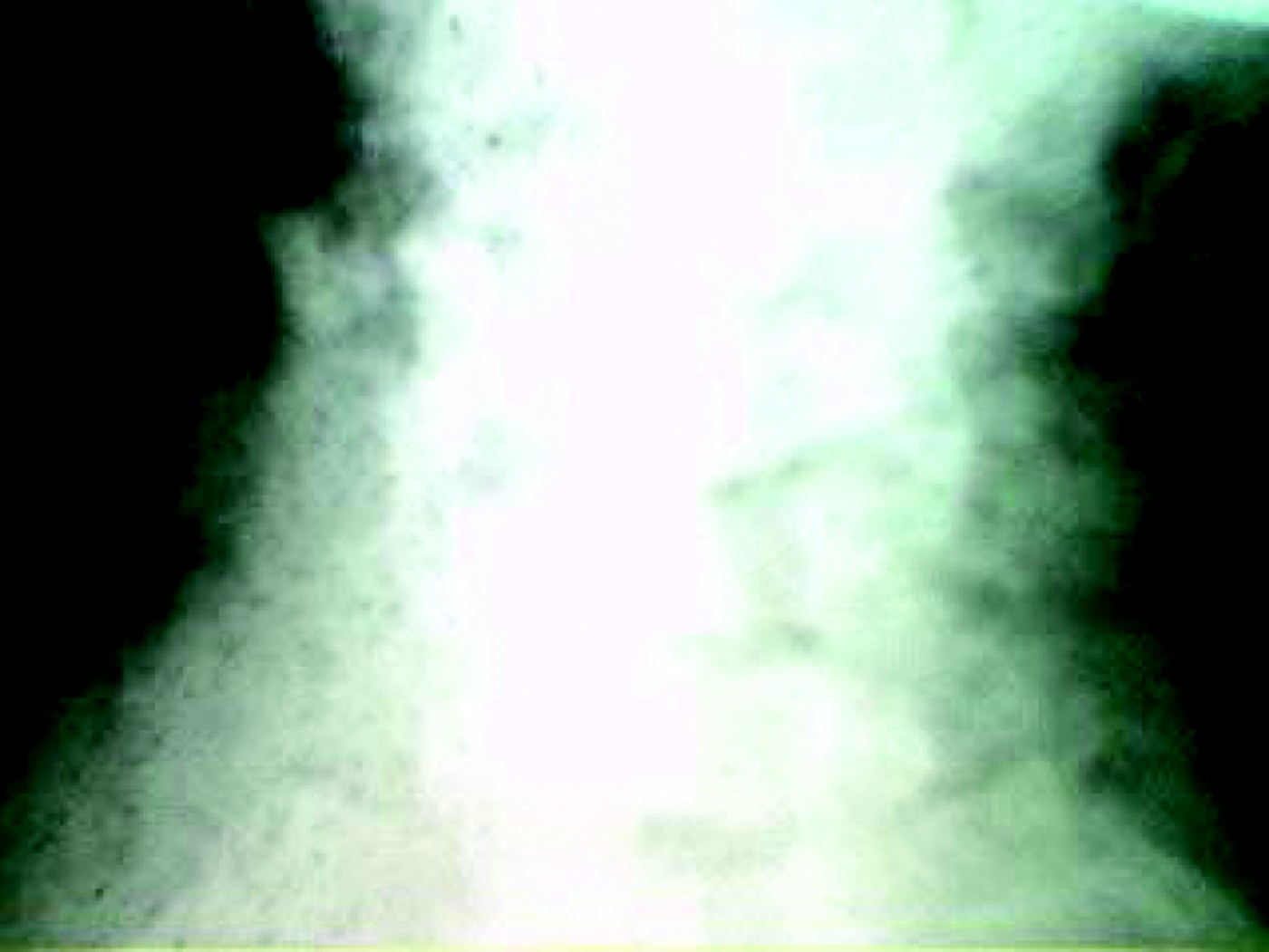
An X-ray of the neck soft tissues showed multiple gas collections (Figure 1). Computed tomography (CT) scanning was not possible due to reasons beyond our control. Human immunodeficiency virus (HIV) and fasting blood glucose tests were negative. The patient's packed cell volume on admission was 47 per cent.
Plain X-ray of neck soft tissues, showing multiple air collections.

The patient was immediately commenced on intravenous ceftazidime 1 g 12-hourly and metronidazole 500 mg 8-hourly.
On the second day of admission, repeated neck tissue aspiration yielded dark-brown, offensive, purulent material which was sent for microbiological analysis. No organism was seen or cultured. A diagnosis of deep neck abscess secondary to poorly managed acute tonsillitis was made, and the patient was scheduled to undergo urgent incision and drainage under general anaesthesia.
However, a CT scan was needed pre-operatively to exclude the possibility of other loculi and mediastinal extension. Unfortunately, this was not forthcoming. The patient deteriorated rapidly, and developed disseminated intravascular coagulation. Her packed cell volume dropped to 27 per cent.
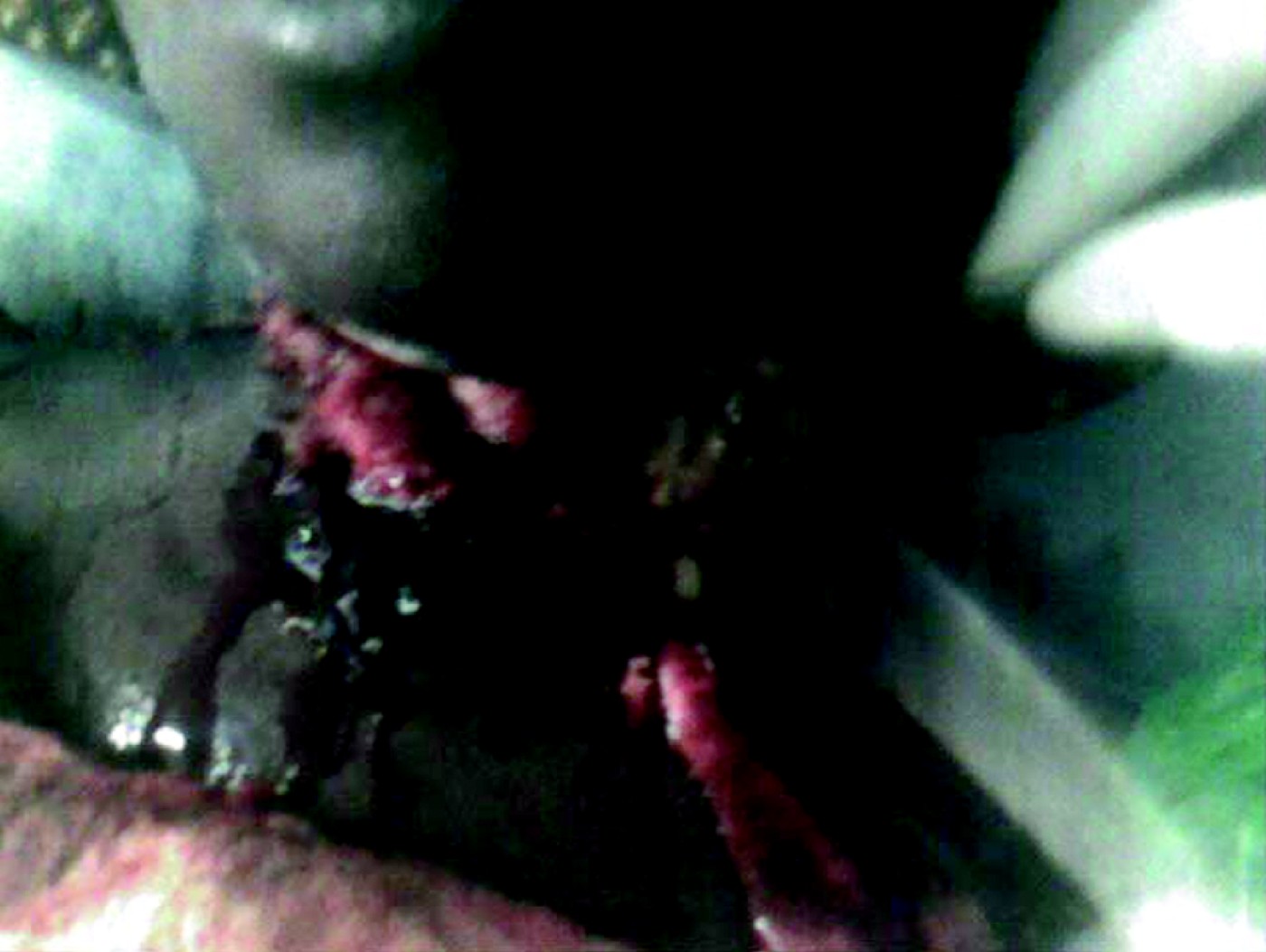
The following day, the patient's anterior neck skin had become gangrenous (Figure 2).
Clinical photograph showing gangrenous anterior neck skin.

She was transfused with fresh whole blood, and received bedside wound debridement under intravenous diazepam and pentazocine anaesthetic cover. Necrotic tissue was excised down to bleeding tissue. The neck muscles appeared to be spared (they bled on contact), except the platysma. A wound biopsy was sent for microbiological analysis, which identified Staphylococcus aureus and klebsiella species. On the microbiologists' recommendation, the patient's ceftazidime was replaced with ciprofloxacine. Aggressive wound cleaning was undertaken with liberal amounts of hydrogen peroxide and honey. Other supportive measures comprised three-hourly feeding with a high-protein liquid diet plus amino acid supplements via a nasogastric tube. This route was chosen as the patient had severe trismus, and intravenous total parenteral nutrition was unavailable. Supplementary oxygen was also given via intranasal catheters (hyperbaric oxygen was not available in our centre). The patient also received regular neck physiotherapy to minimise contracture formation.
On the fourth day of admission, the patient's neck swelling had spread to the upper anterior chest wall, with involvement of the upper half of the left breast. An incision was made just above the left breast to drain the abscess over the chest wall. The cavity was explored, flushed with liberal amounts of hydrogen peroxide and a wick drain left in situ. Further debridements of the necrotic anterior neck wound were performed. All these procedures were undertaken at the bedside under diazepam and pentazocine anaesthetic cover.
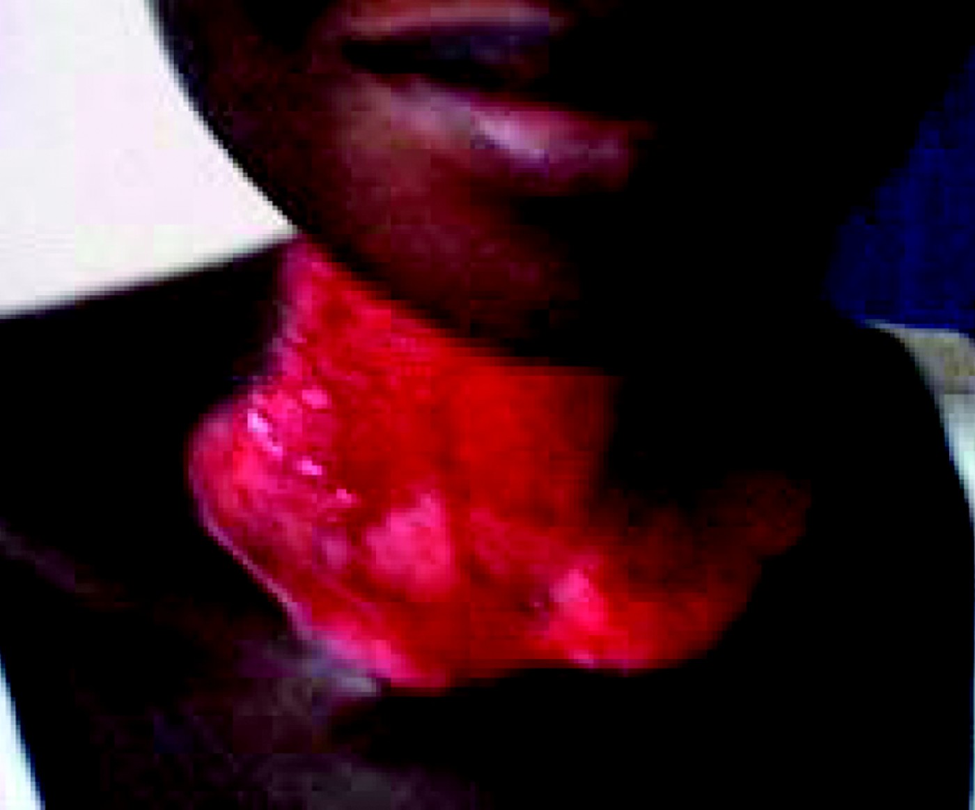
By the second week of aggressive debridement procedures and dressings, the wound was more than 90 per cent granulated and free from obvious purulent discharge (Figure 3). Repeated wound biopsy culture and sensitivity testing yielded the same organisms (S aureus and klebsiella species were sensitive to sparfloxacin, S aureus was resistant to ciprofloxacin, and S aureus and klebsiella species were resistant to ceftazidime and ceftriaxone).
Clinical photograph taken after two weeks of aggressive wound dressing, showing the wound to be well granulated and free of mucopurulent discharge.

After four weeks of debridements, the patient underwent split thickness skin grafting of the anterior neck skin defect, with more than 95 per cent ‘take’.
Discussion
Necrotising fasciitis is a severe and potentially fatal infection of the dermal, fascial and subcutaneous layers of the skin. These infections are marked by the absence of clear local boundaries or palpable limits. This explains the frequent delay in recognising the surgical nature of the infection.
Immunocompromised patients and those suffering from systemic illnesses such as diabetes mellitus are at increased risk of developing this infection.Reference Kantu and Har-El2, Reference Spankus, Flint, Smith and Miller5–Reference Reed and Anand7 In our patient, HIV testing was negative, and fasting and random blood sugar tests were within normal limits. However, necrotising fasciitis may affect previously healthy individuals, and these are often the patients in whom group A β-haemolytic streptococci spread with speed to produce massive tissue infection.Reference Stone and Martin8
In necrotising fasciitis, the causative organism may be a single agent, commonly group A β-haemolytic streptococci or S aureus; alternatively, infection may be polymicrobial, caused by mixed aerobic and anaerobic pathogens. Synergy between the bacterial species contributes to the pathogenesis of polymicrobial fasciitis (this phenomenon has been referred to as synergistic gangrene).Reference Stone and Martin8, Reference McHenry, Brandt, Piotrowske, Jacobs and Malangoni9 Staphylococcus aureus and klebsiella species were cultured from our patient. These organisms may have been introduced by the digital trauma initially inflicted upon the patient in an attempt to rupture the ‘painful swellings on both sides of the throat’.
• Cervical necrotising fasciitis is a fulminant soft tissue infection of the neck
• The condition is usually odontogenic but may in rare cases follow tonsillitis
• A 23-year-old woman presented with a two-week history of fever and an eight-day history of painful neck swelling
• Examination revealed tender, diffuse swelling of the whole neck, and X-ray of the neck soft tissues showed multiple gas collections
• The patient was successfully treated by the bedside, using aggressive debridements and dressings (under local anaesthesia), appropriate intravenous antibiotics, and high protein nutritional support
• Such bedside treatment may obviate the need for general anaesthesia in the treatment of this condition, in a developing country with limited facilities
In most cases of necrotising fasciitis, the pathogen gains entry through a disruption of the epithelium caused by trauma or surgery. Cellulitis initially develops, leading to invasion of the deeper tissues. Clinically, skin erythema and oedema are seen at this stage. Progressive tissue necrosis results in invasion by normal epithelial flora. Continuous bacterial overgrowth and synergy cause a decrease in oxygen tension and the development of local ischaemia, resulting in the proliferation of anaerobic organisms. In four to five days, gangrene is evident, and after eight to ten days necrotic tissue separates from the underlying ischaemic but viable tissue. Staphylococci and streptococci produce extracellular enzymes that damage connective tissue. Bacterial metabolism may also produce insoluble hydrogen, nitrogen, nitrous oxide and hydrogen sulphide gases which result in subcutaneous emphysema.Reference Kantu and Har-El2, Reference Tovi, Fliss and Zirkin6, Reference Stone and Martin8 Although classically associated with clostridial infection, subcutaneous emphysema may also be produced by organisms such as Escherichia coli and klebsiella, peptostreptococcus and bacteroides species.Reference Brook and Frazier10
Consistent CT features of necrotising fasciitis of the head and neck include: diffuse thickening and infiltration of the dermis and subdermis (i.e. cellulitis); diffuse enhancement and/or thickening of the superficial and deep cervical fasciae (i.e. fasciitis); enhancement and thickening of the platysma, sternocleidomastoid muscles or strap muscles (i.e. myositis); and fluid collections in multiple neck compartments.Reference Becker, Zbären, Hermans, Becker, Marchal and Kurt11 Computed tomography scanning facilities may not be available in some parts of developing countries; even so, plain X-ray examination may be extremely useful.
Treatment of cervical necrotising fasciitis involves wide local debridement together with appropriate intravenous antibiotics and other supportive treatment. It is essential that all necrotic tissue be removed;Reference Stone and Martin8 this can be achieved by repeated debridement, especially during dressing changes. Our patient tolerated this on a daily basis, at the bedside under diazepam and pentazocine anaesthetic cover.