INTRODUCTION
Many hydroids are associated with other marine organisms such as algae, seagrasses, sponges, cnidarians, bryozoans, polychaetes, molluscs, crustaceans, echinoderms, tunicates, and fish (e.g. Gili & Hughes, Reference Gili and Hughes1995; Boero & Bouillon, Reference Boero, Bouillon and Rhode2005; Puce et al., Reference Puce, Cerrano, Di Camillo and Bavestrello2008a). The polyps are involved in associations ranging from simple epibiosis to strict symbioses (from mutualism to parasitism). The best-known mutual symbiosis is that of hydroids on the shells of hermit crabs: the hermit crab gives hydroids access to food and the hydroid protects the hermit crab from predators (e.g. Christensen, Reference Christensen1967; Brooks & Mariscal, Reference Brooks and Mariscal1985).
Sessile organisms are often inhabited by various hydroids. Many hydroid species of more than five families have been found on the surfaces or inner walls of the canal systems of sponges (Puce et al., Reference Puce, Calcinai, Bavestrello, Cerrano, Gravili and Boero2005): the hydroids increase the sponges' food supply owing to the water currents produced by the host sponge and may use toxins contained in the sponge tissue to avoid predators such as nudibranchs. The relationship between sponges and hydroids is classified into three types: (1) the hydranth protrudes from the sponge surface and cannot retract into it; (2) the hydranth protrudes from the sponge surface but can retract into the host sponge; and (3) the hydrorhizal system is embedded in the sponge tissue and hydranths do not come out of the sponge (Puce et al., Reference Puce, Calcinai, Bavestrello, Cerrano, Gravili and Boero2005). The last case represents the closest relationship between hydrozoans and sponges. Bryozoans also harbour symbiotic hydrozoans including the six genera Hydranthea, Cytaeis, Octotiara, Halocoryne, Zanclea and Zanclella (Puce et al., Reference Puce, Bavestrello, Di Camillo and Boero2007, Reference Puce, Cerrano, Di Camillo and Bavestrello2008a), and show as wide a range of mutuality levels as sponges.
Although some genera are generalists that live in association with different unrelated groups, almost entire genera or even families are associated with specific groups (Puce et al., Reference Puce, Cerrano, Di Camillo and Bavestrello2008a). The family Zancleidae Russell, 1953, comprises three genera (Halocoryne Hadzi, 1917, Zanclea Gegenbaur, Reference Gegenbaur1857 and Zanclella Boero & Hewitt, 1992) and all species live in association with other benthic organisms. All species of Halocoryne and Zanclella are associated with bryozoa (Boero et al., Reference Boero, Bouillon and Gravili2000; Puce et al., Reference Puce, Cerrano, Boyer, Ferretti and Bavestrello2002, Reference Puce, Cerrano, Di Camillo and Bavestrello2008a). Most Zanclea species are also associated with bryozoa (Z. bomala, Z. divergens, Z. exposita, Z. giancarloi, Z. hirohitoi, Z. polymorpha, Z. protecta, Z. retractilis, Z. sessilis and Z. tipis), although Z. alba live on algae, Z. costata and Z. fanella live on bivalve shells, Z. timida live on octocoral, and Z. gilii and Z. margaritae are associated with scleractinian corals (Gegenbaur, Reference Gegenbaur1857; Hastings, Reference Hastings1930; Calder, Reference Calder1988; Schuchert, Reference Schuchert1996; Boero et al., Reference Boero, Bouillon and Gravili2000; Puce et al., Reference Puce, Cerrano, Boyer, Ferretti and Bavestrello2002, Reference Puce, Di Camillo and Bavestrello2008b; Pantos & Bythell, Reference Pantos and Bythell2010).
Boero et al. (Reference Boero, Bouillon and Gravili2000) described Z. gilii from Papua New Guinea as living in corals, although they did not give a clear description or identification of the coral host species. Recently, Pantos & Bythell (Reference Pantos and Bythell2010) described Z. margaritae, inhabiting the scleractinian coral Acropora muricata (= A. formosa) on the Great Barrier Reef, and this hydroid was not found on any other coral species. From the Sesokojima Island in Okinawa Islands, the southernmost islands in Japan, Yamada & Kubota (Reference Yamada and Kubota1987) reported Zanclea sp. attached to the living stony coral Porites rus and some other coral species at depths of a few metres. However, the details of the hydroid were not described, and no medusa buds were found on the hydroids.
During an ecological study of scleractinian corals on Okinawajima Island, in the Ryukyu Archipelago, we found Zanclea hydroids associated with several scleractinian corals on a shallow reef. Here, we describe this Zanclea species as a new species and discuss an evolutionary trend among coral-symbiotic Zanclea spp. compared to bryozoan-symbiotic species.
MATERIALS AND METHODS
Specimens
Small pieces (3–5 cm long) of Pavona divaricata were collected from reef flats (0.5–1 m) at Bise, Okinawajima Island (26°42′31″N 127°52′58″E) on 9 July 2009. Small pieces of P. divaricata, Pavona venosa and Psammocora contigua were then collected from the same site on 28 June and 8 July 2010. Small pieces of P. venosa and P. contigua were collected from reef flats (0.5–1 m) at Zanpa, Okinawajima Island (26°26′18″N 127°42′40″E) on 24 June and 12 July 2010. Small pieces of P. divaricata, P. venosa and P. contigua were collected from reef flats (0.5–1 m) at Odo, Okinawajima Island (26°05′20″N 127°42′31″E) on 26 July 2010. Each specimen was brought to the laboratory in 300-ml plastic bottles filled with seawater. To collect newly released medusae, each coral species with hydranths was incubated separately in 500-ml plastic jars filled with 0.45-µm-pore filtered seawater (FSW) at room temperature (27–29°C).
Type specimens were deposited in the National Museum of Nature and Science, Tokyo.
Microscopy
Live specimens were used to observe and measure cnidocytes and medusae. Ten or more medusae detached from each coral host species at every collection site were measured within 16 hours after detachment from the parent hydranth. For some photomicrographs, several images were combined to increase the depth of field by using the image post-processing software Helicon Focus Pro 3.79 (Helicon Soft). The capsule size of each cnidocyte and the diameters of medusae were measured from the digital images using ImageJ 1.41 (rebweb. nih. gov).
Histological observation
Small pieces of P. divaricata with Zanclea polyps were fixed in 2.5% glutaraldehyde/0.1 M cacodylate/0.45 M sucrose and stored at 4°C. Specimens were then rinsed with 0.1 M cacodylate/0.45 M sucrose and post-fixed in 1% osmium tetroxide/0.1 M cacodylate for 1.5 hours. After a brief rinse with 50% ethanol, coral skeletons were decalcified with 2.5% acetic acid in FSW. Following dehydration through a graded ethanol series, specimens were embedded in styrene resin. They were sectioned to about 1 µm thickness and stained with 1% toluidine blue for light microscopy. Serial sectioning continued until the desired structures were exposed, after which the resin was removed by soaking in acetone (three times for 1 hour in fresh solvent). After a brief rinse with ethanol, the specimens were immersed in t-butanol and freeze-dried, sputter-coated with gold-palladium, and examined under a scanning electron microscope (SEM; JEOL JSM-6060LV).
RESULTS
TYPE MATERIAL
Holotype: gono-gastrozooids associated with Pavona divaricata, Bise (26°42′31″N 127°52′58″E; Okinawajima Island, Ryukyu Archipelago); water depth ~0.3 m at lowest tide; [NSMT-Co1538]. Collected by M. Hirose, 28 June 2010.
Paratypes: newly released medusa collected on release from parent colony associated with P. divaricata at Bise, 28 June 2010, [NSMT-Co1539]. Collected by M. Hirose, 29 June 2010. Gono-gastrozooids associated with Pavona venosa, Bise (26°42′31″N 127°52′58″E; Okinawajima Island, Ryukyu Archipelago); water depth ~0.3 m at lowest tide; [NSMT-Co1540]. Collected by M. Hirose, 28 June 2010.
Paratypes: newly released medusa collected on release from parent colony associated with P. venosa at Bise, 28 June 2010, [NSMT-Co1541]. Collected by M. Hirose, 29 June 2010. Gono-gastrozooids associated with Psammocora contigua, Bise (26°42′31″N 127°52′58″E; Okinawajima Island, Ryukyu Archipelago); water depth ~0.3 m at lowest tide; [NSMT-Co1543]. Collected by M. Hirose, 28 June 2010.
Paratypes: newly released medusa collected on release from parent colony associated with P. contigua at Bise, 28 June 2010, [NSMT-Co1544]. Collected by M. Hirose, 29 June 2010.
TYPE LOCALITY
Bise, Okinawajima Island, Ryukyu Archipelago.
DIAGNOSIS
Polymorphic Zanclea species associated with living scleractinian coral (Pavona divaricata, P. venosa and Psammocora contigua). Hydrorhiza are surrounded by a perisarc, and grow between the coral skeleton and calicoblastic ectoderm. Newly released medusae are almost spherical, with four perradial exumbrellar nematocyst pouches including stenoteles, and two long marginal tentacles with cnidophores. Nematocyst complement consists of two sizes of stenotele and macrobasic euryteles (both in the hydranth and newly released medusa) and apotrichous euryteles in the cnidophores of the tentacles of newly released medusae.
Description
HYDROID
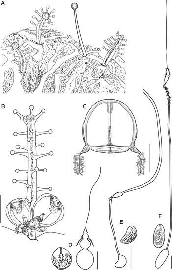
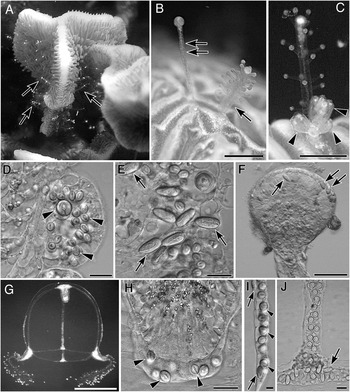
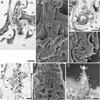
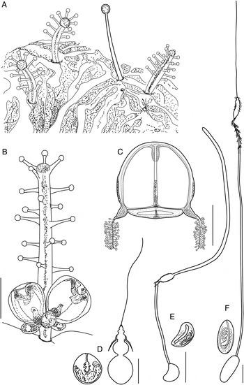
Polymorphic colonies inhabit the tissues of the scleractinian coral Pavona divaricata (Figures 1 & 2A). The hydrorhiza grows between the coral skeleton and the calicoblastic ectoderm (Figure 5A–D). The hydrocaulus and hydrorhiza are surrounded by a perisarc (Figure 5A–D, arrowheads). Gono-gastrozooid hydranths are cylindrical and 0.8–1.9 mm long and 0.1–0.2 mm wide (without capitate tentacles). Five to six oral capitate tentacles on the whitish hypostome surround the mouth, and about 11–22 capitate tentacles in 3–5 rows cover the rest of the hydranth body (Figures 1A–B & 2B–C). Highly retractile dactylozooids (Figure 2B), ~1.2 mm long when fully extended have apical knobs (100–140 µm in diameter) with few apotrichous macrobasic euryteles and many glandular tissues (Figure 2F). One to six medusa buds are found on a single gono-gastrozooid. The medusa buds are on the basal part of the gono-gastrozooid hydranth (Figure 2C). In a nearly matured medusa, the manubrium turns pink and the bell begins pulsating while the medusa is still attached to the hydranth. Following maturation, four exumbrellar pouches become discernible. Large pulsating medusae (about 400 µm in diameter) are sometimes found with small, immature medusae (less than 120 µm in diameter) on the same hydranth (Figure 2C).
Zanclea sango sp. nov. (A) Gastrozooids and dactylozooid emerging from the scleractinian coral Pavona divaricate; (B) gono-gastrozooid; (C) newly released medusa; (D) undischarged and discharged large stenotele from hydroid; (E) undischarged and discharged apotrichous macrobasic eurytele from cnidophores; (F) undischarged and discharged apotrichous macrobasic euryteles from hydroid. Scale bars: (B & C) 0.5 mm; (D, E & F) 10 µm.
Colony, polyps, and newly released medusae of Zanclea sango sp. nov. (A) Field photograph of the hydroid. Many polyps (arrows) emerging from the tissues of the scleractinian coral Pavona divaricata; (B) a gastrozooid (arrow) and dactylozooid (double arrow) on the same coral colony; (C) gono-gastrozooid with medusae at several developmental stages (arrowheads); (D) two sizes of stenoteles (arrowheads) found in the capitate tentacle of the gono-gastrozooid; (E) apotrichous macrobasic euryteles (arrows) found around the mouth; (F) apical knob of dactylozooid (fixed specimen). Arrows indicate apotrichous macrobasic euryteles; (G) newly released medusa; (H) stenoteles (arrowheads) found around mouth; (I) exumbrella pouches containing stenoteles (arrowheads) and apotrichous macrobasic euryteles (arrows); (J) apotrichous macrobasic euryteles (arrow) in the bell margin. Scale bars: (B, C & G) 0.5 mm; (F) 100 µm; (D, E, H & J) 20 µm; (I) 10 µm.
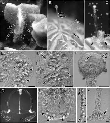
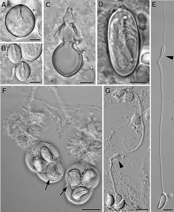
The cnidomes of living specimens were examined to identify cnidocyst types. Two sizes of stenoteles (undischarged ~14 × 13 µm and 8.5 × 6.5 µm) are found in the capitulum of the tentacles (Figures 2D & 3A, B). The size of discharged large stenotele capsule was about 12 × 11 µm and shaft (~12.5 µm long) dilated with three well developing spines (stylets, ~6.3 µm long) (Figure 3C). Apotrichous macrobasic euryteles with the shaft coiled along the main capsule axis (undischarged ~18 × 7.5 µm) are also found in a circle around the mouth and sometime in the ectodermal layer of the basal part of the hydranth (Figures 2E & 3D). The size of discharged capsule was about 16 × 6.5 µm and the dilating end of the long shaft (~140 µm long) is ornamented with spines and a thread protrudes from it (Figure 3E).
Nematocyst of Zanclea sango sp. nov. (A) Large undischarged stenotele from hydroid; (B) small undischarged stenotele from hydroid; (C) large discharged stenotele from hydroid; (D) undischarged apotrichous macrobasic euryteles from hydroid; (E) discharged apotrichous macrobasic euryteles. The dilating end of the long shaft is ornamented with spines (arrowheads) and a thread protrudes from it; (F) cnidophores including undischarged bean-shaped apotrichous macrobasic euryteles (arrows); (G) discharged bean-shaped apotrichous macrobasic euryteles. Arrowhead indicates dilatation of shaft. Scale bars: (A, B, C & D) 5 µm; (E, F & G) 10 µm.
NEWLY RELEASED MEDUSAE
Newly released medusae (526.6–778.0 µm in diameter) are almost spherical, with four perradial exumbrellar nematocyst pouches and four marginal bulbs at the bases of the nematocyst pouches. Two large white conical tentacular bulbs are covered with about 30 cnidophores; the two smaller bulbs have no tentacles. The manubrium with white oral region is contractile and ~200 µm in length when extended (Figures 2G & 3G).
Newly released medusae possess stenoteles of two sizes; small stenoteles are found around the mouth (undischarged ~8.5 × 6.5 µm; Figure 2H), and large stenoteles are present in exumbrellar nematocyst pouches (undischarged ~11 × 10 µm; Figure 2I). Each exumbrellar nematocyst pouch contains 20–30 stenoteles. Apotrichous macrobasic euryteles (undischarged ~18 × 8 µm) are distributed in exumbrellar nematocyst pouches and bells (Figure 2I, J). Bean-shaped apotrichous macrobasic euryteles (undischarged ~8 × 5 µm) are present in the cnidophores of the tentacles (Figure 3F). The size of discharged capsule was about 6.6 × 4.0 µm and shaft dilated distally (Figure 3G).
ETYMOLOGY
The specific name is derived from sango, meaning coral in Japanese, because the hydrozoan colonies are associated with several scleractinian coral species.
REMARKS
This hydrozoan belongs to the genus Zanclea Gegenbaur, Reference Gegenbaur1857, based on the following characters: colonial hydroid, cylindrical gono-gastrozooid with an oral whorl of capitate tentacles and numerous aboral capitate tentacles scattered or in several whorls over the column, medusa buds at the base of the gastrozooid, bell-shaped medusa with four perradial exumbrellar pouches including stenoteles, and two long marginal tentacles with cnidophores containing macrobasic euryteles.
The genus Zanclea currently includes 19 species (Boero et al., Reference Boero, Bouillon and Gravili2000; Puce et al., Reference Puce, Cerrano, Boyer, Ferretti and Bavestrello2002, Reference Puce, Di Camillo and Bavestrello2008b; Pantos & Bythell, Reference Pantos and Bythell2010). Thirteen Zanclea species have monomorphic hydroids, five species have polymorphic hydroids and the hydroid of Z. dubia is unknown. Three of the five polymorphic species are associated with bryozoans. Zanclea sango sp. nov. is a polymorphic hydroid inhabiting corals, and these features are shared with Zanclea gilii, Boero, Bouillon & Gravili, Reference Boero, Bouillon and Gravili2000 and Z. margaritae Pantos & Bythell, Reference Pantos and Bythell2010. Moreover, the two aforementioned species and the present new species have dactylozooids lacking capitate tentacles.
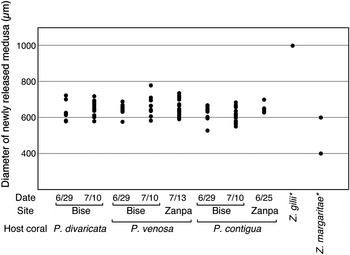
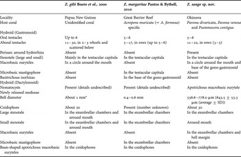
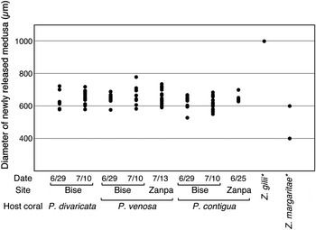
Differences of the character states among the three species are summarized in Table 1. Zanclea margaritae specifically inhabits Acropora muricata (Anthozoa: Scleractinia) on the Great Barrier Reef (Pantos & Bythell, Reference Pantos and Bythell2010); Boero et al. (Reference Boero, Bouillon and Gravili2000) did not identify the coral host species in their original description of Z. gilii. In contrast, colonies of Z. sango sp. nov. were found on at least three scleractinian coral species (Pavona divaricata, P. venosa and Psammocora contigua) in Okinawa. The cnidome of Z. sango sp. nov. is different from those of Z. gilii and Z. margaritae. Additionally, Z. sango sp. nov. does not contain microbasic mastigophores or basitrichous isorhizas in hydroids or newly released medusae, while macrobasic euryteles are found in newly released medusae. The bell diameter of newly released Z. margaritae is 0.4–0.6 mm, approximately half that of Z. gilii (Pantos & Bythell Reference Pantos and Bythell2010). The bell size of newly released Z. sango sp. nov. medusae is 643.5 ± 53.3 µm (average ± SD, N = 91), with maximum and minimum sizes of 778.0 µm and 536.6 µm. In the present species, bell size did not differ significantly among specimens from different coral host species or collection sites (Kruskal–Wallis test, P > 0.05; Figure 4).
Variation in diameter of newly released medusae in Zanclea sango sp. nov. Bell size of Z. gilii and Z. margaritae are cited from Pantos & Bythell (Reference Pantos and Bythell2010). In Z. sango sp. nov., differences in bell sizes were not significant among specimens from different coral host species or different collection sites (Kruskal–Wallis test, P > 0.05).
Character states of Zanclea sango sp. nov. and the other Zanclea species associated with hard corals.
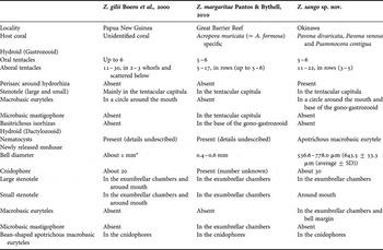
*, estimation by Pantos & Bythell (Reference Pantos and Bythell2010).
Interaction with host
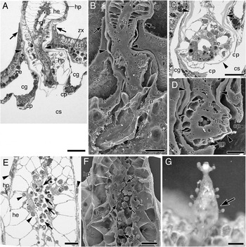
The hydrorhiza of Z. sango sp. nov. grows under the calicoblastic ectoderm of scleractinian corals and is completely covered by host tissues. The base of the hydrocaulus is surrounded by a raised collar of tissue derived from the host coral (Figure 5A, B). At the point where the hydrocaulus passes into the coral tissue, the hydrozoan tissue comes close to the coral epidermal tissue. Inflammatory responses and abnormal growth were not recognized in the tissues of either species in histological examinations. The epidermal layer of the hydrorhiza was thicker (10–15 µm) than that of the hydranth (~8 µm), while the endodermal cells of the hydrorhiza were smaller (10–15 µm) than those of the hydranth (20–40 µm). There were many small particles and much debris in the gastrovascular cavities of gastrozooids (Figure 5E, F). There were some gastrozooids in which the coelenteron (gastrovascular cavity) was expanded by ingested food items (Figure 5G).
Interaction between host coral (Pavona divaricata) and Zanclea sango sp. nov. (A, B) Image pair showing longitudinal section through a gastrozooid emerging from coral tissue (A, resin section; B, scanning electron microscopy (SEM)). The hydrocaulus emerges through a collar of coral (arrows). The base of the hydrocaulus and hydrorhiza are covered by a perisarc (arrowheads); (C, D) image pair showing transversal section of hydrorhiza (C, resin section; D, SEM). Arrowheads indicate perisarcs; (E, F) image pair showing longitudinal section of mid-region of gastrozooid (E, resin section; F, SEM). There are many small particles and debris (arrows) in the gastrovascular cavity. Arrowheads indicate the nuclei of the hydrozoan cells; (G) a gastrozooid. The hydranth is expanded by the ingested food items (arrow). ce, coral endodermis; cg, coral gastrovascular cavity; cs, coral skeleton; cp, coral epidermis; he, hydrozoan endodermis; hg, hydrozoan gastrovascular cavity; hp, hydrozoan epidermis; hr, hydrorhiza; zx, zooxanthella. Scale bars: (G) 0.5 mm; (A & B) 50 µm; (C, D, E & F) 20 µm.
DISCUSSION
Most species of the genus Zanclea live in association with bryozoans, while some species inhabit algae (Z. alba), bivalve shells (Z. costata and Z. fanella), octocorals (Z. timida) and hard corals (Z. gilli and Z. margaritae). Boero et al. (Reference Boero, Bouillon and Gravili2000) proposed an evolutional scenario for the transition of zancleid hydroids from non-symbiotic species to symbiotic hydroids with advanced integration with the host. They presumed that Z. alba, which lives on algae, is the most basal species of Zanclea based on its absence of macrobasic euryteles, less specialized features of the hydroids and medusae, and hydrorhiza creeping on the substrate. Furthermore, Puce et al. (Reference Puce, Cerrano, Boyer, Ferretti and Bavestrello2002) pointed out the importance of the presence or absence of a perisarc around the hydrorhiza. The hydrorhiza is covered by a perisarc in several species that live on algae (Z. alba) and bivalve shells (Z. costata and Z. fanella) (Boero et al. Reference Boero, Bouillon and Gravili2000). Ten Zanclea species are known to associate with bryozoa: six species lack a perisarc and their hydrorhiza is directly covered by the bryozoan skeleton, three species have a perisarc and their hydrorhiza is covered by the skeleton, and Z. exposita is the only species that lacks a perisarc and whose hydrorhiza grows on the bryozoan skeleton (Hastings, Reference Hastings1930; Schuchert, Reference Schuchert1996; Boero et al., Reference Boero, Bouillon and Gravili2000; Puce et al., Reference Puce, Cerrano, Boyer, Ferretti and Bavestrello2002). Puce et al. (Reference Puce, Cerrano, Boyer, Ferretti and Bavestrello2002) hypothesized that the ancestral species of Zanclea had opportunistic associations with some benthic organisms, such as algae or bivalves, and that its hydrorhiza was covered by a perisarc. Later, some species established specific associations with bryozoans, and then the hydrorhiza was lost and directly covered by the bryozoan skeleton. Integration with bryozoans may induce the loss of the perisarc around the hydrorhiza.
Fifteen hydroid species are known to be associated with anthozoans. Among them, three Zanclea species (Z. gilii, Z. margaritae and Z. sango sp. nov.) inhabit scleractinian corals; the other species (Ptilocodium repens Coward, Reference Coward1909; Hydrichthella epigorgia Stechow, Reference Stechow1909; Sarsia medelae Gili et al., Reference Gili, López-González and Bouillon2006; Ralpharia magnifica Watson, Reference Watson1980; Ralpharia coccinea Watson, Reference Watson1984; Ralpharia neira Petersen, Reference Petersen1990; Ralpharia gorgoniae Petersen, Reference Petersen1990; Ralpharia sanctisebastiani da Silveira & Migotto, 1984; Ralpharia parasitica Korotneff, 1887; Asyncoryne philippina Hargitt, 1924; Pteroclava krempfi Billard, 1919; and Zanclea timida Puce et al., Reference Puce, Di Camillo and Bavestrello2008b) are associated with octocorals. Most of the hydroids that inhabit octocorals have a perisarc-covered hydrorhiza that separates the coensarc from the host tissue, while Ptilocodium repens, Z. timida and Hydrichthella epigorgia, associated with the gorgonian Anthoplexaura dimorpha, lack a distinct perisarc sheath (see Gili et al., Reference Gili, López-González and Bouillon2006; Puce et al., Reference Puce, Di Camillo and Bavestrello2008b). Among species that inhabit scleractinian corals, the hydrorhizae of Z. gilii and Z. margaritae are completely embedded in coral tissue and lack perisarcs (Boero et al., Reference Boero, Bouillon and Gravili2000; Pantos & Bethyll, 2010), while that of Z. sango sp. nov has a perisarc. If advanced integration with the host may induce the loss of the perisarc (Puce et al., Reference Puce, Cerrano, Boyer, Ferretti and Bavestrello2002), then Z. sango sp. nov. may be a more basal species among the three Zanclea species that inhabit hard corals. The host specificity of these Zanclea also supports this scenario: Z. sango sp. nov. is associated with at least three coral host species, while Z. margaritae has a specific host, Acropora muricata. On the other hand, Z. gilii lives in unidentified, non-acroporid corals. In the lineage of Zanclea species that inhabit hard corals, the ancestral species is predicted to have been a generalist with a perisarc, whereas advanced species established specific associations with particular host species and lost their perisarc, following the development of the symbiotic relationship.
ACKNOWLEDGEMENTS
The present study was partly supported by grants for Scientific Research (No. 23510296) from the Japan Society for the Promotion of Science and for ‘International Research Hob Project for Climate Change and Coral Reef/Island Dynamics’ from University of the Ryukyus.