




No CrossRef data available.
Neurocognitive and electrophysiological evidence of altered face processing in parents of children with autism: Implications for a model of abnormal development of social brain circuitry in autism
Published online by Cambridge University Press: 01 November 2005
Abstract
Neuroimaging and behavioral studies have shown that children and adults with autism have impaired face recognition. Individuals with autism also exhibit atypical event-related brain potentials to faces, characterized by a failure to show a negative component (N170) latency advantage to face compared to nonface stimuli and a bilateral, rather than right lateralized, pattern of N170 distribution. In this report, performance by 143 parents of children with autism on standardized verbal, visual–spatial, and face recognition tasks was examined. It was found that parents of children with autism exhibited a significant decrement in face recognition ability relative to their verbal and visual spatial abilities. Event-related brain potentials to face and nonface stimuli were examined in 21 parents of children with autism and 21 control adults. Parents of children with autism showed an atypical event-related potential response to faces, which mirrored the pattern shown by children and adults with autism. These results raise the possibility that face processing might be a functional trait marker of genetic susceptibility to autism. Discussion focuses on hypotheses regarding the neurodevelopmental and genetic basis of altered face processing in autism. A general model of the normal emergence of social brain circuitry in the first year of life is proposed, followed by a discussion of how the trajectory of normal development of social brain circuitry, including cortical specialization for face processing, is altered in individuals with autism. The hypothesis that genetic-mediated dysfunction of the dopamine reward system, especially its functioning in social contexts, might account for altered face processing in individuals with autism and their relatives is discussed.
The writing of this paper and the studies reported herein were funded by a grant from the National Institute of Child Health and Human Development (NICHD Grant U19HD34565), which is part of the NICHD Collaborative Program of Excellence in Autism, and a center grant from the National Institute of Mental Health (NIMH Grant U54MH066399), which is part of the NIH STAART Centers Program.
Information
- Type
- Research Article
- Information
- Copyright
- © 2005 Cambridge University Press
Autism is a developmental disorder involving life-long impairments in social interaction and communication. Although evidence for a genetic basis exists, autism risk genes have yet to be found. Evidence for genetic influence in autism is strong, with estimates of heritability ranging from 91 to 93% (Bailey, Le Couteur, Gottesman, Bolton, Simonoff, Yuzda, & Rutter, 1995). Several studies have shown that identical twins are 60–95% concordant for autism (Bailey et al., 1995; Folstein & Rutter, 1977; Ritvo, Freeman, Mason–Brothers, Mo, & Ritvo, 1985; Steffenburg, Gillberg, Hellgren, Andersson, Gillberg, Jakobsson, & Bohman, 1989). Fraternal twins and siblings have a much lower concordance rate, with estimates ranging from 3 to 7% (August, Stewart, & Tsai, 1981; Bailey et al., 1995; Bolton, Macdonald, Pickles, & Rios, 1994; Smalley, Asarnow, & Spence, 1988). The rapid decrease in risk rates from identical twins to siblings and the differential risk rates for male versus female siblings, suggest epistatic effects involving interactions among several genes, estimated to be between 5 and 10 or more (Delong & Dwyer, 1988; Jorde, Hasstedt, Ritvo, Mason–Brothers, Freeman, Pingree, McMahon, Peterson, Jenson, & Mo, 1991; Jorde, Mason–Brothers, Waldmann, & Ritvo, 1990; Pickles, Bolton, Macdonald, Bailey, Le Couteur, Sim, & Rutter, 1995; Risch et al., 1999; Smalley et al., 1988). Several linkage studies have been conducted, which report moderate positive signals on several chromosomes. In general, however, findings have not been strongly consistent across these studies.
One challenging issue for genetic studies is the complex phenotype that comprises the autism syndrome. Not only is the syndrome presentation extremely heterogeneous, but it also involves at least three different symptom domains (social, communication, and restrictive behaviors/flexibility). Furthermore, the autism phenotype appears to extend beyond classic autism to “lesser variant” phenotypes (Rutter, Bailey, Bolton, & Le Couteur, 1993). Numerous studies have shown that relatives of individuals with autism exhibit higher than normal rates of autism-related impairments, referred to as the “broader autism phenotype.” Elevated symptoms have been reported in both parents and siblings (Bailey et al., 1995; Bailey, Phillips, & Rutter, 1996; Baker, Piven, Schwartz, & Patil, 1994; Bolton et al., 1994; Landa, Folstein, & Isaacs, 1991; Landa, Piven, Wzorek, Gayle, Chase, & Folstein, 1992; Narayan, Moyes, & Wolff, 1990; Wolff, Narayan, & Moyes, 1988). For example, Piven, Palmer, Jacobi, Childress, and Arndt (1997), and Piven, Palmer, Landa, Santangelo, Jacobi, and Childress (1997) found that parents of two or more children with autism showed elevated rates of social and communication impairments and stereotyped behaviors. Bolton et al. (1994) reported that 10–20% of siblings exhibit symptoms related to autism, including language, learning, communication, and social impairments.
To date, most genetic studies have characterized the autism phenotype in terms of qualitative discrete diagnoses. However, it is likely that autism susceptibility genes do not cause autism per se, but rather they increase the chance of developing one or more components of the syndrome. In theory, multiple genetically related traits might accumulate to cross a threshold into the full-blown syndrome autism. If so, it will be critical to define these “mind modules,” or endophenotypes, and determine their association with specific genes (Dawson, Webb, Schellenberg, Aylward, Richards, Dager, & Friedman, 2002; Holden, 2003). Such biological or behavioral markers of latent vulnerability to autism are likely not discrete, all or nothing characteristics, but rather they are continuously distributed traits. There have been few genetic studies that have attempted to measure autism-related traits along a continuum. One such study was conducted by Constantino, Davis, Todd, Schindler, Gross, Brophy, Metzger, Shoushtari, Splinter, and Reich (2003) and Constantino, Przybeck, Friesen, and Todd (2000), who developed a questionnaire that captures autism as one continuous trait. They found evidence for a genetic basis of this trait in twin studies (Constantino, Hudziak, & Todd, 2003; Constantino & Todd, 2000; 2003). Dawson, Estes, Munson, Schellenberg, Bernier, and Wijsman (2005) developed a quantitative measure of autism broader phenotype that separately measures several distinct domains of autism symptoms (social motivation, social expressiveness, conversation skills, and restrictive behaviors/flexibility) as well as age of language onset. The Broader Phenotype Autism Symptom Scale (BPASS; Dawson et al., 2005) assesses autism-related traits in both parents and siblings via parent interview about his or her own functioning, or child's functioning, and through direct observation of the parent or child while interacting with the examiner. Nonverbal behaviors, such as eye contact, are assessed via direct observation, whereas behaviors related to restricted activities and routines are assessed via interview. Using BPASS data collected on a sample of 201 autism multiplex families, a genetic investigation of these quantitative traits was conducted (Sung, Dawson, Munson, Estes, Schellenberg, & Wijsman, 2005). Six hundred ninety-four individuals were assessed with the BPASS. Individuals were from nuclear families who had at least two children on the autism spectrum as well as a nonaffected sibling when available. Multivariate polygenic models with ascertainment adjustment to estimate heritabilities and genetic and environmental correlations between the traits were used. Among the traits analyzed, social motivation and restricted activities/flexibility showed the highest heritability (0.19 and 0.16, respectively), suggesting that these traits are most promising for gene mapping. These two traits also showed strong genetic correlation (0.92), suggesting a shared genetic basis for the two traits.
Although these initial studies suggest that quantitative measures of autism symptom-related traits are a promising approach for genetic studies, ultimately, a more refined measure of functional neural trait markers, one that is informed by contemporary affective and social neuroscience, will likely yield greater precision and validity. Studies have identified elevated rates of specific cognitive and language impairments in family members, including impairments on tests of executive function (Hughes, Plumet, & Leboyer, 1999; Koczat, Rogers, Pennington, & Ross, 2002), reading ability (Piven & Palmer, 1997), central coherence (Happe, Briskman, & Frith, 2001), and pragmatic language ability (Landa et al., 1992), but few studies have investigated whether family members show impairments in social and affective processing. The last decade has witnessed an explosion of data and conceptual models on the brain systems that mediate social and affective behavior, which are considered core domains of impairment in autism. Such impairments are believed to reflect contributions of multiple neural systems that support social–affective behavior, including neural systems related to face and emotion processing, perception of biological motion, social motivation, and imitation (Dawson, Webb, et al., 2002). Figure 1 illustrates some the domains of social–affective behavior that are believed to be affected in autism, including (a) affiliative behavior/sensitivity to social reward, (b) motor imitation, and (c) face processing. Impairments in any one neural system (e.g., face recognition) would not be expected to account for the syndrome of autism. Rather, multiple subtle deficiencies in brain systems that support different aspects of social–affective development might act as individual risk factors for autism, analogous to the way that high blood pressure, cholesterol, and obesity increase risk for heart attack. As the number of risk factors increases, the development of complex systems supporting social–affective behavior would be significantly affected, and the probability of developing autism would be increased. If this notion is correct in the case of autism, then it will be important to define and measure underlying neural trait markers. Such markers might allow early detection of risk for autism and help target specific domains for early intervention during periods when the brain systems supporting these domains are developing and plastic.

Candidate traits underlying autism broader phenotype. From “Defining the broader phenotype of autism: Genetic, brain, and behavioral perspectives,” by G. Dawson, S. Webb, G. Schellenberg, E. Aylward, T. Richards, S. Dager, and S. Friedman, 2002, Development and Psychopathology, 14. Copyright 2002 Cambridge University Press. Reprinted with permission.
In this paper, we focus on face processing ability as a potential neural trait marker for susceptibility to autism. Face processing (itself comprised of several subcomponents) is one aspect of complex social brain circuitry that likely contributes to the profound social–affective impairments found in autism. Many of the early social impairments in autism, such as eye contact, joint attention, responses to emotional displays, and face recognition, rely on the ability to attend to and process information from the face (Dawson, Carver, Meltzoff, Panagiotides, McPartland, & Webb, 2002; Dawson, Meltzoff, Osterling, Rinaldi, & Brown, 1998; Dawson, Toth, Abbott, Osterling, Munson, & Estes, 2004; Mundy, Sigman, Ungerer, & Sherman, 1986; Sigman, Kasari, Kwon, & Yirmiya, 1992). Recent behavioral and neuroimaging studies have found reliable evidence for face-processing impairments in individuals with autism. Direct tests of face memory have shown that by middle childhood, children with autism perform worse than mental age and chronological age matched peers on a number of face processing tasks. This includes tests of face discrimination (Tantam, Monagham, Nicholson, & Stirling, 1989), face recognition (Boucher & Lewis, 1992; Boucher, Lewis, & Collis, 1998; Gepner, de Gelder, & de Schonen, 1996; Klin, Sparrow, de Bildt, Cicchetti, Cohen, & Volkmar, 1999), and emotion perception and recognition (Gepner, de Schonen, & Buttin, 1994).
Both positron emission tomography and functional magnetic resonance imaging studies indicate that a portion of the occipitotemporal cortex, the right fusiform gyrus, is more activated in typical individuals during perception of faces than various nonface stimuli, inverted faces, or scrambled faces (Haxby, Horwitz, Ungerleider, Maisog, Pietrini, & Grady, 1994; Haxby, Ungerleider, Clark, Schouten, Hoffman, & Martin, 1999; Hoffman & Haxby, 2000; Kanwisher, McDermott, & Chun, 1997; Kanwisher, Tong, & Nakayama, 1998; McCarthy, Puce, Gore, & Allison, 1997; Puce, Allison, Gore, & McCarthy, 1995; Puce, Allison, Asgari, Gore, & McCarthy, 1996; Sergent, Ohta, & Macdonald, 1992; Wojciulik, Kanwisher, & Driver, 1998). In contrast, individuals with autism have been shown to exhibit irregular and inconsistent patterns of regional activation, which includes areas typically involved in the processing of objects (Pierce, Muller, Ambrose, Allen, & Courchesne, 2001; Schultz, Gauthier, Klin, Fulbright, Anderson, Volkmar, Skudlarski, Lacadie, Cohen, & Gore, 2000). Recently, it has been suggested that this failure to activate the fusiform during face processing might reflect failure to attend to the eye region (Hadjikhani et al., 2004) or the lack of expertise or familiarity with the faces used as stimuli (Aylward, Bernier, Field, Grimme, & Dawson, 2004).
In addition to the lateral fusiform gyrus, other brain regions are important for face processing. The superior temporal sulcus is involved in processing face movements (e.g., the eyes and mouth), and the amygdala is activated when the face is familiar or expressing emotional content (Aylward et al., 2004).
Recognition of familiar faces recruits additional brain regions involved in memory. Faces also evoke a distinct pattern of electrical brain activity. A number of researchers have documented in typical individuals an event-related potential (ERP) component that preferentially activates to faces, which is recorded over the posterior temporal lobe and is greater in the right than the left hemisphere (Bentin, Allison, Puce, Perez, & McCarthy, 1996; Eimer, 1998, 2000a, 2000b, 2000c; George, Evans, Fiori, Davidoff, & Renault, 1996). The N170 component slopes negatively and peaks at approximately 170-ms poststimulus onset. In terms of latency, the N170 is faster to faces and eyes alone than to inverted faces and nonface stimuli. The latency is sensitive to disruptions in early stage processing of faces but is not altered by facial familiarity or recognition processes (e.g., Bentin & Deouell, 2000; Eimer, 2000a). Facial inversion and partial decomposition of faces alters both the latency and amplitude of the component; these effects of inversion are specific to faces (Eimer, 2000b; Rebai, Poiroux, Bernard, & Lalonde, 2001; Rossion, Gauthier, Tarr, Despland, Bruyer, Linotte, & Crommelinck, 2000). Facial movements, such as the eyes looking away and the mouth opening also influence the N170 amplitude (Puce, Smith, & Allison, 2000).
In a series of studies, Dawson and colleagues found that children and adults with autism exhibit abnormal neural responses to faces, as measured by ERP (Dawson, Carver, et al., 2002; Dawson, Webb, et al., 2004; McPartland, Dawson, Webb, Panagiotides, & Carver, 2004; Webb, Dawson, Bernier, & Panagiotides, in press). They found that children with autism as young as 3 years of age exhibit atypical ERPs to faces and facial expressions, but not to objects (Dawson, Carver, et al., 2002; Dawson, Webb, et al., 2004; Webb et al., in press). Typically developing infants exhibit differential ERPs to familiar versus unfamiliar faces and to different emotional expressions by 6–7 months of age (de Haan & Nelson, 1997; Nelson & de Haan, 1996). Three- to 4-year-old children with autism failed to show differential ERPs to familiar versus unfamiliar faces, and to a fearful versus a neutral face, but did show differential ERPs to a familiar versus unfamiliar object. Webb et al. (in press) examined the face-specific ERP component, “developmental N170,” in response to faces and objects from 3- to 4-year-old children with autism spectrum disorder (ASD), typical development, and developmental delay. Children with ASD showed a slower N170 to faces and larger amplitude response to objects compared with the two control groups. Following these children longitudinally, Webb, Dawson, Bernier, and Panagiotides (2005) recently reported that, at age 6 years, children with autism continue to show atypical ERPs to faces. In a separate study of adolescents and adults, it was found that the face-sensitive ERP component, N170, was altered in these older individuals with autism (McPartland et al., 2004). Specifically, compared to cognitively matched typical individuals, high-functioning individuals with autism exhibited slower N170 latencies to faces than furniture, and fail to show a face inversion effect. Similar to findings with 6-year-old children with autism (Webb et al., 2005), scalp topography of the N170 did not show the normal right lateralized pattern, but rather was bilaterally distributed. This ERP study provides evidence that early-stage encoding of faces is disrupted in autism. Taken together, this series of studies suggests a possible life-long pattern of atypical neural responses to faces characterized by a failure to show normal cortical specialization for face processing and slower than normal neural responses to faces.
With the studies reported in this paper, we began our investigation of whether face processing is part of the broader phenotype of autism by examining whether there is any evidence that family members, namely parents, show atypical face processing. At least three previous studies have examined aspects of face processing ability in relatives of individuals with autism. It has been shown that parents and siblings of individuals with Asperger syndrome are worse than expected at the “Eyes Test,” which requires making judgments about emotional state based on photographs of eyes (Baron–Cohen, Wheelwright, & Jolliffe, 1997; Dorris, Espie, Knott, & Salt, 2004). Bolte and Poustka (2003) found that first-degree relatives from multiplex families with autism performed worse on a task requiring judgments about facial affect. Here, we report two studies on face-processing ability in parents of children with autism. The first study examines face-memory ability, compared to verbal and visual–spatial abilities, whereas the second study examines neural indices of face/object processing using ERP.
Study 1: Face Memory in Parents of Children with Autism
Methods
Participants
One hundred forty-three parents of children with idiopathic autism participated. Families were recruited via newspaper articles, parent organizations, NIH announcements, the UW Autism Center Web site, and a network of community service providers. Out of the total sample, 110 parents also had a second child with idiopathic autism, and 33 had a second child with pervasive developmental disorder, not otherwise specified. Participants were 82% Caucasian, 1.4% African American, 2.7% Asian, 4.1% Hispanic/Latino, 1.9% Native American/Native Alaskan, 5.8% biracial, and 2.1% other ethnicity.
Children with autism were diagnosed using NIH Collaborative Program of Excellence in Autism diagnostic criteria established in May of 2003, which combines diagnostic information from the Autism Diagnostic Observation Scale–Generic (Lord, Risi, Lambrecht, Cook, Leventhal, DiLavore, Pickles, & Rutter, 2000), the Autism Diagnostic Interview–Revised (Lord, Rutter, & Le Couteur, 1994), and the clinician's rating of DSM-IV diagnostic criteria for autistic disorder. Exclusionary criteria included having a medical etiology, such as fragile X syndrome, Norrie syndrome, neurofibromatosis, phenylketonuria, or tuberous sclerosis, and failure to have English as the primary language. Absence of fragile X was confirmed through genetic testing.
Neurocognitive tasks
All tasks were age normed on the same sample of 1,250 adults from the general US population (M = 10, SD = 3). The Wechsler Adult Intelligence Scales were used to assess verbal abilities (vocabulary and verbal comprehensive subtests) and visual–spatial abilities (block design and object assembly). The Wechsler Memory Scale—Third Edition (WMS-III): Faces Subtest (Wechsler, 1997) was used to assess immediate recognition memory for faces. In the WMS face memory, 24 faces are presented for a total of 2 s each, and the participant is asked to remember each one. Once the 24 faces have been presented, a second set of 48 faces is presented one at a time. For the immediate memory task, the participant is to indicate if the face is one that he or she was asked to remember. For the delayed memory task, after a 30-min delay a second set of 48 faces is presented one at a time, and participants are asked if the face is one that he or she was asked to remember. All measures were collected in one session.
Results
Mean performance on the three types of tasks is shown in Figure 2. Repeated-measures analyses of variance (ANOVAs) revealed a significant decrement in performance on the face recognition task (M = 10.62, SD = 2.83) relative to the visual spatial tasks (M = 11.88, SD = 2.82), F (1, 142) = 16.17, p < .0001, and the verbal tasks (M = 11.57, SD = 2.67), F (1, 142) = 9.96, p < .01. Twenty-nine percent of the parents had face recognition scores 1 SD (>3 points) lower than the other cognitive tasks; in contrast, only 10.9% of the sample had face recognition scores that were 1 SD higher than the other cognitive tasks. There was no difference in performance related to parent's gender.

Mean verbal, visual spatial, and face recognition abilities of 143 parents of children with autism.
Study 2: Event-Related Brain Potentials to Face and Nonface Stimuli in Parents of Children with Autism
Methods
Participants
Two groups of adults participated in the ERP study. The parent group consisted of 21 parents (11 female) with two children with autism, which was a subset of the parents who participated in the neurocognitive study described above. The mean age for the parent group was 38.5 years (range = 29–52 years). The comparison group consisted of 21 adults (13 female) with no familial history of autism. This group was recruited from postings at the University of Washington Medical Center. The mean age for this group was 38.9 years (range = 28–51 years). Six adults were not included in the analyses due to significant eye movement artifact (one Family Group, five Comparison Group). Exclusionary criteria for both groups included mental illness, significant head trauma or neurological disease, and current use of psychoactive medicines.
ERP paradigm.
Stimuli. Stimuli consisted of gray-scale digital images presented on a computer monitor with a gray background (Bentin et al., 1996; Samaria & Harter, 1994). Images were standardized in terms of size, background color, and mean luminance. The stimulus frames were 520 × 420 pixels and were presented for 300 ms. The experiment involved 250 trials, 55 trials of each of upright faces, inverted faces, upright chairs, inverted chairs, and 30 target stimuli. Scrambled faces were interspersed as target stimuli to control for attention. Subjects were instructed to count the number of target stimuli. Examples of the stimuli used in the experiment are shown in Figure 3.

The stimuli used in the ERP study.
Data collection. The EEGs were recorded in an electrically shielded, sound-attenuated, darkened room. The participant was seated comfortably approximately 75 cm from the computer monitor that delivered the stimuli. A large, threefold screen obscured the back of the monitor and the back part of the room from the participant's view. A 128 lead Geodesic sensor net (Electrical Geodesics Incorporated; Tucker, 1993) was dipped into potassium chloride electrolyte solution, placed on the participant's head, and fitted according to the manufacturer's specifications. The electrodes were evenly spaced and symmetrically covered the scalp from nasion to inion and from left to right ears. Impedances were kept below 40 kΩ.
ERP data were recorded at 250 Hz with a 100-ms baseline and a 1000-ms recording interval. The vertex electrode was used as a reference, and data were rereferenced to an average reference after data collection.
Data editing and reduction. Data were averaged for each subject by stimulus type across trials. Trials with artifacts were excluded from the averages. Each electrode's signal was checked for transits and maximum amplitude. A weighted running average was used with the thresholds set to 150 for transit and 250 for voltage. Running averages are analogous to using a bandpass filter and reject both high-frequency noise and low-frequency drift. This method identifies the slope and rejects sharp transitions in the data. Trials during which eye movement occurred were also excluded. Subjects with less than 10 artifact-free trials were excluded from analysis. Averaged data were transformed to correct for baseline shifts and digitally filtered (low-pass Butterworth 30 Hz) to reduce environmental noise artifacts. Spline interpolation of data from neighboring sites was used to replace data from electrodes for which greater than 25% of the trials were rejected due to artifacts. Participants for whom more than 10 channels required replacement were excluded from further analyses.
Electrodes of interest were selected based on review of the literature and examination of grand averages and individual participant data. The N170 was measured at the inferior right (90, 91, 96, 97) and left (58, 64, 65, 70) posterior temporal electrodes. These lead groups were also deemed most appropriate based on observed magnitude of the N170 at these electrodes and their correspondence to T5 and T6, the electrodes most commonly analyzed in previous studies (e.g., Bentin et al., 1996; Eimer, 1998, 2000a, 2000b, 2000c; Rossion et al., 2000). The layout of the Geodesic Sensor net and the electrodes of interest are shown in Figure 4. The time window for N170 was chosen by visual inspection of the grand average and data for individual participants. The time intervals used extended from 110 to 170 ms after stimulus presentation. Peak and latency to peak were averaged across the specified electrodes within the specified time window and these values were extracted for each participant.

The electrodes used in the ERP analysis.
Neuropsychological tasks. All participants were administered the face recognition subtests from the WMS-III: Immediate and Delayed Memory for Faces (Wechsler, 1997) and the Woodcock Johnson Object Recognition Subtest.
Results
ERP results
Repeated-measures ANOVAs were conducted; Group (Parent, Control) was used as a between-subject factor and stimulus (Face, Object), orientation (Upright, Inverted), and hemisphere (Right, Left) were used as within-subject factors. Greenhouse–Geisser corrections were used. To further investigate face and object processing within group, a repeated-measures ANOVA was conducted separately for each group, with stimulus and hemisphere as within subject factors.
Analysis of N170 amplitude yielded a group by stimulus by hemisphere interaction, F (1, 36) = 4.87, p < .05. Control adults demonstrated the expected larger right hemisphere than left hemisphere N170 to faces (right hemisphere amplitude: M = −6.32 μV, SD = 5.22; left hemisphere amplitude: M = −4.64 μV, SD = 3.27, F (1, 20) = 9.24, p < .01), whereas parents of children with autism demonstrated reduced right hemisphere N170 amplitude to faces, resulting in bilaterally distributed ERPs to faces (right hemisphere amplitude M = −4.05 μV, SD = 2.84; left hemisphere amplitude M = −4.20 μV, SD = 2.85; F (1, 20) = 0.09, ns; see Figure 5).

The event-related brain potentials to face and nonface stimuli in parents of children with autism versus control adults.
Analysis of N170 latency showed a group by stimulus by orientation interaction, F (1, 36) = 3.97, p < .05. Control adults exhibited the expected pattern of faster N170 to upright faces than upright chairs (M latency difference = 10.54 ms, SD = 10.2), F (1, 20) = 22.6, p < .001, whereas parents of children with autism showed no significant difference in N170 latency to upright faces versus upright chairs (M latency difference = 3.60 ms, SD = 12.1), F (1, 20) = 1.87, ns. This is illustrated in Figure 6.

The N170 latency differences between ERP responses to faces versus chairs in parents of children with autism versus control adults.
Neuropsychological test results
Parents of children with autism had significantly lower scores on the Wechsler Face Recognition Test involving a 30-min delay (M = 37.7, SD = 4.5) than controls (M = 40.5, SD = 3, p < .05). Parents of children with autism also had lower scores on the immediate face memory test than control participants, but this difference did not reach statistical significance (p < .10). There were no differences between the groups on the Woodcock Johnson object memory subtest. For control participants only, correlation analyses revealed positive relation between the scores for the WMS Immediate and Delay tasks and amplitude responses to the upright and inverted faces (rs > .44, ps < .05); that is, better performance was associated with larger amplitude N170 to faces. Interestingly, for parents of children with autism, there was no correlation between amplitude and latency of the N170 to faces and face recognition ability.
Discussion
We have argued that progress in genetic research in autism will be facilitated by use of quantitative measures of autism-related traits in addition to qualitative diagnostic measures. In light of the fact that impairments in social–affective behavior are fundamental to autism, we also have argued that measures that quantify functioning of aspects of social brain circuitry might be particularly fruitful. This report describes neurocognitive and neurophysiological evidence of altered face processing in parents of children with autism. Specifically, on standardized neurocognitive tasks, parents of children with autism showed a decrement in face recognition ability relative to their visual spatial (block design and object assembly) and verbal (comprehension and vocabulary) abilities. A third of parents exhibited face recognition scores that were more than 1 SD below their visual spatial scores. Notably, although this significant decrement was found, on average, parents in this sample were functioning at a normal level in their face recognition ability (albeit significantly below what would be expected for their visual spatial and verbal abilities). Thus, clinically, such decrements in face recognition would not have been readily detected. Equally remarkable were findings that parents of children with autism exhibited atypical ERPs to faces but not to nonface stimuli (chairs). Most notably, the pattern of atypical neural response to faces was very similar to what our group found in studies of individuals with autism at a wide range of ages (similar results were found with 3- to 4-year-olds, 6-year-olds, and adolescents and adults with ASD). Examining the face-sensitive ERP component, N170, parents of children with autism failed to show a shorter latency N170 to faces compared to nonface stimuli, and failed to show the expected right hemisphere lateralized pattern. In other words, like children and adults with autism, our data suggest that parents of children with autism show slower than expected neural processing of faces and abnormal cortical specialization for face processing. Based on this evidence, we hypothesize that altered face processing ability might be a functional neural trait marker of genetic susceptibility to autism. Future research will focus on using measures of face recognition for gene mapping studies.
Hypotheses regarding the neural and genetic basis of face recognition impairments in autism
We conclude by discussing hypotheses regarding the neurodevelopmental basis of face processing impairments in autism, and offer speculations regarding the genetic basis of such impairments. We propose a general model of the emergence of social brain circuitry in the first year of life and discuss how the trajectory of normal development of social brain circuitry is altered in autism.
Neural basis of face processing impairments in autism
In normal development, face-processing skills have been hypothesized to rely on innate specialized neural substrates (Kanwisher, 2000). Morton and Johnson (1991) hypothesized that, early on, face processing relies on an innate subcortical system that is replaced by an experience-influenced cortical system around 6 months of age. Nelson (2001) offers a slightly different view. He rejects the notion of an innate face-processing mechanism, and suggests rather that there is the innate potential for cortical specialization for faces. Experience drives domain general mechanisms, such as visual processing in the inferior temporal cortex, to become domain specific mechanisms (Karmiloff–Smith, 1998; Nelson, 2001).
In the case of autism, at least two alternatives can be offered to explain face-processing impairments. The first possibility is that a basic perceptual/cognitive impairment might exist, for example, in general abilities that are critical in face processing, such as the ability to perceptually bind features of a stimulus (Dawson, Webb, et al., 2002), or to form prototypes (Klinger & Dawson, 2001; Strauss, 2004), or in a specific neural mechanism that is specialized for face processing, namely, the fusiform gyrus. A primary perceptual deficit potentially would have broader effects on other aspects of social brain circuitry, especially those aspects that rely on face perception, such as joint attention, interpretation of emotional expression, and even speech perception.
The second hypothesis, referred to as the social motivation hypothesis, posits face-processing deficits are not fundamental, but rather are secondary to a primary impairment in social motivation/affective tagging of social relevant stimuli (Dawson, Carver, et al., 2002; Dawson, Webb, & McPartland, 2005; Grelotti, Gauthier, & Schultz, 2002; Waterhouse, Fein, & Modahl, 1996). The evidence for a social motivation impairment in autism comes primarily from clinical observation. Diagnostic criteria include “a lack of spontaneous seeking to share enjoyment, interests, or achievements with other people” and “lack of social or emotional reciprocity.” Dawson, Hill, Galpert, Spencer, and Watson (1990) found that preschool age children with autism during social interaction were less likely to smile when looking at their mothers. Others have shown that young children with autism are less likely to express positive emotion during joint attention episodes (Kasari, Sigman, Mundy, & Yirmiya, 1990). As discussed earlier in this paper, we now have evidence that the social motivation trait has a genetic basis in autism, based on data derived from the BPASS, which was administered to both affected children and family members. The social motivation trait on the BPASS reflected the degree to which an individual enjoys spending time with others, prefers spending time with people, and feels comfortable in social situations.
According to the social motivation hypothesis, reduced social motivation results in less time spent paying attention to faces as well as to all other social stimuli, such as the human voice, hand gestures, and so on. Previously, we have hypothesized that social motivational impairments in autism are related to a difficulty in forming representations of the reward value of social stimuli (Dawson, Carver, et al., 2002). One of the primary neural systems involved in processing reward information is the dopamine system (Schultz, 1998). Dopaminergic projections to the striatum and frontal cortex, particularly the orbitofrontal cortex, are critical in mediating the effects of reward on approach behavior. Formation of representations of reward value in the orbitofrontal cortex is dependent on input from the basolateral amygdala (Schoenbaum, Setlow, Saddoris, & Gallagher, 2003). The dopamine reward system is activated in response to social rewards, including eye contact (Kampe, Frith, Dolan, & Frith, 2001). Gingrich, Liu, Cascio, Wang, and Insel (2000) showed that dopamine D2 receptors in the nucleus accumbens are important for social attachment in voles. We (Dawson, Munson, Estes, Osterling, McPartland, Toth, Carver, & Abbott, 2002) reported that the severity of joint attention impairments in young children with autism is strongly correlated with performance on neurocognitive tasks that tap the medial temporal lobe–orbitofrontal circuit (e.g., object discrimination reversal). We hypothesize that dysfunction of the dopamine reward system, especially its functioning in social contexts, might account for impairments in social motivation found in autism.
Oxytocin and its relation to the dopamine reward system
Waterhouse et al. (1996) hypothesized that, in autism, impaired oxytocin system function flattens social bonding and affiliation. The elegant work of Insel and colleagues allows further speculation regarding the potential genetic basis for impairments in social motivation and face recognition (see Insel & Fernald, 2004, for a full discussion of this hypothesis). Insel (1997) has discussed the role of peptides, specifically, oxytocin and vasopressin, in the modulation of the dopamine reward circuit in social contexts (Pedersen, Caldwell, Walker, Ayers, & Mason, 1994). In this way, these peptides are particularly important for linking social signals to reinforcement pathways. Several animal studies have shown that vasopressin and oxytocin play important roles in facilitating “social memory.” For example, oxytocin knockout mice show a profound and specific deficit in social memory (Ferguson, Young, Hearn, Insel, & Winslow, 2000; Ferguson, Young, & Insel, 2002; Nishimori, Young, Guo, Wang, Insel, & Matzuk, 1996; Winslow & Insel, 2002). These knockout mice studies support the notion that social memory has a neural basis that is distinct from other forms of memory. Interestingly, it has been shown that oxytocin acts in the medial amygdala during the initial exposure to a familiar conspecific to facilitate social recognition. Both oxytocin and vasopressin appear to facilitate a range of social behaviors, including social affiliation (Witt, Winslow, & Insel, 1992), maternal behavior (Pedersen et al., 1994), and social attachment (Insel & Hulihan, 1995; Winslow, Hastings, Carter, Harbaugh, & Insel, 1993). These peptides may operate on social behavior through their influence on the mesocorticolimbic dopamine circuit. Specifically, Insel and Fernald (2004) suggests that a circuit linking the anterior hypothalamus to the ventral tegmental area and the nucleus accumbens may be especially important for mediating sensitivity to social reward in the context of social interaction. We hypothesize that the face-recognition impairments found in individuals in autism and the decrement in face recognition seen in relatives, described in this paper, might stem from reduced reward value (“emotional tagging”) of faces. Like Insel, O'Brien, and Leckman (1999), we speculate that this might be related to abnormalities in oxytocin and/or vasopressin, which in turn, are important for modulating the dopamine reward pathway, specifically in the context of social interactions. What evidence do we have that abnormalities in oxytocin and vasopressin might be involved in autism? Modahl, Green, Fein, Morris, Waterhouse, Feinstein, and Levin (1998) found that plasma concentration of oxytocin is reduced in children with autism. Kim, Young, Gonen, Veenstra–VanderWeele, Courchesne, Courchesne, Lord, Leventhal, Cook, and Insel (2002) found a nominally significant transmission disequilibrium between an AVPR1A microsatellite and autism. AVPR1A is a V1a receptor in the brain that has been shown to mediate action of vasopressin.
Emergence of social brain circuitry in the first year of life
In Figure 7, we propose a developmental model for the normal emergence of social brain circuitry during early infancy, stressing the key role of the reward system in the development of social brain circuitry. In the model, drawing upon the work of Insel and colleagues, modulation of the dopamine reward circuit by oxytocin is important for shaping the infant's early preference for social stimuli and attention to such stimuli. In normal development, neonates display a particular attraction to people, especially to the sounds, movements, and features of the human face (Maurer & Salapatek, 1976; Morton & Johnson, 1991). Infants as young as 5 months of age are sensitive to even very small deviations in eye gaze during social interactions with adults, smiling and attending less when eyes are averted (Symons, Hains, & Muir, 1998). In these earliest stages, the infant's orienting behavior is involuntary rather than intentional. Later emerging aspects of social cognition likely depend on this very early propensity to devote particular attention to faces (Rochat & Striano, 1999). Active volitional orienting to a social stimulus, such as head turning when name is called, typically emerges by 5–7 months of age. As early as 6 months of age, typically developing infants have been shown to match the direction of mother's head turn to a visible target (Morales, Mundy, & Rojas, 1998).

The role of social reward in the emergence of social brain circuitry in the first years of life.
By 7 months of age, the typical infant spontaneously and intentionally orients to naturally occurring social stimuli in his/her environment. We hypothesize that this occurs, in part, because the infant anticipates pleasure (reward) to be associated with such stimuli (Dawson, Toth, et al., 2004). This type of interaction involves activation of the reward circuit, including parts of the prefrontal regions, such as the orbital prefrontal cortex, that are involved in forming reward representations. With increasing experience with faces and voices, which occurs in the context of social interactions, cortical specialization for faces and linguistic stimuli occurs, involving the fine-tuning of perceptual systems. Furthermore, areas specialized for the processing of social stimuli, such as the fusiform gyrus and superior temporal sulcus, become increasingly integrated with regions involved in reward circuitry (e.g., amygdala), as well as regions involved in actions and attention (cerebellum, prefrontal/cingulate cortex). As a result, more complex social brain circuitry emerges supporting more complex behaviors, such as disengagement of attention, joint attention, intentional communication, and delayed imitation.
Implications for autism
One of the earliest symptoms is a lack of “social orienting” (Dawson et al., 1998; Dawson, Toth, et al., 2004). This reduced attention to social stimuli would result in a failure to become an expert face and language processor (Dawson, Webb, & McPartland, 2005; Grelotti et al., 2002). Because experience drives cortical specialization (Nelson, 2001), reduced attention to faces and speech would result in a failure of specialization of regions that typically mediate face and language processing, and would be reflected in decreased cortical specialization and abnormal brain circuitry for face processing resulting in slower information processing speed (e.g., N170 latency).
The abnormal trajectory for brain development in autism is not caused by a simple lack of exposure to face and language. Infants with autism, like typically developing infants, are held, talked to, and fed by their parents during face to face interactions. However, if the infant with autism does not find such interactions inherently interesting or rewarding, then the infant might not be actively attending to the face and voice or perceiving the face within a larger social/affective context. Recent research by Kuhl, Tsao, and Liu (2003) suggests that simple exposure to language does not necessarily facilitate the development of brain circuitry specialized for language. Instead, language needs to be experienced by the infant within a social interactive context for speech perception to develop. It has been demonstrated that, early in life, infants are capable of discerning differences among the phonetic units of all languages, including native- and foreign-language sounds. Between 6 and 12 months of age, the ability to discriminate foreign-language phonetic units declines as the brain becomes proficient at speech perception (Kuhl, Andruski, Chistovich, Chistovich, Kozhevnikova, Ryskina, Stolyarova, Sundberg, & Lacerda, 1997). Kuhl et al. (2003) investigated whether it was possible to reverse this decline in foreign-language phonetic perception by exposing American infants to native Mandarin Chinese speakers. These investigators found that reversal in the decline of foreign-language phonetic perception was achievable, but only if exposure to speech occurred in the context of interpersonal interaction. An experimental condition in which infants were exposed to the same speech stimuli via audiotapes did not affect the development of speech perception for nonnative language. In the case of autism, because the child is not actively attending to the facial and speech stimulation with the social context, their early exposure to speech and faces might not facilitate face and speech perception. A recent study supports this notion. In a sample of 3- to 4-year-old children with autism, Kuhl, Coffey–Corina, Padden, and Dawson (2004) found that listening preferences in children with autism differed dramatically from those of typically developing children. Children with autism preferred listening to mechanical-sounding auditory signals (signals acoustically matched to speech referred to as “sine-wave analog”) rather than speech (motherese). The preference for the mechanical-sounding auditory signal was significantly correlated with lower language ability, more severe autism symptoms, and abnormal ERPs to speech sounds. Children with autism who preferred motherese were more likely to show differential ERPs to different phonemes, whereas those who preferred the mechanical-sounding auditory signal showed no differences between ERP waveforms in response to two different syllables. We hypothesize that a failure to affectively tag social stimuli as relevant, and the resultant failure to attend to such stimuli, impedes cortical specialization for face and language brain regions. Perceptual fine-tuning of such stimuli and the formation of representations of social stimuli are impeded. More complex behaviors requiring integration of social stimuli with coordinated and intentional movements and volitional attention, such as disengagement of attention and joint attention, fail to emerge.
Conclusions: Implications for Defining Broader Phenotype of Autism
The model presented above suggests that the genetic basis of face-processing impairments as part of the broader phenotype of autism might be related to underlying abnormalities in social motivation. If so, ultimately, phenotypic measures more directly related to social motivation might be more fruitful for genetic studies. Alternatively, there might be two pathways to face processing impairments that are part of the broader autism phenotype: one related to a fundamental perceptual impairment, and another secondary to a social motivational impairment. Clearly, we are still in our infancy in understanding the complex biological and behavioral phenotype of autism. It will be important to cast a wide net in studying potential autism broader phenotype traits.
Furthermore, it should be noted that there is no reason to believe that face processing and social motivation impairments would be specific to autism. Several other disorders have been shown to be associated with face-processing impairments, including Turner syndrome, schizophrenia, Alzheimer disease, and developmental prosopagnosia (Bolte & Poustka, 2003; Giannakopoulos, Gold, Duc, Michel, Hof, & Bouras, 2003; Herrmann, Ellgring, & Hallgatter, 2004; Kuntsi, Coleman, Campbell, & Skuse, 2003). In contrast, face-processing abnormalities have not been found to be associated with dyslexia or Williams syndrome (Rüsseler, 2003; Tager–Flusberg, Plesa–Skwerer, Faja, & Joseph, 2003). When genes are found that are associated with face processing, it would be of interest to compare such populations so that common and distinct genetic risk factors can be defined for these different populations.
By defining the genetically mediated risk factors for autism, we might eventually be able to detect risk for autism very early in infancy. Understanding such risk factors will also target relevant domains for early behavioral and/or biological interventions when the brain systems are emerging and most plastic. Using this strategy, it is hoped that long term outcome in autism might eventually be improved.
References
REFERENCES
August, G. J.,
Stewart, M. A., &
Tsai, L.
(1981).
The incidence of cognitive disabilities in the siblings of autistic
children.
British Journal of Psychiatry
138,
416–422.Google Scholar
Aylward, E.,
Bernier, R.,
Field, K.,
Grimme, A., &
Dawson, G.
(2004).
Normal activation of fusiform gyrus in adolescents and adults with
autism during viewing of familiar faces.
Presented at the International Meeting for Autism Research,
Sacramento, CA.
Bailey, A.,
Le Couteur, A.,
Gottesman, I.,
Bolton, P.,
Simonoff, E.,
Yuzda, E., &
Rutter, M.
(1995).
Autism as a strongly genetic disorder: Evidence from a British twin
study.
Psychological Medicine
25,
63–77.Google Scholar
Bailey, A.,
Phillips, W., &
Rutter, M.
(1996).
Autism: Toward an integration of clinical, genetic,
neuropsychological, and neurobiological perspectives.
Journal of Child Psychology & Psychiatry & Allied
Disciplines
37,
89–126.Google Scholar
Baker, P.,
Piven, J.,
Schwartz, S., &
Patil, S.
(1994).
Brief report: Duplication of chromosome 15q11-13 in two individuals
with autistic disorder.
Journal of Autism Development Disorder
24,
529–535.Google Scholar
Baron–Cohen, S.,
Wheelwright, S., &
Jolliffe, T.
(1997).
Is there a “language of the eyes”? Evidence from normal
adults and adults with autism or Asperger syndrome.
Visual Cognition
4,
311–332.Google Scholar
Bentin, S.,
Allison, T.,
Puce, A.,
Perez, E., &
McCarthy, G.
(1996).
Electrophysiological studies of face perception in humans.
Journal of Cognitive Neuroscience
8,
551–565.Google Scholar
Bentin, S., &
Deouell, L.
(2000).
Structural encoding and identification in face processing: ERP
evidence for separate mechanisms.
Cognitive Neuropsychology
17,
35–54.Google Scholar
Bolte, S., &
Poustka, F.
(2003).
The recognition of facial affect in autistic and schizophrenic
subjects and their first-degree relatives.
Psychological Medicine
33,
907–915.Google Scholar
Bolton, P.,
Macdonald, H.,
Pickles, A., &
Rios, P.
(1994).
A case–control family history study of autism.
Journal of Child Psychology and Psychiatry
35,
877–900.Google Scholar
Boucher, J., &
Lewis, V.
(1992).
Unfamiliar face recognition in relatively able autistic
children.
Journal of Child Psychology and Psychiatry
33,
843–859.Google Scholar
Boucher, J.,
Lewis, V., &
Collis, G.
(1998).
Familiar face and voice matching and recognition in children with
autism.
Journal of Child Psychology and Psychiatry
39,
171–181.Google Scholar
Constantino, J. N.,
Davis, S. A.,
Todd, R. D.,
Schindler, M. K.,
Gross, M. M.,
Brophy, S. L.,
Metzger, L. M.,
Shoushtari, C. S.,
Splinter, R., &
Reich, W.
(2003).
Validation of a brief quantitative measure of autistic traits:
Comparison of the social responsiveness scale with the Autism Diagnostic
Interview–Revised.
Journal of Autism Development and Disorder,
33,
427–433.Google Scholar
Constantino, J. N.,
Hudziak, J. J., &
Todd, R. D.
(2003).
Deficits in reciprocal social behavior in male twins: Evidence for a
genetically independent domain of psychopathology.
Journal of the American Academy of Child Adolescent
Psychiatry
42,
458–467.Google Scholar
Constantino, J. N.,
Przybeck, T.,
Friesen, D., &
Todd, R. D.
(2000).
Reciprocal social behavior in children with and without pervasive
developmental disorders.
Journal of Developmental and Behavioral Pediatrics
21,
2–11.Google Scholar
Constantino, J. N., &
Todd, R. D.
(2000).
Genetic structure of reciprocal social behavior.
American Journal of Psychiatry
157,
2043–2045.Google Scholar
Constantino, J. N., &
Todd, R. D.
(2003).
Autistic traits in the general population: A twin study.
Archives of General Psychiatry
60,
524–530.Google Scholar
Dawson, G.,
Carver, L. J.,
Meltzoff, A. N.,
Panagiotides, H.,
McPartland, J., &
Webb, S. J.
(2002).
Neural correlates of face recognition in young children with autism
spectrum disorder.
Child Development
73,
700–717.Google Scholar
Dawson, G.,
Estes, A.,
Munson, J.,
Schellenberg, G.,
Bernier, R., &
Wijsman, E.
(2005).
Quantitative assessment of autism symptoms in children with autism
and their parents: Broader Phenotype Autism Symptom Scale.
Unpublished manuscript.
Dawson, G.,
Hill, D.,
Galpert, L.,
Spencer, A., &
Watson, L
(1990).
Affective exchanges between young autistic children and their
mothers.
Journal of Abnormal Child Psychology
18,
335–345.Google Scholar
Dawson, G.,
Meltzoff, A.,
Osterling, J.,
Rinaldi, J., &
Brown, E.
(1998).
Children with autism fail to orient to naturally occurring social
stimuli.
Journal of Autism and Developmental Disorders
28,
479–485.Google Scholar
Dawson, G.,
Munson, J.,
Estes, A.,
Osterling, J.,
McPartland, J.,
Toth, K.,
Carver, L., &
Abbott, R.
(2002).
Neurocognitive function and joint attention ability in young
children with autism spectrum disorder.
Child Development
73,
345–358.Google Scholar
Dawson, G.,
Toth, K.,
Abbott, R.,
Osterling, J.,
Munson, J., &
Estes, A.
(2004).
Defining the early social attention impairments in autism: Social
orienting, joint attention, and responses to emotions.
Developmental Psychology
40,
271–283.Google Scholar
Dawson, G.,
Webb, S.,
Carver, L.,
Panagiotides, H., &
McPartland, J.
(2004).
Young children with autism show atypical brain responses to fearful
versus neutral facial expressions.
Developmental Science
7,
340–359.Google Scholar
Dawson, G.,
Webb, S., &
McPartland, J.
(2005).
Understanding the nature of face processing impairment in autism:
Insights from behavioral and electrophysiological studies.
Developmental Neuropsychology
27,
403–424.
[Special Issue on Autism]Google Scholar
Dawson, G.,
Webb, S.,
Schellenberg, G.,
Aylward, E.,
Richards, T.,
Dager, S., &
Friedman, S.
(2002).
Defining the broader phenotype of autism: Genetic, brain, and
behavioral perspectives.
Development and Psychopathology
14,
581–611.Google Scholar
de Haan, M., &
Nelson, C. A.
(1997).
Recognition of the mother's face by 6-month old infants: A
neurobehavioral study.
Child Development
68,
187–210.Google Scholar
Delong, G. R., &
Dwyer, J. T.
(1988).
Correlation of family history with specific autistic subgroups:
Aspergers and bipolar affective disease.
Journal of Autism and Developmental Disorders
18,
593–600.Google Scholar
Dorris, L.,
Espie, C. A. E.,
Knott, F., &
Salt, J.
(2004).
Mind-reading difficulties in the siblings of people with
Asperger's syndrome: Evidence for a genetic influence in the abnormal
development of a specific cognitive domain.
Journal of Child Psychology and Psychiatry,
45,
412.Google Scholar
Eimer, M.
(1998).
Does the face specific component reflect the activity of a
specialized eye processor?
NeuroReport
9,
2945–2948.Google Scholar
Eimer, M.
(2000a).
Effects of face inversion on the structural encoding and recognition
of faces: Evidence from event-related brain potentials.
Cognitive Brain Research
10,
145–158.Google Scholar
Eimer, M.
(2000b).
Event related potentials distinguish processing stages involved in
face perception and recognition.
Clinical Neurophysiology
111,
694–705.Google Scholar
Eimer, M.
(2000c).
The face-specific N170 component reflects late stages in the
structural encoding of faces.
Cognitive Neuroscience
11,
2319–2324.Google Scholar
Ferguson, J. N.,
Young, L. J.,
Hearn, E.,
Insel, T. R., &
Winslow, J.
(2000).
Social amnesia in mice lacking the oxytocin gene.
Nature Genetics
25,
284–288.Google Scholar
Ferguson, J. N.,
Young, L. J., &
Insel, T. R.
(2002).
The neuroendocrine basis of social recognition.
Frontiers in Neuroendocrinology
23,
200–224.Google Scholar
Folstein, S., &
Rutter, M.
(1977).
Infantile autism: A genetic study of 21 twin pairs.
Journal of Child Psychology and Psychiatry
18,
297–321.Google Scholar
George, N.,
Evans, J.,
Fiori, N.,
Davidoff, J., &
Renault, B.
(1996).
Brain events related to normal and moderately scrambled
faces.
Cognitive Brain Research
4,
65–76.Google Scholar
Gepner, B.,
de Gelder, B., &
de Schonen, S.
(1996).
Face processing in autistics: Evidence for a generalized
deficit?
Child Neuropsychology
2,
123–139.Google Scholar
Gepner, B.,
de Schonen, S., &
Buttin, C.
(1994).
Face processing in young autistic children.
Infant Behavior and Development,
17,
661.Google Scholar
Giannakopoulos, P.,
Gold, G.,
Duc, M.,
Michel, J. P.,
Hof, P. R., &
Bouras, C.
(2003).
Impaired processing of famous faces in Alzheimer's disease is
related to neurofibrillary tangle densities in the prefrontal and anterior
cingulate cortex.
Dementia and Geriatric Cognitive Disorders
11,
336–341.Google Scholar
Gingrich, B.,
Liu, Y.,
Cascio, C.,
Wang, Z., &
Insel, T. R.
(2000).
Dopamine D2 receptors in the nucleus accumbens are important for
social attachment in female prairie voles.
Behavioral Neuroscience
114,
173–183.Google Scholar
Grelotti, D.,
Gauthier, I., &
Schultz, R.
(2002).
Social interest and the development of cortical face specialization:
What autism teaches us about face processing.
Developmental Psychobiology
40,
213–225.Google Scholar
Hadjikhani, N.,
Joseph, R. M.,
Snyder, J.,
Chabris, C. F.,
Clark, J.,
Steele, S.,
McGrath, L.,
Vangel, M.,
Aharon, I.,
Feczko, E.,
Harris, G. J., &
Tager–Flusberg, H.
(2004).
Activation of the fusiform gyrus when individuals with autism
spectrum disorder view faces.
NeuroImage
22,
1141–1150.Google Scholar
Happe, F.,
Briskman, J., &
Frith, U.
(2001).
Exploring the cognitive phenotype of autism: Weak central conference
in parents and sibling of children with autism: I. Experimental
test.
Journal of Child Psychology and Psychiatry
42,
299–307.Google Scholar
Haxby, J. V.,
Horwitz, B.,
Ungerleider, L. G.,
Maisog, J. M.,
Pietrini, P., &
Grady, C. L.
(1994).
The functional organization of human extrastriate cortex: A PET-rCBF
study of selective attention to faces and locations.
Journal of Neuroscience
14,
6336–6353.Google Scholar
Haxby, J.,
Ungerleider, L.,
Clark, B.,
Schouten, J.,
Hoffman, E., &
Martin, A.
(1999).
The effect of face inversion on activity in human neural systems for
face and object perception.
Neuron
22,
189–199.Google Scholar
Herrmann, M. J.,
Ellgring, H., &
Hallgatter, A. J.
(2004).
Early-stage face processing dysfunction in patients with
schizophrenia.
American Journal of Psychiatry
16,
915–917.Google Scholar
Hoffman, E., &
Haxby, J.
(2000).
Distinct representations of eye gaze and identity in the distributed
human neural system for face perception.
Nature Neuroscience
3,
80–84.Google Scholar
Hughes, C.,
Plumet, M. H., &
Leboyer, J.
(1999).
Towards a cognitive phenotype for autism: Increased prevalence of
executive dysfunction and superior spatial span amongst siblings of
children with autism.
Journal of Child Psychology and Psychiatry
40,
705–718.Google Scholar
Insel, T. R.
(1997).
A neurobiological basis of social attachment.
American Journal of Psychiatry
154,
726–735.Google Scholar
Insel, T. R., &
Hulihan, T. J.
(1995).
A gender-specific mechanism for pair bonding: Oxytocin and partner
preference formation in monogamous voles.
Behavioral Neuroscience
109,
782–789.Google Scholar
Insel, T. R., &
Fernald, R. D.
(2004).
How the brain processes social information: Searching for the social
brain.
Annual Review of Neuroscience
27,
697–722.Google Scholar
Insel, T. R.,
O'Brien, D. J., &
Leckman, J. F.
(1999).
Oxytocin, vasopressin, and autism: Is there a connection.
Biological Psychiatry
45,
145–157.Google Scholar
Jorde, L. B.,
Hasstedt, S. J.,
Ritvo, E. R.,
Mason–Brothers, A.,
Freeman, B. J.,
Pingree, C.,
McMahon, W. M.,
Peterson, B.,
Jenson, W. R., &
Mo, A.
(1991).
Complex segregation analysis of autism.
American Journal of Human Genetics
49,
932–938.Google Scholar
Jorde, L. B.,
Mason–Brothers, A.,
Waldmann, R., &
Ritvo, E. R.
(1990).
The UCLA-University of Utah epidemiology survey of autism:
Genealogical analysis of familial aggregation.
American Journal of Medical Genetics
36,
85–88.Google Scholar
Kampe, K.,
Frith, C. D.,
Dolan, R. J., &
Frith, U.
(2001).
Attraction and gaze—The reward value of social stimuli.
Nature,
413,
589.Google Scholar
Kanwisher, N.
(2000).
Domain specificity in face perception.
Nature Neuroscience
3,
759–763.Google Scholar
Kanwisher, N.,
McDermott, J., &
Chun, M. M.
(1997).
The fusiform face area: A module in human extrastriate cortex
specialized for face perception.
Journal of Neuroscience
17,
4302–4311.Google Scholar
Kanwisher, N.,
Tong, F., &
Nakayama, K.
(1998).
The effect of face inversion on the human fusiform face area.
Cognition
68,
B1–B11.Google Scholar
Karmiloff–Smith, A.
(1998).
Development itself is the key to understanding developmental
disorders.
Trends in Cognitive Sciences
2,
389–399.Google Scholar
Kasari, C.,
Sigman, M.,
Mundy, P., &
Yirmiya, N.
(1990).
Affective sharing in the context of joint attention interactions of
normal, autistic, and mentally retarded children.
Journal of Autism and Developmental Disorders
20,
87–100.Google Scholar
Kim, S. J.,
Young, L. J.,
Gonen, D.,
Veenstra–VanderWeele, J.,
Courchesne, R.,
Courchesne, E.,
Lord, C.,
Leventhal, B. L.,
Cook, E. H., Jr., &
Insel, T. R.
(2002).
Transmission disequilibrium testing of arginine vasopressin receptor
1A (AVPR1A) polymorphisms in autism.
Molecular Psychiatry
7,
503–507.Google Scholar
Klin, A.,
Sparrow, S.,
de Bildt, A.,
Cicchetti, D.,
Cohen, D., &
Volkmar, F.
(1999).
A normed study of face recognition in autism and related
disorders.
Journal of Autism and Developmental Disorders
29,
499–508.Google Scholar
Klinger, L., &
Dawson, G.
(2001).
Prototype formation in children with autism and Down
syndrome.
Development and Psychopathology
13,
111–124.Google Scholar
Koczat, D. L.,
Rogers, S. J.,
Pennington, B. F., &
Ross, R. G.
(2002).
Eye movement abnormality suggestive of a spatial working memory
deficit is present in parents of autistic probands.
Journal of Autism and Developmental Disorders
32,
513–518.Google Scholar
Kuhl, P. K.,
Andruski, J. E.,
Chistovich, I. A.,
Chistovich, L. A.,
Kozhevnikova, E. V.,
Ryskina, V. L.,
Stolyarova, E. I.,
Sundberg, U., &
Lacerda, F.
(1997).
Cross-language analysis of phonetic units in language addressed to
infants.
Science
277,
684–686.Google Scholar
Kuhl, P. K.,
Coffey–Corina, S.,
Padden, D., &
Dawson, G.
(2004).
Links between social and linguistic processing of speech in
preschool children with autism: Behavioral and electrophysiological
evidence.
Developmental Science
7,
19–30.Google Scholar
Kuhl, P. K.,
Tsao, F. M., &
Liu, H. M.
(2003).
Foreign-language experience in infancy: Effects of short-term
exposure and social interaction on phonetic learning.
Proceedings of the National Academy of Sciences of the United States
of America
100,
9096–9101.Google Scholar
Kuntsi, L. K.,
Coleman, J.,
Campbell, R., &
Skuse, D.
(2003).
Face and emotion recognition deficits in Turner syndrome: A possible
role of X-linked genes in amygdala development.
Neuropsychology
17,
39–49.Google Scholar
Landa, R.,
Folstein, S., &
Isaacs, C.
(1991).
Spontaneous narrative discourse performance of parents of autistic
individuals.
Journal of Speech and Hearing Research
34,
1339–1345.Google Scholar
Landa, R.,
Piven, J.,
Wzorek, M. M.,
Gayle, J. O.,
Chase, G. A., &
Folstein, S. E.
(1992).
Social language use in parents of autistic individuals.
Psychological Medicine
22,
245–254.Google Scholar
Lord, C.,
Risi, S.,
Lambrecht, L.,
Cook, E. H.,
Leventhal, B. L.,
DiLavore, P. C.,
Pickles, A., &
Rutter, M.
(2000).
The Autism Diagnostic Observation Schedule–Generic: A standard
measure of social and communication deficits associated with the spectrum
of autism.
Journal of Autism and Developmental Disorders
30,
205–223.Google Scholar
Lord, C.,
Rutter, M., &
Le Couteur, A.
(1994).
Autism Diagnostic Interview—Revised: A revised version of a
diagnostic interview for caregivers of individuals with possible pervasive
developmental disorders.
Journal of Autism and Developmental Disorders
24,
659–685.Google Scholar
Maurer, D., &
Salapatek, P.
(1976).
Developmental changes in the scanning of faces by young
infants.
Child Development
47,
523–527.Google Scholar
McCarthy, G.,
Puce, A.,
Gore, J. C., &
Allison, T.
(1997).
Face specific processing in the human fusiform gyrus.
Journal of Cognitive Neuroscience
9,
604–609.Google Scholar
McPartland, J.,
Dawson, G.,
Webb, S.,
Panagiotides, H., &
Carver, L.
(2004).
Event-related brain potentials reveal anomalies in temporal
processing of faces in autism spectrum disorder.
Journal of Child Psychology and Psychiatry
45,
1235–1245.Google Scholar
Modahl, C.,
Green, L.,
Fein, D.,
Morris, M.,
Waterhouse, L.,
Feinstein, C., &
Levin, H.
(1998).
Plasma oxytocin levels in autistic children.
Biological Psychiatry
43,
270–277.Google Scholar
Morales, M.,
Mundy, P., &
Rojas, J.
(1998).
Gaze following and language development in six-month-olds.
Infant Behavior and Development
21,
373–377.Google Scholar
Morton, J., &
Johnson, M. H.
(1991).
CONSPEC and CONLERN: A two process theory of infant face
recognition.
Psychological Review
2,
164–181.Google Scholar
Mundy, P.,
Sigman, M.,
Ungerer, J., &
Sherman, T.
(1986).
Defining the social deficits of autism: The contribution of
nonverbal communication measures.
Journal of Child Psychology and Psychiatry
27,
657–669.Google Scholar
Narayan, S.,
Moyes, B., &
Wolff, S.
(1990).
Family characteristics of autistic children: A further
report.
Journal of Autism and Developmental Disorders
20,
523–535.Google Scholar
Nelson, C. A.
(2001).
The development and neural bases of face recognition.
Infant and Child Development
10,
3–18.Google Scholar
Nelson, C. A., &
de Haan, M.
(1996).
A neurobiological approach to the recognition of facial expressions
in infancy. In
J. A. Russell (Ed.),
The psychology of facial expression (pp.
176–204).
Cambridge:
Cambridge University Press.
Nishimori, K.,
Young, L. J.,
Guo, Q.,
Wang, Z.,
Insel, T. R., &
Matzuk, M. M.
(1996).
Oxytocin is required for nursing but is not essential for
parturition or reproductive behavior.
Proceedings of the National Academy of Science of the United States
of America
93,
11699–11704.Google Scholar
Pedersen, C. A.,
Caldwell, J. O.,
Walker, C.,
Ayers, G., &
Mason, G. A.
(1994).
Oxytocin activates the postpartum onset of rat maternal behavior in
the ventral tegmental and medial preoptic areas.
Behavioral Neuroscience
108,
1163–1171.Google Scholar
Pickles, A.,
Bolton, P.,
Macdonald, H.,
Bailey, A.,
Le Couteur, A.,
Sim, C.-H, &
Rutter, M.
(1995).
Latent-class analysis of recurrence risks for complex phenotypes
with selection and measurement error: A twin and family history study of
autism.
American Journal of Human Genetics
57,
717–726.Google Scholar
Pierce, K.,
Muller, R.,
Ambrose, J.,
Allen, G., &
Courchesne, E.
(2001).
Face processing occurs outside the fusiform “face area”
in autism: Evidence from functional MRI.
Brain
124,
2059–2073.Google Scholar
Piven, J., &
Palmer, P.
(1997).
Cognitive deficits in parents from multiple-incidence autism
families.
Journal of Child Psychology and Psychiatry
38,
1011–1021.Google Scholar
Piven, J.,
Palmer, P.,
Jacobi, D.,
Childress, D., &
Arndt, S.
(1997).
Broader autism phenotype: Evidence from a family history study of
multiple-incidence autism families.
American Journal of Psychiatry,
154,
185–190.Google Scholar
Piven, J.,
Palmer, P.,
Landa, R.,
Santangelo, S.,
Jacobi, D., &
Childress, D.
(1997).
Personality and language characteristics in parents from
multiple-incidence autism families.
American Journal of Medical Genetics
74,
398–411.Google Scholar
Puce, A.,
Allison, T.,
Asgari, M.,
Gore, J. C., &
McCarthy, G.
(1996).
Differential sensitivity of human visual cortex to faces,
letterstrings, and textures: A functional MRI study.
Journal of Neuroscience
16,
5205–5215.Google Scholar
Puce, A.,
Allison, T.,
Gore, J. C., &
McCarthy, G.
(1995).
A functional MRI study of face perception in extrastriate
cortex.
Journal of Neurophysiology
74,
1192–1199.Google Scholar
Puce, A.,
Smith, A., &
Allison, T.
(2000).
ERPs evoked by viewing facial movements.
Cognitive Neuropsychology
17,
221–239.Google Scholar
Rebai, M.,
Poiroux, S.,
Bernard, C., &
Lalonde, R.
(2001).
Event-related potentials for category-specific information during
passive viewing of faces and objects.
International Journal of Neuroscience
106,
209–226.Google Scholar
Risch, N.,
Spiker, D.,
Lotspeich, L.,
Nouri, N.,
Hinds, D.,
Hallmayer, J.,
Kalaydjieva, L.,
McCague, P.,
Dimiceli, S.,
Pitts, T.,
Nguyen, L.,
Yang, J.,
Harper, C.,
Thorpe, D.,
Vermeer, S.,
Young, H.,
Hebert, J.,
Lin, A.,
Ferguson, J.,
Chiotti, C.,
Wieseslater, S.,
Rogers, T.,
Salmon, B.,
Nicholas, P.,
Petersen, P. B.,
Pingree, C.,
McMahon, W.,
Wong, D. L.,
Cavallisforza, L. L.,
Kraemer, H. C., &
Myers, R. M.
(1999).
A genomic screen of autism: Evidence for a multilocus
etiology.
American Journal of Human Genetics
65,
493–507.Google Scholar
Ritvo, E. R.,
Freeman, B. J.,
Mason–Brothers, A.,
Mo, A., &
Ritvo, A.
(1985).
Concordance for the syndrome of autism in 40 pairs of afflicted
twins.
American Journal of Psychiatry
142,
74–77.Google Scholar
Rochat, P., &
Striano, T.
(1999).
Emerging self-exploration by 2-month-olds.
Developmental Science,
2,
206–218.Google Scholar
Rossion, B.,
Gauthier, I.,
Tarr, M.,
Despland, P.,
Bruyer, R.,
Linotte, S., &
Crommelinck, M.
(2000).
The N170 occipito-temporal component is delayed and enhanced to
inverted faces but not to inverted objects: An electrophysiological
account of face-specific processes in the human brain.
Cognitive Neuroscience
11,
69–74.Google Scholar
Rüsseler, J.
(2003).
Recognition memory for unfamiliar faces does not differ for adult
normal and dyslexic readers: An event-related brain potential
study.
Clinical Neurophysiology
114,
1285–1291.Google Scholar
Rutter, M.,
Bailey, A.,
Bolton, P., &
Le Couteur, A.
(1993).
Autism: Syndrome definition and possible genetic mechanisms.
In
R. Plomin &
G. E. McClearn (Eds.),
Nature, nurture and psychology (pp.
269–284).
Washington, DC:
American Psychological Association.
Samaria, F., &
Harter, A.
(1994, December).
Parameterisation of a stochastic model for human face
identification.
Presented at the 2nd IEEE Workshop on Applications of Computer
Vision, Sarasota, FL.
Schoenbaum, G.,
Setlow, B.,
Saddoris, M. P., &
Gallagher, M.
(2003).
Encoding predicted outcome and acquired value in orbitofrontal
cortex during cue sampling depends upon input from basolateral
amygdala.
Neuron
39,
731–733.Google Scholar
Schultz, R.,
Gauthier, I.,
Klin, A.,
Fulbright, R.,
Anderson, A.,
Volkmar, F.,
Skudlarski, P.,
Lacadie, C.,
Cohen, D., &
Gore, J.
(2000).
Abnormal ventral temporal cortical activity during face
discrimination among individuals with autism and Asperger syndrome.
Archives of General Psychiatry
57,
331–340.Google Scholar
Schultz, W.
(1998).
Predictive reward signal of dopamine neurons.
Journal of Neurophysiology
80,
1–27.Google Scholar
Sergent, J.,
Ohta, S., &
Macdonald, B.
(1992).
Functional neuroanatomy of face and object processing: A positron
emission tomography study.
Brain
115,
15–36.Google Scholar
Sigman, M.,
Kasari, C.,
Kwon, J., &
Yirmiya, N.
(1992).
Responses to the negative emotions of others by autistic, mentally
retarded, and normal children.
Child Development
63,
796–807.Google Scholar
Smalley, S. L.,
Asarnow, R. F., &
Spence, A.
(1988).
Autism and genetics.
Archives of General Psychiatry
45,
953–961.Google Scholar
Steffenburg, S.,
Gillberg, C.,
Hellgren, L.,
Andersson, L.,
Gillberg, I.,
Jakobsson, G., &
Bohman, M.
(1989).
A twin study of autism in Denmark, Finland, Iceland, Norway, and
Sweden.
Journal of Child Psychology and Psychiatry
30,
405–416.Google Scholar
Strauss, M.
(2004, May).
Facial expertise: The perception and recognition of faces by
infants, preschoolers, and adults with autism. In
Current research and hypotheses about the face processing abilities
of individuals with autism.
Paper presented at the International Meeting for Autism
Research. Sacramento, CA.
Sung, Y. J.,
Dawson, G.,
Munson, J.,
Estes, A.,
Schellenberg, G. D., &
Wijsman, E.
(2005).
Genetic investigation of quantitative traits related to autism: Use
of multivariate polygenic models with ascertainment adjustment
American Journal of Human Genetics
76,
68–81.Google Scholar
Symons, L.,
Hains, S., &
Muir, S.
(1998).
Look at me: 5-month-old infant's sensitivity to very small
deviations in eye-gaze during social interactions.
Infant Behavior and Development
21,
531–536.Google Scholar
Tager–Flusberg, H.,
Plesa–Skwerer, D.,
Faja, S., &
Joseph, R. M.
(2003).
People with Williams syndrome process faces holistically.
Cognition
89,
11–24.Google Scholar
Tantam, D.,
Monagham, L.,
Nicholson, J., &
Stirling, J.
(1989).
Autistic children's ability to interpret faces: A research
note.
Journal of Child Psychology and Psychiatry
30,
623–630.Google Scholar
Tucker, D. M.
(1993).
Spatial sampling of head electrical fields: The geodesic sensor
net.
Electroencephalography and Clinical Neurophysiology
87,
154–163.Google Scholar
Waterhouse, L.,
Fein, D., &
Modahl, C.
(1996).
Neurofunctional mechanisms in autism.
Psychological Review
103,
457–489.Google Scholar
Webb, S. J.,
Dawson, G.,
Bernier, R., &
Panagiotides, H.
(2005).
Recognition memory in children with autism spectrum disorder:
Developmental change in ERP responses.
Unpublished raw data.
Webb, S. J.,
Dawson, G.,
Bernier, R., &
Panagiotides, H.
(in press).
ERP evidence of atypical face processing in young children with
autism.
Journal of Autism and Developmental Disorders.
Wechsler, D.
(1997).
Wechsler Adult Intelligence Scale
(3rd ed.).
San Antonio, TX:
Psychological Corp.
Winslow, J. T.,
Hastings, N.,
Carter, C. S.,
Harbaugh, C. R., &
Insel, T. R.
(1993).
A role for central vasopressin in pair bonding in monogamous prairie
voles.
Nature
365,
545–548.Google Scholar
Winslow, J. T., &
Insel, T. R.
(2002).
The social deficits of the oxytocin knockout mouse.
Neuropeptides
36,
221–229.Google Scholar
Witt, D. M.,
Winslow, J. T., &
Insel, T. R.
(1992).
Enhanced social interactions in rats following chronic, centrally
infused oxytocin.
Pharmacology, Biochemistry, and Behavior
43,
855–861.Google Scholar
Wojciulik, E.,
Kanwisher, N., &
Driver, J.
(1998).
Covert visual attention modulates face specific activity in the
human fusiform gyrus: fMRI study.
Journal of Neurophysiology
79,
1574–1578.Google Scholar
Wolff, S.,
Narayan, S., &
Moyes, B.
(1988).
Personality characteristics of parents of autistic children: A
controlled study.
Journal of Child Psychology and Psychiatry
29,
143–153.Google Scholar
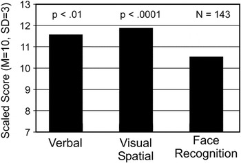
Mean verbal, visual spatial, and face recognition abilities of 143 parents of children with autism.
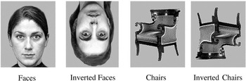
The stimuli used in the ERP study.
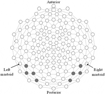
The electrodes used in the ERP analysis.
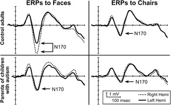
The event-related brain potentials to face and nonface stimuli in parents of children with autism versus control adults.
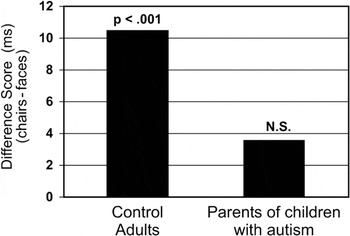
The N170 latency differences between ERP responses to faces versus chairs in parents of children with autism versus control adults.
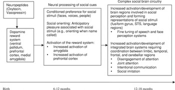
The role of social reward in the emergence of social brain circuitry in the first years of life.
 You have
Access
- 208
- Cited by