

INTRODUCTION
Nemopilema nomurai Kishinouye is one of the biggest rhizostome jellyfish attaining a bell diameter of 2 m and wet weight of over 200 kg, and is endemic in the Bohai Sea, Yellow Sea, East China Sea and Japan Sea (Uchida, Reference Uchida1954; Omori & Kitamura, Reference Omori and Kitamura2004; Wu et al., Reference Wu, Li, Li and Cheng2008; Uye, Reference Uye2008). The massive blooms of this species have become annual events fsince 2002, although they were very rare, once per ca. 40 years, during the 20th Century (Kishinouye, Reference Kishinouye1922; Shimomura, Reference Shimomura1959; Yasuda, Reference Yasuda2003; Kawahara et al., Reference Kawahara, Uye, Ohtsu and Iizumi2006; Uye, Reference Uye2008). It is thought that environmental changes in Chinese coastal waters such as eutrophication, overfishing and global warming are the causes of the recent massive blooms of N. nomurai (Uye, Reference Uye2008). Medusae originating in the Yellow Sea and East China Sea are transported offshore by the extension of the Changjian River low-salinity water mass, and drift northward to the Japan Sea by the Tsushima Current (Nishimura, Reference Nishimura1961; Kawahara et al., Reference Kawahara, Uye, Ohtsu and Iizumi2006; Reizen & Isobe, Reference Reizen and Isobe2006; Yoon et al., Reference Yoon, Yang, Shim and Kang2008). The drifting medusae clogged set nets and caused serious damage to coastal fisheries of Japan (Yasuda, Reference Yasuda2003). Since the drifting medusae have mature oocytes and sperm follicles (Toyokawa et al., Reference Toyokawa, Shimizu, Sugimoto, Nishiuchi and Yasuda2010), there is concern that they reproduce and establish new populations in the Japanese coastal waters (Uye, Reference Uye2008) like other rhizostome species such as Rhopilema nomadica Galil in the Mediterranean (Lotan et al., Reference Lotan, Ben-Hillel and Loya1992) and Phyllorhiza punctata von Lendenfeld in the Gulf of Mexico (Graham et al., Reference Graham, Martin, Felder, Asper and Perry2003).
The life cycle of N. nomurai consists of an alternation between sexual pelagic medusa and asexual sessile scyphistoma (Kawahara et al., Reference Kawahara, Uye, Ohtsu and Iizumi2006), and the latter stage is important for the population dynamics of this species because scyphistomae are perennial and increase in number by asexual reproduction (Willcox et al., Reference Willcox, Moltschaniwskyj and Crawford2008). However, because of its extraordinary body size, N. nomurai may have a huge fecundity (in the order of 108 eggs per medusa, unpublished data). For this reason, sexual reproduction may also be important for the population explosion as well as expanding the geographical range of this species.
Nemopilema nomurai is a gonochoristic jellyfish having a ribbon-like ovary and testis at the bottom of pleated interradial gastric pouches which protrude beneath the umbrella. The process of sexual reproduction in N. nomurai, from fertilization to the settlement of scyphistomae, has been described in our previous paper (Ohtsu et al., Reference Ohtsu, Kawahara, Ikeda and Uye2007). Our major findings were as follows: (1) intact medusae have immature ovaries or testes, while medusae physically damaged or stranded on the seashore around Oki Island, south-western Japan Sea, during October–December, have relatively mature gonads; (2) the maturation of gonads in immature medusae is accelerated after they have been physically damaged, and is completed within several days; and (3) spawning is induced by exposure to light after the gonads have been placed in darkness; male spermiation proceeds about 1 hour prior to female ovulation, which occurs 1.5 hours after exposure to light.
However, the histological background of neither the rapid maturation of oocytes and sperm follicles nor the spawning was revealed in our previous paper. Oocytes of semaeostome and rhizostome jellyfish associate with the specialized ovarian epithelial cells, called trophocytes, which support the vitellogenesis of oocytes (Eckelbarger & Larson, Reference Eckelbarger and Larson1988, Reference Eckelbarger and Larson1992; Pitt & Kingsford, Reference Pitt and Kingsford2000; Adonin et al., Reference Adonin, Podgornaya, Matveev and Shaposhnikova2009). A question is how surrounding tissues of oocytes support the rapid vitellogenesis in N. nomurai. The spermatogenesis of rhizostome jellyfish has not been well studied, so that it is unclear whether supporting epithelial cells like trophocytes occur in sperm follicles. Furthermore, there is little information about the spawning of gametes in scyphozoans.
We therefore examined the structural changes of the ovary and testis during artificially induced gonadal maturation and spawning using light microscopy and transmission electron microscopy in order to understand the histological bases of the rapid maturation of gonads and the spawning of N. nomurai.
MATERIALS AND METHODS
Medusae of Nemopilema nomurai (Rhizostomeae, Cnidaria) were collected off Kamo, Oki Island (36°10′N 133°16′E), from 15 October to 9 December in 2007, when the surface temperature decreased from 24 to 19°C. Six (3 males and 3 females) medusae (75–130 cm bell diameter) which looked morphologically intact and swam actively were captured with a scoop net (mouth diameter: 1 m) from a boat and towed slowly to the pier of the Oki Marine Biological Station, where they were arrested in a net (1.5 m in diameter and 1 m in depth) to accelerate the maturation of their gonads (Ohtsu et al., Reference Ohtsu, Kawahara, Ikeda and Uye2007). A piece of the genital organ (10 cm × 10 cm) was dissected daily from each captive medusa and photographed with an Olympus digital camera as a part of a stereo microscope. The long axis diameter of oocytes (111–442 images per daily sample) in the ovaries was measured to the nearest of 0.1 µm using an image analyser (ImageJ, version 1.43u, NIH). To determine whether the oocyte diameter increased after the arrest of medusae, data were analysed with one-way ANOVA and linear regression analysis. The area of sperm follicles (62–248 images per daily sample) of male medusae was also measured and the daily change was analysed with the same method as that in oocyte maturation.
The dissected genital organ was cut into small pieces (1 cm × 1 cm) containing gonads and mesoglea and was then immediately fixed with a fixative of 2.5% glutaraldehyde and 2% formaldehyde in 0.1 M phosphate buffer containing 0.4 M sodium chloride overnight at room temperature for subsequent observations using light and electron microscopy. In 2–5 days of arrest in a net, when the gonads matured so as to be ready for spawning (see Ohtsu et al., Reference Ohtsu, Kawahara, Ikeda and Uye2007), the dissected pieces of gonads were exposed to light to induce spawning and placed in polystyrene vessels (13.5 cm diameter and 5.5 cm height) containing 200 ml of filtered seawater at 23°C. Thereafter, the pieces of gonads were fixed at 5 minute intervals for the first 30 minutes and 10 minute intervals for the next 1.5 hours during the spawning.
The fixed specimens were rinsed with 0.1 M phosphate buffer three times and then postfixed with 1% osmium tetraoxide in the buffer for 2 hours under ice-cold conditions. These tissues were dehydrated with ethanol (50%, 70%, 90% and 100%), and propylene oxide. Finally, the dehydrated tissues were embedded in Quetol 651 resin (Nissin EM Co. Ltd, Japan) at 60°C for 24 hours. For light microscopy, thick sections (≈500 nm in thickness) were cut on the Leica EM UC6rt ultramicrotome using a glass knife. These sections were mounted on a slide glass, stained with 0.1% toluidine blue on a heat block and then observed under an Olympus BX51 light microscope. For transmission electron microscopy, thin sections (≈90 nm in thickness) were cut on the same ultramicrotome using a diamond knife. These sections were directly mounted on a copper grid. They were stained with 0.2% oolong tea extract (Nissin EM Co. Ltd., Japan) in 0.02 M phosphate buffer, followed by Farmy's 0.1% lead citrate, and then examined with a JEOL JEM 1200 transmission electron microscope.
The development of oocytes was divided into five stages on the basis of fine structures as described by Eckelbarger & Larson (Reference Eckelbarger and Larson1988). Twenty to 33 oocytes at each developmental stage were randomly selected from the thick sections of ovaries in the three females and the maximum long axis length of oocytes among serial sections was measured as their diameter under an Olympus BX50 light microscope.
RESULTS
Maturation of oocyte
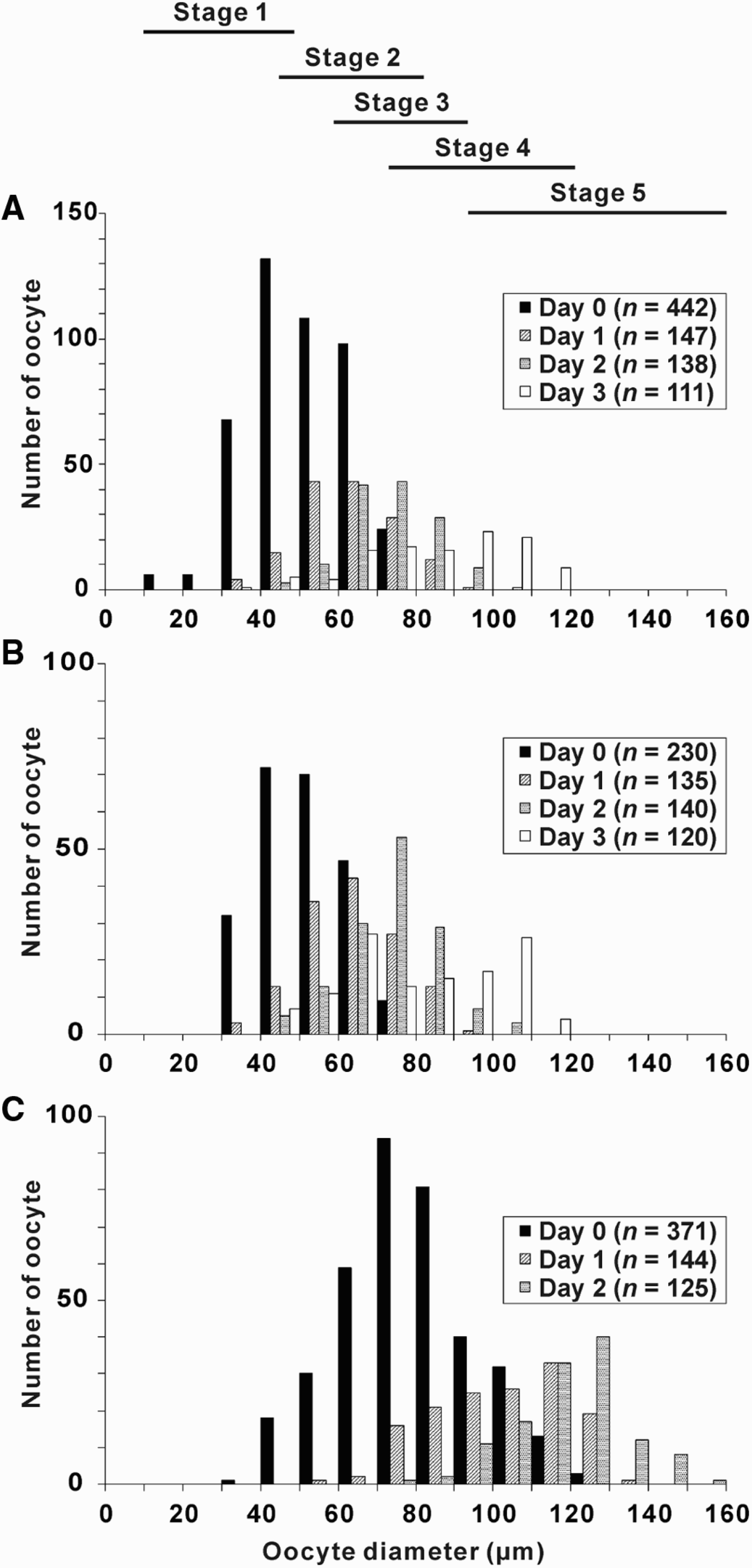
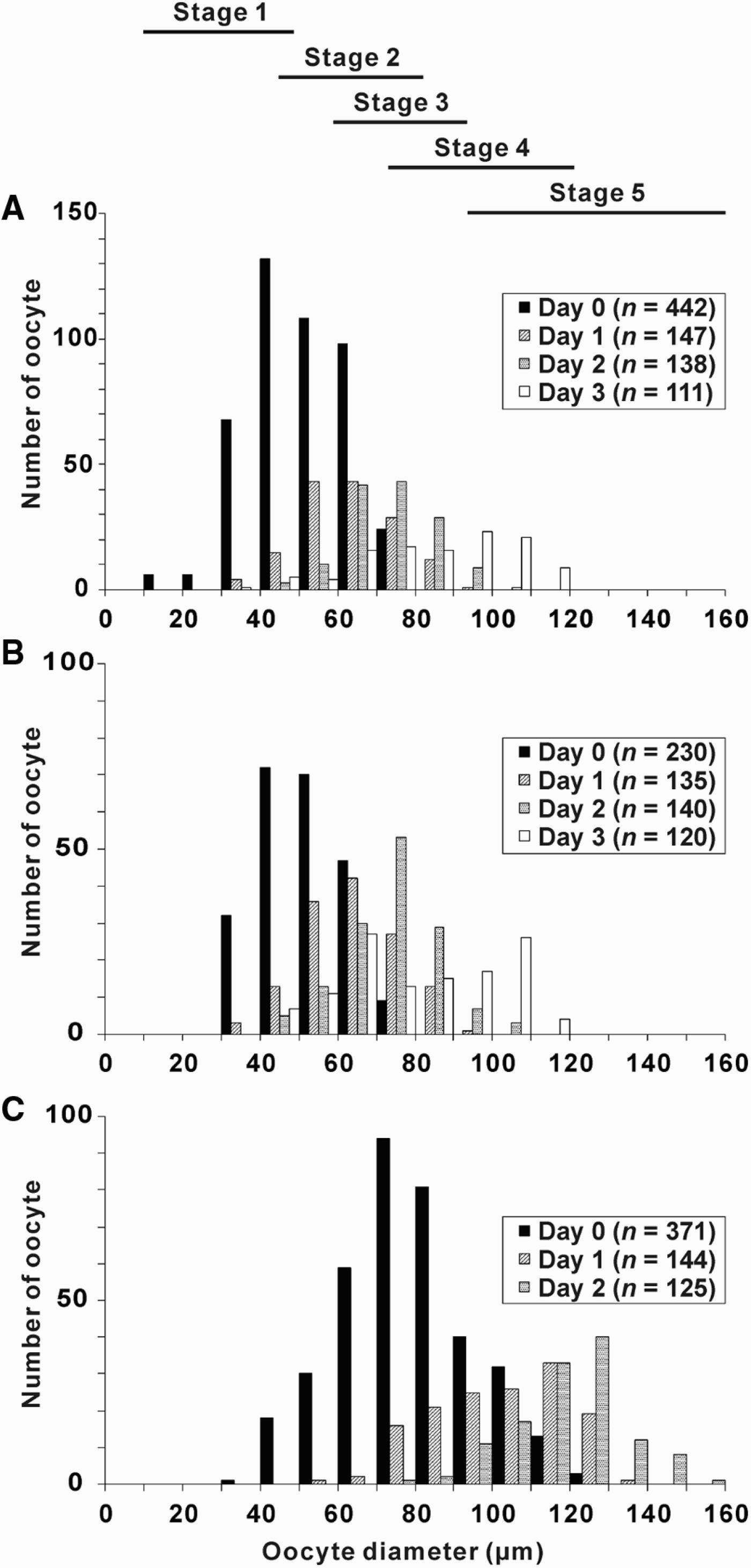
The two female medusae (both 85 cm bell diameter) captured in October and one female (130 cm bell diameter) captured in December had immature ovaries containing many developing oocytes whose mean diameter was 41.4, 42.1 and 69.1 µm, respectively. The maturation of these oocytes was accelerated after the arrest of medusae; the diameter of oocytes significantly increased (one-way ANOVA, P < 0.01) (Figure 1). A linear regression analysis showed that the increment rate of the mean oocyte diameter was 11.5, 9.7 and 19.5 µm per day, respectively, and there was no significant difference among the three females. Mature oocytes appeared 3–5 days after the arrest.
Daily change in oocyte diameter distribution after arresting three female medusae of Nemopilema nomurai. (A, B) Oocytes of medusae captured in October 2007; (C) captured in December 2007. Horizontal bars indicate the size-range of each maturation stage; n, number of oocytes measured.
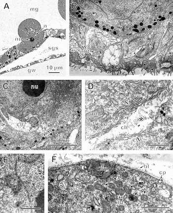
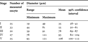
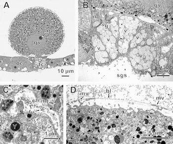
At the first stage, a primary oocyte arises from the ovarian epithelium of endodermal origin, protruding into the ovarian mesoglea (Figure 2A). This early vitellogenic oocyte (mean diameter: 35 µm; Table 1) has a nucleus, including a single nucleolus, in the subgenital sinus side of the ooplasm (Figure 2A). Some ovarian epithelial cells associated with the oocyte became a specialized cell type, which consisted of trophocytes, having microvilli on the side of the subgenital sinus, and Golgi complexes and vesicles in the cytoplasm (Figure 2B). These vesicles fuse with each other to form a large mass. The oocyte also maintains contact with other ovarian epithelial cells around the trophocytes. The ooplasm contains many mitochondria, rough endoplasmic reticulum which is well developed and forms the parallel arrays around the nucleus, and intraooplasmic channels associated with rough endoplasmic reticulum (Figure 2C, D). These channels are lined with a membrane and inside include small vesicular materials (Figure 2D). Larger channels occur more frequently around the nucleus of the oocyte and smaller ones are scattered throughout the entire ooplasm (Figure 2C, F). Small yolk bodies (0.2–2 µm) begin to be formed in the ooplasm near the trophocytes (Figure 2B, C). Densely stained aggregations of granular materials form distinctive patches around the nucleus of the oocyte (Figure 2E). A thin basal lamina covers the entire surface of the oocyte and is continuous with that underlying the adjacent epithelial cells (Figure 2F). No microvilli occur on the surface of the oocyte. Lamellated membranes occur near the oolemma and a few coated pits appear on the surface of the oocyte (Figure 2F).
Fine structure of the ovary at the first stage of oocyte maturation in Nemopilema nomurai. (A) Light microscopy of an oocyte arising from the ovarian epithelium; (B) trophocytes associated with the early vitellogenic oocyte. Vesicles, Golgi complexes, and mitochondria are seen in the cytoplasm of trophocytes; (C) rough endoplasmic reticulum and intraooplasmic channel around the nucleus of the oocyte; (D) magnified view of the intraooplasmic channel with rough endoplasmic reticulum and vesicular materials (asterisk) in the channel; (E) a patch of electron-dense granular materials around the nucleus of the oocyte; (F) coated pit and lamellated membrane at the surface area of the oocyte. Many mitochondria (arrowheads) occur in the cytoplasm. bl, basal lamina; ch, intraooplasmic channel; cp, coated pit; dg, electron-dense granular materials; g, Golgi complex; gw, genital wall; lm, lamellated membrane; mg, mesoglea; mt, mitochondria; mv, microvilli; n, nucleus; nm, nuclear membrane; nu, nucleolus; oe, ovarian epithelium; rer, rough endoplasmic reticulum; sgs, subgenital sinus; tc, trophocytes; v, vesicles; Y, yolk bodies.

Size of oocytes at each maturation stage in three females.
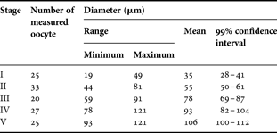
At the second stage of maturation, the oocytes have mean diameter of 55 µm (Table 1) and keep depositing yolk bodies in the ooplasm adjacent to the trophocytes, which well develop and have many vesicles (Figure 3A, B). The oocyte moves toward the mesoglea of the ovary, keeping close contact only with the trophocytes (Figure 3A, B). At this stage, nascent yolk bodies occur near Golgi complexes in the ooplasm (Figure 3C). Small perivitelline spaces with fibrous materials inside are sparsely observed on the surface area of the oocyte, and coated pits frequently appear on the oolemma (Figure 3D).
An oocyte and trophocytes at the second stage of oocyte maturation in Nemopilema nomurai. (A) Light microscopy of an oocyte and trophocytes. Densely-stained granules in the ooplasm adjacent to the trophocytes are yolk bodies; (B) trophocytes associated with the oocyte; (C) nascent yolk bodies (double arrowheads) formed near the Golgi complex in the ooplasm; (D) perivitelline space including fibrous materials (asterisk) at the surface area of the oocyte. Coated pits occur on the oolemma. bl, basal lamina; ch, intraooplasmic channel; cp, coated pit; g, Golgi complex; mg, mesoglea; mv, microvilli; n, nucleus; nu, nucleolus; oe, ovarian epithelium; tc, trophocytes; v, vesicle; Y, yolk bodies.

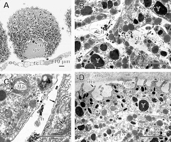
At the third stage of oocyte maturation (mean diameter: 78 µm; Table 1), the nucleus of the oocyte becomes a large germinal vesicle whose diameter reaches 1/3 that of the oocyte (Figure 4A). The trophocytes develop further but begin to dissociate from the oocyte, resulting in clefts that appear at the junction with the oocyte (Figure 4B). High electron-dense flocculent materials occur in the intraooplasmic channels (Figure 4C). Small yolk bodies are distributed over the entire ooplasm (Figure 4A, D). Some yolk bodies include many electron-dense granules (Figure 4C). Microvilli grow and perivitelline spaces appear throughout the surface of oocyte (Figure 4D). Large clusters of fibrous materials occur in the perivitelline space (Figure 4D).
An oocyte and trophocytes at the third stage of oocyte maturation in Nemopilema nomurai. (A) Light microscopy of an oocyte and trophocytes. The nucleus of the oocyte becomes a germinal vesicle; (B) trophocytes filled with many vesicles. Clefts (arrowheads) appear between the trophocytes and the oocyte; (C) intraooplasmic channel containing flocculent materials. Vesicular materials (asterisk) also occur in the channel. Some of the yolk bodies include electron-dense granules; (D) enlarged perivitelline space including fibrous materials (double asterisks). Small microvilli are seen in the perivitelline space. bl, basal lamina; ch, intraooplasmic channel; fu, flocculent materials; g, Golgi complex; gv, germinal vesicle; mt, mitochondria; mv, microvilli; n, nucleus; nu, nucleolus; oe, ovarian epithelium; rer, rough endoplasmic reticulum; sgs, subgenital sinus; tc, trophocytes; v, vesicle; Y, yolk bodies.

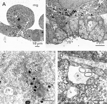
At the fourth stage of maturation, the oocyte (mean diameter: 93µm; Table 1) is completely separated from the trophocytes that include large vesicles (Figure 5A). The intraooplasmic channels form a wide network extending throughout the entire oocyte (Figure 5A, B). The width of a channel is larger around the germinal vesicle than in the peripheral area of the oocyte (Figure 5A, B & D). Golgi complexes become abundant in the ooplasm (Figure 5C). Yolk bodies largely increase in number and show an uneven distribution in the ooplasm, being more abundant around the germinal vesicle (Figure 5A). Many microvilli develop on the surface of the oocyte and a wide perivitelline space appears between the basal lamina and the oolemma (Figure 5D).
An oocyte and trophocytes at the fourth stage of oocyte maturation in Nemopilema nomurai. (A) Light microscopy of an oocyte and trophocytes. They are completely separated by the gap (arrowheads). The long and narrow lucent area in the oocyte presents intraooplasmic channels. Note the uneven distribution of the densely stained yolk bodies; (B) highly branched intraooplasmic channels with flocculent materials around the germinal vesicle. Ooplasm is filled with a number of yolk bodies with various electron densities; (C) magnified view of the intraooplasmic channel associated with rough endoplasmic reticulum and Golgi complexes; (D) perivitelline space (asterisk) and microvilli on the surface of the oocyte. bl, basal lamina; ch, intraooplasmic channel; fu, flocculent materials; g, Golgi complex; gv, germinal vesicle; mg, mesoglea; mv, microvilli; nu, nucleolus; oe, ovarian epithelium; rer, rough endoplasmic reticulum; tc, trophocytes; Y, yolk bodies.

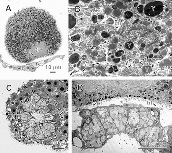
At the final stage of maturation, an oocyte fully matures to have a mean diameter of 106 µm (Table 1), and possesses a large germinal vesicle (≈30 µm diameter) including a single distinct nucleolus (Figure 6A). The ooplasm is full of many yolk bodies with various electron densities (Figure 6B). Golgi complexes and oval mitochondria are abundant in the ooplasm, and endoplasmic reticulum is also present (Figure 6B). The intraooplasmic channels remain in the ooplasm but have shrunk in comparison with those of the oocyte at the fourth stage of maturation (Figures 5B & 6B). The trophocytes form a large mass (≈50 µm in diameter) consisting of 10–15 triangular-prismatic cells which are radially arranged (Figure 6C, D). The nuclei of the trophocytes are located in the periphery of the mass (Figure 6D). The cytoplasm of trophocytes is occupied by vesicles (Figure 6D). The basal lamina covers the entire surface of the oocyte, and occurs in the gap between the oocyte and trophocytes (Figure 6D).
An oocyte and trophocytes at the final stage of oocyte maturation in Nemopilema nomurai. (A) Light microscopy of a mature oocyte and trophocytes; (B) magnified view of ooplasm; (C) cross-section of trophocytes. Arrowheads indicate the boundary between trophocytes and ovarian epithelial cells; (D) tangential section of trophocytes adjacent to a mature oocyte. The oocyte and trophocytes are separated by the gap (asterisk) including a basal lamina. bl, basal lamina; ch, intraooplasmic channels; g, Golgi complexes; gv, germinal vesicle; mt, mitochondria; n, nucleus; nu, nucleolus; oe, ovarian epithelium; rer, rough endoplasmic reticulum; tc, trophocytes; v, vesicles; Y, yolk bodies.

The medusae had oocytes at the first and second stages of maturation just after they had been captured. Following artificial induction of maturation, oocytes at the third maturation stage appeared in the ovary from the second day of arrest in the net. Oocytes at the fourth stage occurred from the third and fourth day, and mature oocytes were observed from the fourth and fifth days (Figure 1). The spawning of oocytes was artificially induced by light exposure 3–5 days after the arrest. Thus, the duration of each maturation stage was about one day when the medusae were arrested in the net as the temperature ranged from 19 to 24°C.
Maturation of sperm follicle
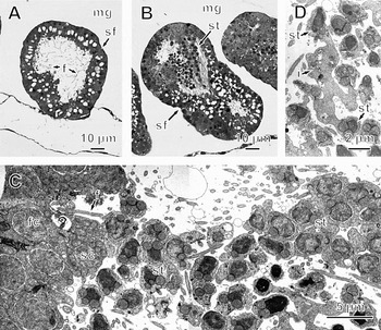
In the immature testis of the male medusa caught in October (75 cm bell diameter), there were very few spermatozoa in the sperm follicles although many flagella were seen (Figure 7A). Spermatogenic cells appeared in the sperm follicles from the fourth day of the arrest in the net, indicating the beginning of spermatogenesis (Figure 7B). Sperm follicles were filled with spermatids that became mature from the fifth day following the arrest, and spawning was then observed from the sixth day of the arrest. On the other hand, spermatogenesis had already started and a number of spermatids occurred in the sperm follicles of two male medusae captured in December (130 cm bell diameter). The spawning of these two males occurred from the third day of the arrest. The spermatogenesis occurred in synchrony among sperm follicles in the testis of each medusa. However, there was no clear relationship between the size of sperm follicles and the days after the arresting in the three medusae.
Maturation of sperm follicle in Nemopilema nomurai. (A) Light microscopy of cross-section of an immature sperm follicle in the testis of a male medusa; (B) light microscopy of a sperm follicle where spermatogenesis takes place. Many spermatids fill the sperm follicle; (C) magnified view of the sperm follicle during spermatogenesis; (D) intercellular bridge between developing spermatids. f, flagellum; fc, follicle cell; i, intercellular bridge; mg, mesoglea; sc, spermatocyte; sf, sperm follicle; st, spermatids.

In the sperm follicle where spermatogenesis takes place, mitochondria-rich cells consisting of spermatocytes appear adjacent to the follicle cells, and many spermatids occur inside of the sperm follicle (Figure 7C). The nuclei of spermatids in the central area of the sperm follicle are denser than those in the peripheral part (Figure 7C). The cytoplasm of spermatids fuse with each other through the intercellular bridge during spermatogenesis, and the bridge appears to stretch at the late stage of spermatogenesis (Figure 7D).
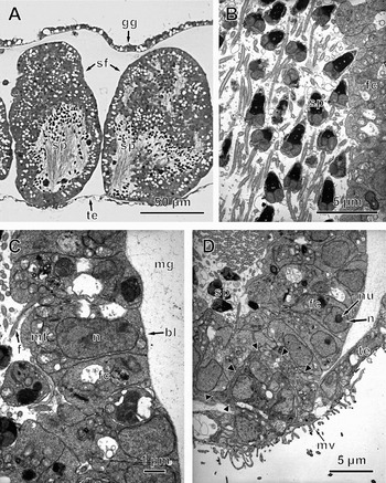
The mature sperm follicle has an irregular shape, having long axis of ≈120 µm (Figure 8A). The sperm follicle is filled with spermatozoa possessing a long conical head containing a nucleus, spherical mitochondria, and a flagellum (Figure 8B). The head of the spermatozoa has a tendency to be directed outward in the follicles, with the tails directed centrally. The follicle cell has a relatively large nucleus with a distinct nucleolus and many mitochondria. The flagellum originates from some of the follicle cells (Figure 8C, D). Like the oocyte, the sperm follicle is surrounded by a thin basal lamina continuous with that underlying the testicular epithelium, although there are no microvilli on the outer surface of follicle cells (Figure 8C). The sperm follicle is associated with the specialized testicular epithelial cells, which have microvilli on the subgenital sinus side and include no vesicle in the cytoplasm, unlike the trophocytes in the ovary (Figure 8D). These microvilli-rich epithelial cells invaginate into the sperm follicle as they plug it.
Fine structure of mature sperm follicles in Nemopilema nomurai. (A) Light microscopy of cross-section of mature sperm follicles; (B) mature free spermatozoa in a sperm follicle; (C) follicle cells. A flagellum originates from a follicle cell; (D) associated site of the sperm follicle with the testicular epithelium. Arrowheads indicate the border between microvilli-rich testicular epithelial cells and the sperm follicle. bl, basal lamina; f, flagellum; fc, follicle cell; gg, gonadal gastrodermis; mg, mesoglea; mt, mitochondria; mv, microvilli; n, nucleus; nu, nucleolus; sf, sperm follicles; sp, spermatozoa; te, testicular epithelium.

Structural changes of gonads during spawning
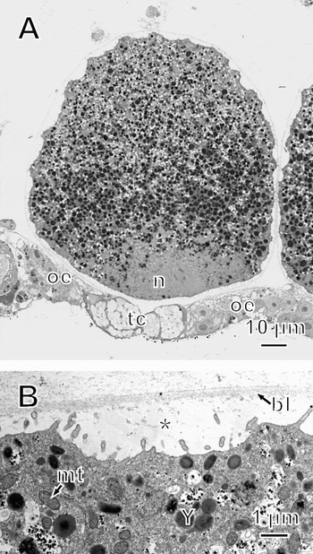
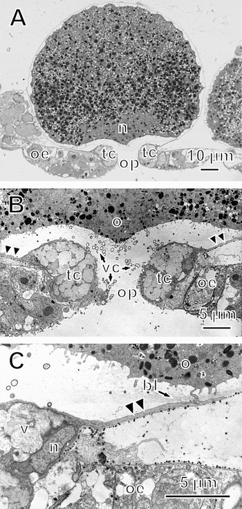
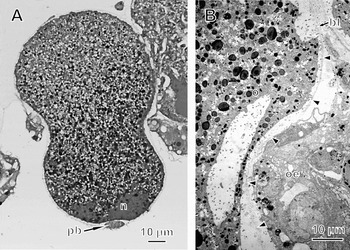
In spawning of the female (ovulation), no significant histological change occurs for the first 60 minutes after the mature ovary is exposed to light. The breakdown of the germinal vesicle, indicating the reinitiation of meiosis, occurs 60–70 minutes after the light exposure, and the broken nucleus disperses in the ooplasm at the side of the subgenital sinus (Figure 9A). At the same time, the microvilli on the surface of the oocyte diminish and the oolemma undulates entirely, resulting in a widening of the perivitelline space (Figure 9B). After the breakdown of the germinal vesicle (≈80 minutes after light exposure), an ovulation pit opens at the centre of the mass of trophocytes and the oocyte projects toward the pit (Figure 10A). Trophocytes stretch toward the oocyte, forming the fine extension of the cytoplasm (Figure 10B, C). In the ovulation pit between the separating trophocytes, many small spherical vesicular materials are observed (Figure 10B). At 90–100 minutes after the light exposure, the ovulation pit gradually enlarges and the oocyte then passes through the pit in a peanut-like distorted form (Figure 11A). A single polar body occurs near the broken nucleus, indicating that the maturation division continues (Figure 11A). The basal lamina is separated from the ovarian mesoglea and keeps surrounding the oocyte during the ovulation (Figure 11B). Finally, the oocyte is liberated from the ovary into the subgenital sinus. No structural change occurs in immature oocytes even when they are exposed to light.
Ovary of Nemopilema nomurai 70 minutes after light exposure. (A) Light microscopy of an oocyte and trophocytes. The nucleus breaks down and disperses in the ooplasm near the trophocytes. Note the undulation of the surface of the oocyte; (B) magnified view of the surface area of the oocyte. Microvilli diminish and the perivitelline space (asterisk) widens. bl, basal lamina; mt, mitochondria; n, nucleus; oe, ovarian epithelium; tc, trophocytes; Y, yolk bodies.

An oocyte and trophocytes of Nemopilema nomurai 80 minutes after light exposure. (A) Light microscopy of an oocyte projecting into the ovulation pit; (B) magnified view of the ovulation pit. Small vesicular materials occur near the ovulation pit; (C) fine extension from the trophocyte (double arrowheads). bl, basal lamina; n, nucleus; o, oocyte; oe, ovarian epithelium; op, ovulation pit; tc, trophocyte; v, vesicles; vc, vesicular materials.

An oocyte of Nemopilema nomurai 90 minutes after light exposure. (A) Light microscopy of an oocyte showing a peanut-like form during ovulation; (B) magnified view of the lateral side of the oocyte. Basal lamina (arrowheads) separates from the ovarian mesoglea and covers the oocyte. bl, basal lamina; n, nucleus; o, oocyte; oe, ovarian epithelium; pb, polar body.

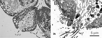
In spawning of the male (spermiation), microvilli-rich epithelial cells associated with the sperm follicle dissociate from each other to form a spermiation pit about 5 minutes after the testis has been exposed to light (Figure 12A, B). The spermatozoa are shed through the pit into the subgenital sinus (Figure 12B). The spermiation pits mainly occurred during 5–20 minutes after the light exposure although they were observed until 1 hour after.
Spermiation in Nemopilema nomurai after the testis is exposed to light. (A) Light microscopy of sperm follicle shedding spermatozoa through the spermiation pit 5 minutes after light exposure; (B) magnified view of the spermiation pit. f, flagellum; sf, sperm follicle; sp, spermatozoa; spp, spermiation pit; te, testicular epithelium.

DISCUSSION
Maturation of oocytes
A huge fecundity (in the order of 108 eggs, unpublished data) and the induction of rapid (within several days) gonadal maturation from immature stage to fully mature one after medusae are physically damaged (Figure 1; see also Ohtsu et al., Reference Ohtsu, Kawahara, Ikeda and Uye2007) characterize the sexual reproduction of Nemopilema nomurai. The large fecundity is not only owing to large medusa size (maximum weight: over 200 kg; Uchida, Reference Uchida1954; Omori & Kitamura, Reference Omori and Kitamura2004) but also heavily folded structure of the ovary, which can increase the surface area so enormously as to carry as many eggs as possible (Ohtsu et al., Reference Ohtsu, Kawahara, Ikeda and Uye2007). This electron microscopic study confirms our previous finding on the rapid oocyte maturation (Ohtsu et al., Reference Ohtsu, Kawahara, Ikeda and Uye2007) and reveals the process much more in detail based on ultrastructural features of the oocytes and trophocytes.
The oocyte of N. nomurai carries yolk bodies, rough endoplasmic reticulum, intraooplasmic channels, coated pits, and Golgi complexes, as have already been found in Aurelia aurita (Linnaeus) (Semaeostomeae) and Stomolophus meleagris L. Agassiz (Rhizostomeae) (Eckelbarger & Larson, Reference Eckelbarger and Larson1988, Reference Eckelbarger and Larson1992), indicating that the fine structure of oocyte is basically the same among semaeostome and rhizostome jellyfish. The morphological features of trophocytes is also the same among semaeostome and rhizostome jellyfish since the trophocytes of N. nomurai are similar to those of A. aurita, Pelagia noctiluca (Forsskål) (Semaeostomeae), Discomedusa lobata Claus (Semaeostomeae), and S. meleagris in intimate association with young oocyte, microvilli formation on the subgenital sinus side, and deposition of yolk bodies in ooplasm adjacent to trophocytes (Eckelbarger & Larson, Reference Eckelbarger and Larson1988, Reference Eckelbarger and Larson1992; Avian & Rottini Sandrini, Reference Avian and Rottini Sandrini1991). These results suggest that the trophocytes transfer nutrients from the subgenital sinus to the oocytes for the vitellogenesis in N. nomurai as suggested in the above-mentioned species (Eckelbarger & Larson, Reference Eckelbarger and Larson1988, Reference Eckelbarger and Larson1992; Avian & Rottini Sandrini, Reference Avian and Rottini Sandrini1991).
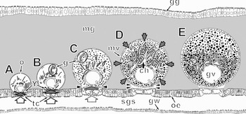
On the basis of these fine structural features of oocyte and trophocytes, Figure 13 schematically summarizes the morphological changes of the oocyte with maturation and the assumed process of nutrient uptake by the oocyte in N. nomurai. Important features of oocyte maturation in N. nomurai are: (1) oocyte forms yolk bodies using nutrient which is transported from the subgenital sinus by the associating trophocytes at the first and second maturation stage; and (2) oocyte obtains nutrient from surrounding mesoglea through coated pits and developed microvilli and forms yolk bodies with Golgi complexes and rough endoplasmic reticulum associating with intraooplasmic channels in the ooplasm from the third stage when the oocyte separates from the trophocytes. The whole vitellogenic process is completed within 5 days at 19–24°C. The function of the trophocytes can be important at least in the early vitellogenic stage for the rapid yolk formation of oocyte.
Schematic diagram of the source of nutrients at various oocyte maturation stages of Nemopilema nomurai. (A) First stage. Trophocytes take nutrients from the subgenital sinus and transfer them to the oocyte, which forms yolk bodies in the ooplasm adjacent to the trophocytes; (B) second stage. Yolk deposition takes place in the oocyte adjacent to the trophocytes and near the Golgi complexes; (C) third stage. The oocyte begins to separate from the trophocytes (arrowheads), and the transfer of nutrients by trophocytes gradually decreases (broken-line arrows); (D) fourth stage. The oocyte completely dissociates from the trophocytes (double arrowheads), but takes nutrients from the surrounding mesoglea through the developed microvilli located over the surface of the oocyte. Intraooplasmic channels form the wide networks in the ooplasm; (E) fifth stage. The oocyte becomes mature and is filled with a number of yolk bodies. Blank arrows indicate the uptake of nutrients from the subgenital sinus to the trophocytes, and filled arrows indicate the transfer of nutrients from the trophocytes to oocytes (large arrows) or from surrounding mesoglea (small arrows). ch, intraooplasmic channel; g, Golgi complex; gg, gonadal gastrodermis; gv, germinal vesicle; gw, genital wall; mg, mesoglea; mv, microvilli; o, oocyte; oe, ovarian epithelium; sgs, subgenital sinus; tc, trophocytes; Y, yolk bodies.

Maturation of sperm follicles
During spermatogenesis, spermatocytes arise near the wall of the sperm follicle and the maturation takes place in the cavity of follicles (Figure 7C). The wall of the sperm follicle includes flagellated cells, indicating that the sperm follicle of N. nomurai includes spermatogonia as in other cnidarians (Goffredo et al., Reference Goffredo, Telò and Scanabissi2000; Morandini & Silveira, Reference Morandini and Silveira2001; Gaino et al., Reference Gaino, Bo, Boyer and Scoccia2008). Therefore, the immature sperm follicle may be ready to form spermatocytes and spermatozoa. Because a spermatocyte is divided into four spermatids after the meiosis (Lyke & Robson, Reference Lyke and Robson1975), spermatozoa could be produced from spermatocytes provided by the follicle cells in an exponential fashion, resulting in the rapid accumulation of spermatozoa. The developing spermatids keep the connection with each other through the intercellular bridge, as observed in a hydromedusa (Roosen-Runge & Szollosi, Reference Roosen-Runge and Szollosi1965) and anthozoans (Lyke & Robson, Reference Lyke and Robson1975; Gaino et al., Reference Gaino, Bo, Boyer and Scoccia2008). The elongation of intercellular bridge in the late spermatogenic stage (Figure 7D), possibly indicates that the spermatids lose their excess cytoplasm and become free spermatozoa (Goffredo et al., Reference Goffredo, Telò and Scanabissi2000).
The testicular epithelial cells associate with a sperm follicle and bear microvilli on the subgenital sinus side as observed with trophocytes in the ovary. However, these microvilli-rich cells hardly have vesicles, Golgi complexes or endoplasmic reticulum and are therefore in marked contrast with the trophocytes (Figure 8D). These microvilli-rich cells may convey nutrients from the subgenital sinus to the follicle cells, but their function would be less important than that of the trophocytes (Wedi & Dunn, Reference Wedi and Dunn1983). The follicle cells do not have microvilli on the outer surface (Figure 8C), suggesting that they could take less nutrients from the surrounding mesoglea than the oocyte. Because the spermatozoon is a simple cell without a reserve of nutrients (Figure 8B), it is possible that the sperm follicle acquires sufficient nutrients for spermatogenesis from the microvilli-rich epithelial cells.
Mechanisms of spawning
The oocyte of N. nomurai seems to be compressed by surrounding tissues during ovulation (Figure 11A), but there are no follicle cells around the developed oocyte (Figure 6A). Epitheliomuscular cells incorporate the contractile process of ovulation in hydra (Schroeder & Talbot, Reference Schroeder and Talbot1985). Since isolated pieces of N. nomurai ovary have occasional contraction (Ohtsu et al., Reference Ohtsu, Kawahara, Ikeda and Uye2007) the contraction of ovarian tissue such as ovarian epithelium may involve the escape of the oocyte from the ovary. Furthermore, the oocyte modifies its own shape after light exposure (Figures 9A & 10A), indicating that an amoeboid movement of the oocyte is also involved in ovulation. The trophocytes also modify their structures during ovulation as described by Avian & Rottini Sandrini (Reference Avian and Rottini Sandrini1991), and vesicular materials near the separating trophocytes seem to be broken pieces of cytoplasm or extracellular matrix (Figure 10B). However, it is uncertain whether the structural changes are caused by the trophocytes or the oocyte. The basal lamina surrounding the oocyte separates from the ovarian mesoglea and overlays the oocyte during ovulation (Figure 11B). A vitelline membrane surrounds the spawned and unfertilized oocyte of N. nomurai (Ohtsu et al., Reference Ohtsu, Kawahara, Ikeda and Uye2007) although the oocyte does not have surrounding follicle cells producing vitelline membrane in the ovary (Figure 6A). These phenomena might suggest that the basal lamina surrounding the oocyte is modified to develop into the vitelline membrane.
In comparison with ovulation, spermiation is a simple process involving the sperm follicle opening at the attachment site with the testicular epithelium (Figure 12A, B). Scott & Harrison (Reference Scott and Harrison2009) suggested that sperms were released through the specialized testicular epithelium, called trophonemata, associated with sperm follicles in the anthozoan Entacmaea quadricolor (Rüppel & Leukert), but its histological mechanism was not apparent. Our study clearly demonstrates that the sperms liberate from the spermiation pits formed by the separation of microvilli-rich cells of sperm follicle (Figure 12A, B). No sperm follicles with opening spermiation pit were observed 1.5 hours after the light exposure (unpublished data). Furthermore, isolated pieces of testes can repeatedly spawn spermatozoa successive for several days in N. nomurai (Ohtsu et al., Reference Ohtsu, Kawahara, Ikeda and Uye2007). These findings indicate that the spermiation pit closes after the spermiation and the spermatogenesis takes place again in the sperm follicle.
The time course of spawning is largely different between the male and female. Spermiation takes place about 1 hour prior to ovulation, which occurs 1.5 hours after the light exposure (Ohtsu et al., Reference Ohtsu, Kawahara, Ikeda and Uye2007). This difference is possibly caused due to the mechanism of ovulation being more complicated than that of spermiation. The priority of spermiation is due to the fact that it takes several tens of minutes for the sperm to be active after it is spawned (Ohtsu et al., Reference Ohtsu, Kawahara, Ikeda and Uye2007). The timing of spawning is precisely controlled in response to light depending on the physiological characteristics of each gamete. These strategies of gametogenesis and spawning in N. nomurai would enhance the success of fertilization and would leave many more offspring.
ACKNOWLEDGEMENTS
We would like to thank Dr M. Kawahara (Hiroshima University) and Mr M. Nishizaki (Shimane University) for assistance with the collection of jellyfish and experiments, and Mrs K. Koike (TEM service, Hiroshima University) for help with transmission electron microscopy. This research was supported by research grants from the Japan Society for the Promotion of Science (No. 20780138) and the Ministry of Agriculture, Forestry, and Fisheries, Japan.