Fungal infections are uncommon in children but can cause significant morbidity and mortality in hospitalised patients. Reference Suleyman and Alangaden1,Reference Brissaud, Guichoux, Harambat, Tandonnet and Zaoutis2 Risk factors for fungal blood infections and fungal endocarditis include prior cardiac surgery, presence of pacemakers and implantable cardioverter–defibrillators, central venous catheters, prolonged use of broad-spectrum antibiotics, immunocompromised state, and diabetes. Reference Antinori, Ferraris and Orlando3–Reference Varghese and Sobel5 Fungal endocarditis has been classically described as associated with dense heterogenous vegetations. However, several case reports have described more subtle changes in the myocardium and papillary muscles in adults and some children with clinical diagnoses of endocarditis and/or myocarditis, Reference Harris, Ananth Narayan and Orchard6,Reference Donal, Abgueguen and Coisne7 including one study that demonstrated localised fungal infections of the myocardium on autopsy of adult patients. Reference Atkinson, Connor, Robinowitz, McAllister and Virmani8 In addition, our echocardiography laboratory observed multiple paediatric patients without clear vegetation, but who had single or multiple areas of focal myocardial brightenings within the myocardium and who were also identified to have systemic fungal infection (with fungal infection identified after the echocardiogram in at least one case). The primary aim of this study was to determine whether these atypical echocardiographic changes occur more frequently in hospitalised patients with fungal infections of the blood and/or urine compared to a control group of hospitalised patients without fungal infections. The secondary aim was to determine whether patients with fungal infection and echocardiographic changes have worse outcomes compared to patients with fungal infection but no echocardiographic changes. We hypothesised that echocardiographic evidence of myocardial involvement is more common in patients with fungal infections compared to patients without fungal infections and that patients with fungal infection and significant echocardiographic findings are more ill than fungal patients without significant echocardiographic changes. Although the inclusion of fungal urinary tract infections leads to a more heterogenous population than limiting our population to those with fungal bloodstream infections, it may help elucidate potential pathophysiologic mechanisms for these echocardiographic findings.
Material and methods
Study population
The University of Michigan Institutional Review Board approved this study. In this retrospective case-control study, the Fungal Group consisted of patients with positive blood, urine, or vascular catheter tip cultures and the Control Group was matched 1:1 by age, hospital service (i.e., Cardiology, Gastrointestinal, Haematology/Oncology), hospital location, and need for extracorporeal membranous oxygenation support prior to echocardiogram. Potential study patients for the Fungal Group were identified by the Infection Prevention & Epidemiology program at University of Michigan and by query of a microbiological database, searching for all hospitalised patients at C.S. Mott Children’s Hospital with positive blood, urine, and/or catheter tip cultures over a 5-year period (between January 2010 and January 2015). Patients were included in the Fungal Group if at least one echocardiogram was performed near the time of positive culture (defined as occurring from 1 week prior to fungal culture up to hospital discharge date).
Potential Control Group patients were identified by query of the institutional echocardiographic database for echocardiograms performed on all inpatients during the study period. Exclusion criteria for Control Group patients were the presence of any positive fungal cultures. Control patients were then matched to Fungal Group patients by age, hospital service, and hospital location (Intensive care unit versus specific floor service) at time of echocardiogram. As it is not yet understood whether severe haemodynamic instability could be involved in the pathophysiology of these myocardial changes, patients were also matched based on whether or not they had required extracorporeal membranous oxygenation support prior to the echocardiogram during the same hospitalisation.
Data collection
Data regarding infectious history (fungal and bacterial culture sites and treatment), hospital course, and survival status were collected through review of internal medical records. The primary admitting diagnosis for each patient was recorded (e.g., cardiac, respiratory, infectious, oncologic, etc.). The presence or absence of cardiac defects was also documented. A vasoactive-inotropic support score at the time of the echocardiogram was calculated for each patient using the following equation: vasoactive-inotropic support score = dopamine dose (µg/kg/minute) + dobutamine dose (µg/kg/minute) + 100 × epinephrine dose (µg/kg/minute) + 10 × milrinone dose (µg/kg/minute) + 10,000 × vasopressin dose (U/kg/minute) + 100 × norepinephrine (µg/kg/minute). The vasoactive-inotropic support score is a measure of the haemodynamic instability of the patient at the time of echocardiogram, with higher scores indicating a higher degree of haemodynamic instability compared to lower scores. Reference Gaies, Gurney and Yen9,Reference Wernovsky, Wypij and Jonas10
Echocardiographic evaluation
Two independent experienced echocardiographers (GJE and STO) reviewed up to two echocardiograms for each patient. The readers were blinded to the Fungal/Control Group status for each echocardiogram. For patients in the Fungal Group, the echocardiograms reviewed were those closest to the time of the positive fungal culture result. For patients in the Control Group, the echocardiograms reviewed were the first performed at the matched location/service and the next echocardiogram performed closest to 1 week later.
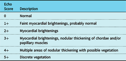
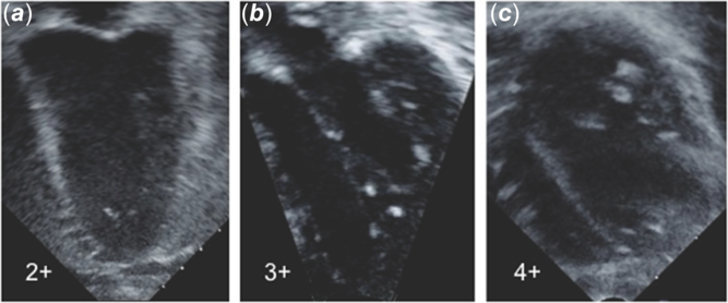
Each echocardiographic study was scored by both readers using 5-point scale based on severity of findings, as described in Table 1 with examples shown in Figure 1 and two cine echo loops (Supplementary Videos S1 and S2). In cases where the two readers differed in scoring by greater than 1, a third experienced echocardiographer (MDN), also blinded to Fungal/Control Group status, was asked to score the studies as well. For patients with two echocardiograms available, the more conservative lower echocardiographic score was chosen and included in the statistical analysis. A “positive” echo score was defined as an echo score ≥ 2+, indicating findings more than myocardial brightening alone.
Description of echo scores

Examples of Echo Scores (2+ to 4+). ( a ) Example of 2+ score with myocardial brightenings; ( b ) Example of 3+ score with myocardial brightenings and nodular thickening of the papillary muscles and chordae; ( c ) Example of 4+ score with multiple areas of nodular thickening of the papillary muscles and chordae with possible vegetation on mitral valve chordae.

The echocardiography machine used for each study was also recorded. The vast majority of studies were performed on a Philips IE33 (94%; Philips North America Corporation, Andover, MA). The remainder were performed on a GE Vivid (GE Healthcare, General Electric Company, Boston, MA) in 4.5% of studies, Siemens Accuson (Siemens Corporation, Washington, DC) in 0.8% of studies, and Philips EPIQ (Philips North America Corporation, Andover, MA) in 0.8% of studies. All echocardiographic data images were digitally stored on Syngo Dynamics (Siemens Medical Solutions USA, Inc., Malvern, PA) and were analysed and scored in the Michigan Congenital Heart Center echocardiography reading room at C.S. Mott Children’s Hospital.
Outcomes
The primary outcome was “positive” echo scores (echo score of ≥ 2+). Secondary outcomes included clinical outcomes among patients in the Fungal Group, such as mortality, hospital and intensive care unit length of stay, and need for extracorporeal membranous oxygenation support.
Statistical analysis
Demographic and hospital course data between the Fungal and Control groups were compared using Chi-square test or Fisher’s exact test for categorical variables and Wilcoxon rank sum test for continuous variables. Difference in “positive” versus “negative” echocardiographic findings between the Fungal and Control groups was examined using McNemar’s test. Patient demographics and outcome data between “positive” versus “negative” echocardiographic findings in the Fungal Group patients were compared using Chi-square test, Fisher’s exact test, or Wilcoxon rank sum test, as appropriate. All data were analysed using the SAS Version 9.4 (SAS Institute, Inc., Cary, NC). A p-value < 0.05 was considered statistically significant.
Results
Study population
During the study period, a total of 177 patients with positive fungal blood, urine, and/or catheter tip cultures were identified. Among those, 71 patients had echocardiograms performed near the time of positive fungal culture. Of the 71 patients with echocardiograms available, 3 patients had echocardiograms of technically limited quality that could not be scored and were therefore excluded. One additional patient with positive fungal cultures was excluded from the study population, as an appropriately matched control was not available. The final study cohort included 67 patients in the Fungal Group with 67 matched control patients.
Demographics
The median age of the overall study cohort was 1.5 years (range from less than 1 month to 21.2 years). The Fungal and Control Groups were well matched; median age at hospital admission was 1.6 years (interquartile range 0.3–9.5) in the Fungal Group versus 1.4 years (interquartile range 0.3–8.6) years in the Control Group (p = 0.92). Twelve patients in each group were mechanically supported by extracorporeal membranous oxygenation prior to the echocardiogram.
Among patients in the Fungal Group, the site of positive culture was blood in 44 patients (65.7%), urine in 29 patients (43.3%), and vascular catheter tip in 7 patients (10.5%). Several patients had more than one source of positive fungal cultures (6 patients had positive blood and urine fungal cultures, and 7 patients had positive catheter tip and urine fungal cultures). The most common fungal species were Candida albicans (13 cultures), Candida lusitaniae (10 cultures), Candida parapsilosis (12 cultures), and “yeast” not further speciated (33 cultures). Of the 67 Fungal Group patients, 50 patients (74.6%) received at least one course of anti-fungal treatment and the infectious disease team was consulted in 46 patients (68.7%). Urinalysis data was not collected to confirm the presence of urinary tract infection; however, all 29 patients with positive fungal urine cultures were treated with antifungals, indicating clinical suspicion for fungal urinary tract infection.
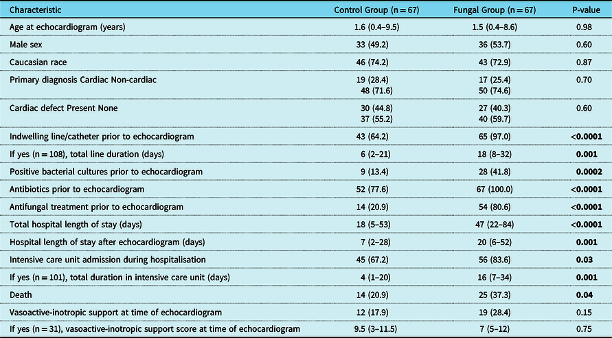
Additional demographic and hospital course information between the two groups is presented in Table 2. Notably, the two groups had similar percent of patients with an underlying cardiac defect and similar number of patients requiring vasoactive support at time of echocardiogram. Not surprisingly, the Fungal Group had significantly higher number of patients with indwelling lines, bacterial infections, treatment with antibacterial and antifungal medications, intensive care unit admission, and had longer duration of intensive care unit and hospital length of stay compared to the Control Group patients. The Fungal Group had more patients with underlying gastrointestinal diagnosis such as short-gut syndrome (20.9 versus 11.9% in the Control Group) and genetic/metabolic diagnoses (7.5 versus 0% in the Control Group), which often require indwelling central lines for nutrition or metabolic supplementation. Antifungal therapy was used prior to echocardiogram in 54/67 (80.6%) of Fungal Group patients compared to 14/67 (20.9%) of Control Group patients (p < 0.0001). There was a higher mortality rate in the Fungal Group (37.3 versus 20.9% in the Control Group, p = 0.04, Table 2). There were 10 deaths among the patients in the Fungal Group who had “positive” echo scores; autopsy was declined in all of these patients.
Demographics and hospital course of control versus fungal group patients*

* Data are presented as N (%) or median (IQR). P-value was from Chi-square test for categorical variables and Wilcoxon rank sum test for continuous variables
Echocardiographic findings
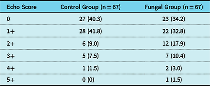
There was 91% agreement (difference in echo score of ≤1) between the two echocardiographers. Fungal Group patients had echocardiograms performed within median 3 days (interquartile range 2–9 days) of positive fungal culture. Control Group patients had an echocardiogram performed at a median of 3 days (interquartile range 1–17 days) from time of admission. Table 3 summarises the echocardiographic scores of both groups. Interestingly, many studies (25.4% of all studies evaluated) received an echo score of 2+ or greater and only one study received an echo score of 5+. In total, 33% of the Fungal Group had an echo score of 2+ or greater, which was a significantly higher proportion than 18% in the Control Group (p = 0.04, Table 4). A subgroup analysis comparing patients with fungal bloodstream infections (excluding patients with urine cultures only) to their matched controls (n = 44 cases and 44 matched controls) showed no significant difference in “positive” echo scores between groups (13/44 “positive” scores in the fungal blood culture group versus 8/44 in matched controls, p = 0.19).

Comparison of “positive” and “negative” echo scores between groups *

* Data are presented as N (%). P-value was from McNemar’s test
Outcomes among fungal group patients
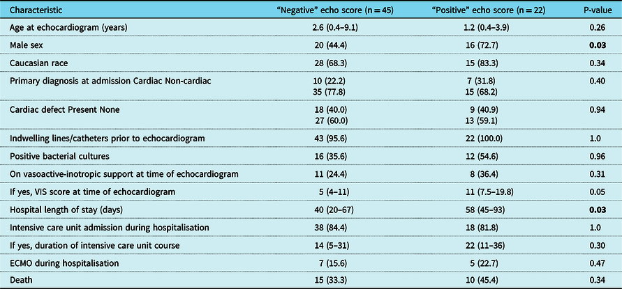
In the Fungal Group, patients with “positive” versus “negative” echo scores were of similar age and both groups had a similar number of patients with an underlying cardiac defect (Table 5). Males were more likely to have “positive” echo scores (p = 0.03). Importantly, there was no significant difference in the number of positive bacterial cultures between patients with “positive” versus “negative” echo scores. There was more Candida parapsilosis in the “positive” echo group (31.8 versus 11.1%, p = 0.049), otherwise the fungal species were similar between groups. In five of the patients in the Fungal Group, apparent echocardiographic abnormalities preceded positive culture results by 1–3 days. Duration of antifungal treatment was similar in “positive” and “negative” echo groups (median 5 versus 6 days, p = 0.045). Clinical outcomes among “positive” and “negative” echo groups are shown in Table 5. Of note, hospital length of stay was significantly longer in patients with “positive” echo scores (p = 0.03).
Univariate comparison of demographics and clinical course for patients with “positive” versus “negative” echo score in the fungal group*

* Data are presented as N (%) or median (IQR). P-value was from Chi-square test or Fisher’s exact test for categorical variables and Wilcoxon rank sum test for continuous variables
ECMO = extracorporeal membranous oxygenation
ICU = intensive care unit
VIS = vasoactive-inotropic support
Discussion
This is the first known study to date investigating whether subtle myocardial changes such as myocardial and papillary muscle brightening with nodular thickening (rather than classic vegetations) are associated with fungal infections in a paediatric population. Prior literature on myocardial changes observed on echocardiogram in association with fungal infections has been limited to case reports and small series of patients with “hyperechoic heterogenous myocardial texture” in addition to classic cardiac vegetations. Reference Harris, Ananth Narayan and Orchard6-Reference Atkinson, Connor, Robinowitz, McAllister and Virmani8 We found that one-third of patients with systemic fungal infection have focal echo brightening and/or papillary muscle nodular thickening, significantly higher than those without fungal infection. Among the Fungal Group patients, males were more likely to have “positive” echo findings. As expected, the patients with fungal infections had a higher proportion of bacterial infections. However, there was no difference in proportion of bacterial infections among the Fungal Group patients with “positive” versus “negative” echo findings, indicating that these myocardial changes are unlikely to be related to presence of bacterial infection alone. Interestingly, there was a higher proportion of Candida parapsilosis infection, typically the second most common fungal cause of endocarditis after Candida albicans, in the group with myocardial changes. Reference Giuliano, Guastalegname and Russo11 This finding may be related to the ability of this species to create a biofilm on medical devices. Reference Silva, Negri and Henriques12
It is notable that the inclusion of patients with fungal urine cultures in addition to those with blood-related infections created heterogeneity within the study population. The study population was somewhat small and inclusion of the urinary tract infection group was necessary to appropriately power the study to detect important differences. While this is an important limitation, the inclusion of the fungal urinary infection group may also provide insight to potential mechanisms for the observed myocardial changes. The presence of myocardial changes in the fungal urinary tract group suggests an etiology related to a systemic response to fungus (e.g., inflammatory or auto-immune processes), rather than due to localised infection. The location of many myocardial brightening and thickening within the “watershed distribution” areas of myocardial blood flow suggests a possible perfusion-related contribution. If these findings are related to an inflammatory process in areas of limited perfusion, the observed presence of “positive” myocardial changes in 12 of the control patients for other perfusion or inflammatory reasons would not be unexpected. Alternatively, patients with fungal urinary tract infections may experience transient fungemia not documented by cultures that leads to haematogenous spread to the myocardium.
Unfortunately, no pathologic data were available in this study as autopsies were declined for the patients in the Fungal Group who had died. However, a pathologic evaluation of 60 adult patients with “fungal infections of the heart” at time of autopsy by Atkinson et al seems to provide clarification. Reference Atkinson, Connor, Robinowitz, McAllister and Virmani8 They identified characteristic fungal morphologies in histologic sections and showed that the myocardium was the most common site of mycotic infections (41/60 patients). Similar to our study findings, 27 of these patients did not have classic vegetations present and the majority of patients identified were males (42/60 patients). The authors describe the myocardial findings as ranging from microscopic abscesses to transmural involvement of the myocardium. Histologic examination of the myocardium was variable, with some showing neutrophilic infiltration, others with neutrophils and lymphocytes, and others without inflammatory cells present. Interestingly, when clinical data of these patients were reviewed, only 43% of the total study population had documented positive fungal cultures pre-mortem, suggesting that the cardiac findings might have been due to unrecognised or transient fungemia with haematogenous seeding from a distant infected site. No echocardiographic data were available, but pathologic findings from this study by Atkinson et al in combination with our study’s echocardiographic findings suggest that fungal cardiac involvement may be more common than previously thought when the diagnosis is based on positive blood cultures or presence of classic vegetations alone.
Patients with both fungal infection and significant myocardial changes had a longer hospital length of stay, suggesting potential prognostic implications of these echocardiographic findings. In addition, some patients were found to have significant echocardiographic abnormalities prior to the positive fungal culture result. Therefore, we suggest that clinicians may consider earlier echocardiogram in patients with concern for fungal infections. Furthermore, if these echocardiographic findings are present in a hospitalised patient with known risk factors for fungal infection, clinicians may consider empiric antifungal therapy while awaiting culture results.
The temporal progression of these subtle echocardiographic findings is not yet known. In a case report of a preterm male neonate with bowel perforation, severe sepsis, and positive blood cultures for Candida parapsilosis, echocardiographic images showed “bright areas within the septal myocardium” that progressed over 2 weeks, then regressed following treatment for Candida infection. Reference Harris, Ananth Narayan and Orchard6 Given the retrospective nature of this study, we were unable to control the timing of echocardiograms or whether follow-up studies were performed. A prospective study in which echocardiograms are performed on patients with fungal infections at specific time intervals may elucidate the clinical significance of these early findings as well as help to gain a better understanding of whether these represent acute and/or chronic myocardial changes during and following fungal infections. Only then can we determine to what extent these myocardial changes should alter therapeutic decisions.
Conclusions
Echocardiographic changes including focal myocardial brightening and papillary muscle thickening appear to be associated with the presence of fungal bloodstream and urinary tract infections, and patients with these myocardial changes have a longer hospital length of stay compared to fungal patients without these echo findings. These data suggest that echocardiographic assessment of patients with fungal infections may help supplement other prognostic indicators and increase clinical suspicion of a fungal infection in patients with known risk factors. The pathophysiology and temporal progression of these myocardial changes are not yet delineated but warrant further investigation.
Supplementary material
To view supplementary material for this article, please visit https://doi.org/10.1017/S1047951120003674
Acknowledgements
None.
Financial support
This research did not receive any specific grant from funding agencies in the public, commercial, or not-for-profit sectors.
Conflicts of interest
None.
Ethical standards
None.