




No CrossRef data available.
Article contents
DNA-binding activity in the excretory–secretory products of Trichinella pseudospiralis (Nematoda: Trichinelloidea)
Published online by Cambridge University Press: 28 November 2001
Abstract
A novel DNA-binding peptide of Mr∼30 kDa was documented for the first time in the excretory–secretory (E–S) products of the infective-stage larvae of Trichinella pseudospiralis. Larvae recovered from muscles of infected mice were maintained for 48 h in DMEM medium. E–S products of worms extracted from the medium were analysed for DNA-binding activity by the electrophoretic mobility shift assay (EMSA). Multiple DNA-protein complexes were detected. A comparison of the Mr of proteins in the complexes indicated that they could bind to the target DNA as a dimer, tetramer or multiples of tetramers. Site selection and competition analysis showed that the binding has a low specificity. A (G/C-rich)-gap-(G/T-rich)-DNA sequence pattern was extracted from a pool of degenerate PCR fragments binding to the E–S products. Results of immunoprecipitation and electrophoretic mobility supershift assay confirmed the authenticity of the DNA-binding protein as an E–S product.
Information
- Type
- Research Article
- Information
- Copyright
- © 2002 Cambridge University Press
INTRODUCTION
Trichinella is a unique pathogenic nematode which can multiply within mammals or birds (Ko & Mak, 1999). The new-born larvae (NBL) (released by the female worms in the intestine) migrate to the striated muscles where they reorganize the tissues into a new type of syncitium known as the ‘nurse cell complex’. Each complex consists of a cytoplasmic region with numerous hypertrophic myonuclei (HN), small nuclei/cells, endoplasmic reticulum, secretory vesicles, mitochondria, and a cavity containing 1–4 worms (Despommier, 1975; Lee & Shivers, 1987; Li, Chung & Ko, 1999). The function of the complex is to sustain the growth and development of the larvae. The expression of structural and regulatory muscle genes are down-regulated and numerous proteins normally not expressed in muscles are detected in the infected cells. In Trichinella spiralis infection, the complex is encapsulated by collagenous fibres but in T. pseudospiralis infection, encapsulation is absent. The 2 trichinellids are morphologically similar but are biologically distinct. The former species mainly occurs in mammals whereas the latter parasitizes both birds and mammals.
The mechanism of host cell reorganization is unknown. In a freeze-fracture study, Lee & Shivers (1987) suggested that the reorganization of myofibres is probably triggered by secretions from T. spiralis. Trichinella E–S products induced extensive lesions in myocytes under both in vivo and in vitro conditions (Leung & Ko, 1997; Lee et al. 1991). E–S products of both T. spiralis and T. pseudospiralis induced type IV collagen synthesis in host fibroblasts (Haehling, Niederkorn & Stewart, 1995). In our earlier study based on immunocytolocalization (using polyclonal/monoclonal antibodies), parasitic epitopes were observed inside the HN in the nurse cell. The antigens in the HN were originated from the stichocytes of worms. The finding indicates that parasitic molecules interact with the host cells at the nuclear level. The parasitic molecule may be involved in controlling the transcription and/or translation of the host nuclei (Lee et al. 1991). Despommier et al. (1990) also observed a positive reaction for parasitic epitopes in the HN.
Ko et al. (1994) and Ko & Mak (1999) proposed a working hypothesis suggesting that the invasion of muscles by Trichinella elicits 2 major independent events i.e. a general degenerative/regenerative response in muscles and specific change(s) in the genomic expression of myonuclei. The 2 events are mediated by a wide spectrum of specific effector molecules, which initiate the infectious process, destroy the existing host environment and construct a new parasitic habitat. The effector molecules occur in the E–S products of the 1st-stage larvae.
Vassilatis et al. (1992) cloned a cDNA encoding the T. spiralis-derived 43 kDa secreted glycoprotein. They suggested that either this protein, or a closely related family member, is secreted into muscles and then translocated into the HN. However, a later study by Jasmer et al. (1994) failed to show the binding of p43 to the nucleoplasm of the HN. Nevertheless, we have good grounds to believe that the E–S products of Trichinella larvae may consist of DNA-binding proteins. In a preliminary study in our laboratory, Leung (1995) observed the binding of E–S products of T. spiralis to both murine and bovine genomic DNA in the filter-binding assay. However, the assay yielded inconsistent results when the experiment was repeated several times. Therefore, the present study was undertaken to provide more concrete evidence of DNA-binding protein in the E–S products of Trichinella by employing more sensitive techniques. An attempt was also made to determine the specificity and molecular mass of the protein.
MATERIALS AND METHODS
Preparation of excretory–secretory products
Infective-stage larvae of T. pseudospiralis were recovered from the skeletal muscles of experimentally infected ICR mice by the standard pepsin digestion method. The larvae were incubated at a density of 10000 larvae/ml DMEM medium for 48 h at 37 °C, supplemented with antibiotics and complete protease inhibitor (Boehringer Mannheim). E–S products were concentrated 100-fold by ultrafiltration using a PM-10 membrane (Amicon). For the control study, freshly isolated larvae were inactivated thrice by alternatively placing in liquid nitrogen and a water bath at 37 °C. The larvae were then maintained in the culture medium as above.
Electrophoretic mobility shift assay (EMSA)
Samples of 0.25–2.5 μg of E–S products were allowed to react for 10 min at room temperature with 10 ng of a 5′-32P-end-labelled double-stranded DNA fragment, CTGCCAGAGATCTGATATCG(N)20 GCAGGCCTACTAGTTCCAGG, in binding buffer (40 mM Tris, pH 8.0, 50 mM NaCl, 10% glycerol, 2.5 mM MgCl2, 1 mM ZnCl2, 100 mM pyrophosphate, 0.1 mM DTT) containing 25 ng poly (dI.dC).
For the competition-binding assay, 10–500 ng poly (dI.dC) or poly (dA.dT) was added to the reaction mixture simultaneously with 10 ng of labelled probe. The mixture was incubated for 10 min with 1 μg E–S products. The reaction products were separated by polyacrylamide gel electrophoresis (PAGE) on 8% native gels in 0.5× Tris-borate buffer (pH 8.0). The gel was then dried and the DNA-protein complexes were detected by autoradiography.
Random binding site selection
The single-stranded oligonucleotide CTGCCAGAGATCTGATATCG(N20) GCAGGCCTACTAGTTCCAGG was converted into a double-stranded form by 4 cycles of PCR amplification (94 °C denaturation, 55 °C annealing, 68 °C extension), using [γ-32P]ATP labelled forward primer (CTGCCAGAGATCTGATATCG) and reverse primer (CCTGGAACTAGTAGGCCTGC). The double-stranded oligonucleotide was purified by native PAGE. Initially, 100 ng DNA was used in the first round of selection in EMSA binding buffer. DNA-protein complexes were eluted in 0.5 M ammonium acetate, 1 mM EDTA, 0.1% SDS, 10% methanol and 50 μg/ml of proteinase K. The eluate was treated with phenol twice, and amplified by 6 cycles of PCR reaction. The amplified products were purified by native PAGE. Then 100 ng of products were labelled with [γ-32P]ATP and T4 polynucleotide kinase. The labelled double-stranded oligonucleotide was used in the subsequent rounds of selection. The resultant fourth round-selected fragments were subcloned into pGEM-T-vector (Promega).
Sequence alignment
In total, 45 independent clones containing the site-selected DNA fragments were sequenced by the ALFExpress automated sequencing system (Amersham). The sequences were first aligned by the sequence logo technique (Schneider, 1996) and then further analysed by manual alignment.
Immunoprecipitation and electrophoretic mobility supershift assay
Polyclonal anti-E–S antibodies were immobilized onto Protein A agarose (Pierce) at room temperature as described (Mak & Ko, 1999). Pre-immune serum was used as control. Antibody–Protein A agarose was washed with 20 mM Tris, pH 7.0 buffer before 20 μg E–S products were added. After 1 h, the Protein A–antibody–antigen complexes were removed from the E–S products by centrifugation at 300 g for 10 min. Interactions between the pre-cleared E–S products and 32P-labelled site-selected oligonucleotide were analysed by EMSA.
In the supershift assay, anti-E–S antibodies were first purified by Protein-A agarose chromatography. The labelled DNA probe from the fourth round of selection was exposed to the E–S products for 10 min. Antibodies were added to the reaction mixture which was kept on ice for 15 min. The resulting DNA–protein complexes were resolved by PAGE in 8% native gels. After drying, the gels were exposed to an X-ray film.
UV photo-crosslinking
A modified nucleotide 5-bromo 2′ deoxyuridine 5′-triphosphate and radioactive [α-32P] dCTP were introduced into the site-selected double-stranded DNA. This was done with 6 cycles of PCR, using the fourth round-selected DNA fragment and the forward and reverse primer set. E–S products (containing 0.5 or 2.5 μg protein) were mixed with 10 ng labelled DNA. EMSA was then performed. The DNA–protein complexes in the native gel were cross-linked by irradiating with 3 J of 260 nm wavelength ultraviolet light (UV cross-linker, Hoefer). The complexes identified by autoradiography were excised from the gel. After mixing with 25 μl of Laemmli buffer containing 5% SDS, the samples were heated for 5 min at 70 °C. After electrophoresis, the gel was dried and exposed to an X-ray film.
RESULTS
DNA-binding affinity
The ability of the E–S products of the infective-stage larvae of T. pseudospiralis to bind to the double-stranded oligonucleotides containing 20 random bases was studied by EMSA (Fig. 1A). A strong retarded band (indicating binding activity) was observed when E–S products from living larvae were used.

Fig. 1. Results of electrophoretic mobility shift assay (EMSA) using excretory–secretory (E–S) products of the infective-stage larvae. (A) Sample from Trichinella pseudospiralis. Lane 1, probe alone. Five μl of concentrated E–S from active larvae (lane 2) and inactivated larvae (lane 3) were incubated with 10 ng radio-isotope labelled oligonucleotide in binding buffer. DNA–protein complexes were resolved on a non-denaturing 8% polyacrylamide gel and detected by autoradiography. (B) Trichinella pseudospiralis E–S DNA-binding protein formed multiple DNA–protein complexes in EMSA. Samples of 0.25–2.5 μg of E–S products were incubated with labelled oligonucleotide. DNA–protein complexes were resolved on non-denaturing 8% polyacrylamide gels and detected by autoradiography. Complexes were labelled as A1, A2 and A3, with decreasing electrophoretic mobility.
The affinity of binding was studied by employing different concentrations of E–S products in EMSA (Fig. 1B). When 0.25 μg of E–S product was used, a DNA–protein complex (A1) was detected. However, when the sample concentration was increased to 1 or 2.5 μg, not only the amount of free probe was decreased, but slower migrating DNA–protein complexes (A2 and A3) appeared.
With increasing concentration of E–S products, the mobility of the predominant migrating species was decreased. This indicates that at high concentrations, the protein may bind to the target sequence as multimers.
The molecular mass(Mr) was calculated by the following equation (Bading, 1988)

where m stands for the migration distance of free probe, m′ is the migration distance of the DNA–protein complex, and K = 100 kDa for 8% polyacrylamide gels.
The calculated Mr values of A1, A2 and A3 were 54.5, 125 and 240 kDa, respectively. Supposing the protein binds to DNA as integral units, the bound protein in A2 and A3 is a multiple of A1. Assuming the fastest migrating species is a complex containing 1 ‘unit’ of DNA-binding protein, then A2 and A3 probably would contain 2 and 4 units, respectively.
Competition EMSA
To determine the DNA-binding specificity of the E–S protein, increasing concentrations of the non-specific competitor poly (dI.dC) or poly (dA.dT) were added to the reaction. When the ratio of protein to the target DNA was decreased, multimers of lower molecular mass bound to the DNA. Figure 2 shows that introducing an equal amount of poly (dI.dC) reduced the intensity and increased the mobility of the retarded band. Poly(dA.dT) competed with the labelled oligonucleotide less efficiently than poly(dI.dC). A 5-fold excess of poly(dI.dC), or a 10-fold excess of poly(dA.dT) could completely eliminate the signals of the retarded bands. This indicates that the E–S protein has a low DNA-binding specificity.

Fig. 2. Results of competition electrophoretic mobility shift assay (EMSA) using 1 μg E–S products of Trichinella pseudospiralis. The assay was carried out with 10 ng radio-isotope labelled oligonucleotide in binding buffer with increasing concentration (10–500 ng) of unlabelled non-specific competitor poly (dI.dC) and poly (dA.dT), DNA–protein complexes were resolved by 8% polyacrylamide gel electrophoresis and autoradiography.
Selection of DNA-binding sites and analysis of site-selected sequences
Samples of 100 ng oligonucleotides (containing all possible combinations of 20 nucleotide randomized regions) were used in the selection experiment. The PCR-amplified DNA after each round of selection was used in EMSA. Figure 3 shows that E–S products have higher affinity for the oligonucleotide pool isolated after 4 rounds of selection than the starting population. This reflects that although the DNA-binding specificity is low, there is some degree of sequence-specific interaction between the protein and its target DNA.

Fig. 3. Results of binding of selected oligonucleotide populations to the DNA-binding protein in the E–S products of Trichinella pseudospiralis. 100 ng of the 60 bp oligonucleotide populations isolated from the indicated rounds of selection were end-labelled with 32P. The selected probes were incubated with 0.5 μg of the E–S products and the electrophoretic mobility shift assay (EMSA) was undertaken. DNA–protein complexes were resolved in 8% polyacrylamide gel electrophoresis and autoradiography.
After 4 rounds of selection, DNA fragments were cloned into a plasmid vector and sequenced. Figure 4A summarizes the sequences of 45 independent clones. Multiple alignment programmes such as Clustal W, Matind and Pretty, which were used to study the consensus sequences, failed to reveal any specific conserved pattern. Using the information analysis and sequence logo technique, the sequence pattern bound by the protein and the frequency of bases at each position were determined. Figure 4B shows a GC-rich pentameric and a GT-rich tetrameric region at the end of the sequences. After splitting the 20 mer into two 10 mer sequences, the sequence logo analysis again indicated a GC and GT rich sequence pattern separated by a gap (Fig. 4C). Since the stack height of the signal was less than 1 bit of information, the protein was probably only in contact with the minor groove of DNA.

Fig. 4. DNA sequencing. (A) DNA sequences of oligonucleotides isolated after 4 rounds of binding-site selection. The 60 bp oligonucleotide population (with the random 20 bp sequences located at the centre) from round 4 of selection with E–S products of Trichinella pseudospiralis was cloned into pGEM-T. Clones were chosen at random for DNA sequence analysis. The 20 bp sequences were numbered 1–45. Selected sequences were analysed by the sequence logo technique. (B) The predicted pattern of selected sequences by aligning the 20 bp sequences. (C). The predicted pattern of selected sequences by aligning the 10 bp sequences obtained by splitting the selected sequences.
Since the protein recognized GC and GT-rich regions, any GC-rich sequences in the selected clones were searched and analysed manually. All 45 oligonucleotides contained 1 or 2 heptameric sequences with a GC content over 70%. The GC-rich sequences can be grouped into 2 main categories: SSSS (70%) or SSNSS (18%) (S = G or C residue; N can be any of the 4 residues). From the 45 sequences, heptamers with the highest GT content were searched and 43 (with a GT content over 70%) were located. Of the GT heptamers, 50% showed 3 or more constitutive T residues. The GC and GT-rich sequences were separated by 0–3 intervening residues. The data suggest that the DNA-binding protein favours the (G/C rich)-gap-(G/T rich) sequence pattern.
Authenticity of DNA-binding activity in E–S products
The main purpose of the freezing/thawing and immunoprecipitation experiments was to determine whether the detected DNA-binding activity was a real E–S component or a leaky by-product of somatic proteins. Examination under the microscope indicated that after freezing and thawing, more than 90% of the larvae were killed. The concentration of the E–S products of the treated larvae was only about 25% of the untreated sample (data not shown). Figure 1A shows that after treatment, DNA-binding activity was abolished. Such activity was only observed in untreated samples. The authenticity of DNA-binding activity was further studied by immunoprecipitation using antiserum raised against Trichinella E–S products. Pre-incubation of the E–S products with homologous antiserum completely eliminated the DNA-binding activity (Fig. 5A). The immunoprecipitation data confirm that the DNA-binding activity is an authentic component of the E–S products and not due to leakage of the somatic tissues.
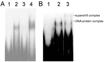
Fig. 5. Results of experiments showing that the DNA-binding protein is an authentic E–S product of Trichinella pseudospiralis (TpES). (A) Activity of DNA-binding protein was neutralized by anti-E–S antibodies in immunoprecipitation. E–S products were pre-incubated with various antibodies before being applied to electrophoretic mobility shift assay (EMSA). Pre-cleared E–S products were incubated with radio-isotope labelled oligonucleotide pool obtained from the 4th round of site selection in binding buffer. DNA–protein complexes were resolved by 8% polyacrylamide gel electrophoresis and autoradiography. Lane 1, probe alone; lane 2, TpES+control antibodies; lane 3, TpES+anti-TpES antibodies; lane 4, TpES without treatment. (B) Treatment with antibody led to supershift effect. A supershifted band was shown in EMSA when the E–S product was incubated with anti-E–S antibodies. Lane 1, probe alone; lane 2, TpES+control IgG; lane 3, TpES+anti-TpES IgG.
A slight reduction in DNA-binding activity was observed when the E–S products were pre-incubated with normal rabbit serum (Fig. 5A). This was likely to be due to the non-specific binding of the E–S products to Protein-A-agarose. However, the decrease was insignificant as compared to those of the experimental samples.
An experiment was undertaken to determine whether attaching purified IgG antibody to the DNA-binding complex would produce a supershift (by retarding mobility in EMSA). Figure 5B shows that adding normal rabbit IgG did not affect the mobility of the DNA-protein complexes. However, when antibodies against E–S products of T. pseudospiralis were used, a slower migrating and supershifted band was observed. The intensity of the band was also lighter. The observations further confirmed that the DNA-binding protein is an authentic E–S product.
Determination of molecular mass of DNA-binding protein by UV-cross-linking
The EMSA data show that the migration of DNA-protein complexes was slower, when the concentration of E–S products was increased. The retarded bands could be regarded as multiples of a DNA-binding ‘unit’. To determine the molecular mass of the DNA-binding ‘unit’, E–S products were first tagged by cross-linking to the 5′-end-32P labelled site-selected target DNA (containing 5-bromo-2′-deoxyuridine-5′-triphosphate). After EMSA and UV irradiation, the DNA–protein complexes were visualized by autoradiography. Cross-linked complexes A1 and A3 were resolved in a 10% SDS-polyacrylamide gel. Autoradiography showed that an E–S product of Mr 30 kDa, corresponding to the E–S DNA-binding monomer, occurred in both A1 and A3 samples (Fig. 6). This indicates that both the fast and slow migrating complexes probably were due to oligomerization of a 30 kDa DNA-binding protein. The calculated molecular mass showed that the DNA-protein complexes A1, A2 and A3 were a dimer, tetramer and octamer, respectively. Radioactive signal with the same mobility was not detected in the sample containing free DNA.

Fig. 6. Determination of molecular mass of DNA-binding peptide of Trichinella pseudospiralis by UV cross-linking. A 30 kDa band (arrow) was identified. E–S product was incubated with radio-isotope labelled 4th round-selected oligonucleotide (containing 5-bromo-2-deoxyuridine-5′-triphosphate). DNA–protein complexes were resolved by 8% polyacrylamide gel electrophoresis and then UV irradiated. The products were detected by autoradiography. DNA–protein complexes A1 and A3 were isolated and the products were resolved by 10% SDS–PAGE and detected by autoradiography. Lane 1, probe alone; lane 2, A3 complexes; lane 3, A1 complexes.
DISCUSSION
The results of the present study clearly demonstrate the presence of DNA-binding protein activity in the E–S products of the infective-stage larvae of T. pseudospiralis. The novel 30 kDa protein is the first reported secretory multimeric complex dsDNA-binding protein. The protein does not appear to target a very specific DNA sequence for binding. Similar activities were also observed in the E–S products of T. spiralis (data not shown) but the results were not as consistent as those of T. pseudospiralis. This is probably due to the presence of a strong activity of double-stranded endonuclease in the E–S product of the former species (Mak & Ko, 1999). Such activity invariably interfered with the DNA-binding experiments. In the present experiments, steps were taken to suppress endonuclease activities by adding an inhibitor, Zn2+, to the samples. A more detailed discussion is given below.
Recently, we reported that the E–S products of T. pseudospiralis and T. spiralis contain single-stranded endonuclease with Mr 55–60 (Mak, Chung & Ko, 2000). In addition, a double-stranded endonuclease of 30 kDa was also detected in the E–S products of the latter species (Mak & Ko, 1999). Although some DNA-binding proteins e.g. the 30 kDa protein of larval Anopheles stephensi and the EcoRV endonucleases are known to bind and cleave DNA (Gakhar, Singh & Shandilya, 2000; Martin et al. 1999), the DNA-binding peptide identified in the present study is likely not to be an endonuclease. Firstly, its molecular mass is distinctly smaller than that of the single-stranded endonuclease. Secondly, the double-stranded endonuclease activity usually occurs at a very low level in the E–S products of T. pseudospiralis. In the present study, a strong DNA-binding activity was detected in the E–S products. However, it is possible that the requirements for the two activities are different and that the same peptide encodes both the DNA-binding and cleavage activity on different domains. Nevertheless, further work, especially on the structure of the protein, is required to confirm this interpretation.
The presence of a DNA-binding protein in the E–S products of Trichinella will add another dimension to the understanding of host–parasite interactions at the nuclear level and the pathogenesis of intracellular parasitism. The finding suggests that during development, the larva of Trichinella can actively secrete molecules which target the host cell DNA. Therefore, the host cell colonization strategy of the nematode appears to be similar to that of viruses and other micro-organisms. However, since DNA-binding protein has not been reported previously in parasitic organisms, one can only speculate on its possible functions.
Our finding shows that the DNA-binding protein (p30) has a relatively small molecular mass and it is not one of the well-known immunodominant E–S antigens specific to Trichinella. DNA-binding activity of the E–S products has not been studied previously although attempts have been made by some authors to elucidate the parasitic epitopes in the HN. Vassilatis et al. (1992) cloned a specific 43 kDa antigen which is a glycoprotein belonging to the basic helix-loop-helix (bHLH) DNA-binding protein family. Members of the same family include myogenic transcription factors. However, a subsequent study using antibodies failed to observe the binding of p43 to the HN (Jasmer et al. 1994). We have not yet undertaken an in vivo study on p30 because, at present, it is difficult to obtain sufficient working material for cloning or purification.
Since p30 occurs in both T. pseudospiralis and T. spiralis, it may play a crucial role in the reorganization of host cells by trichinellids. It is probably the key molecule which targets the DNA of HN. The lack of specificity of the protein may facilitate the binding to the DNA of myonuclei of all mammalian or avian species. The action of p30 may be a precursor of a chain of cascade reprogramming reactions e.g. the cleavage of host DNA, shutting-off host genes, altering genomic expression, arresting host cell cycles etc (see Ko & Mak, 1999; Mak & Ko, 1999; Mak, Chung & Ko, 2000). The end result is the construction of a new habitat which can sustain the parasite in a hostile environment.
Similar to some DNA-binding proteins of other micro-organisms, p30 of Trichinella may also have multiple functions. Multimeric complex dsDNA-binding proteins, which interact with the target sequences in the DNA minor groove with a low degree of sequence specificity, are known to play important roles in the fundamental process of DNA functions, including replication and transcription (Bewley, Gronenborn & Clore, 1998) and the protection of genomic DNA (Serrano, Salas & Hermoso, 1993). For example, histone H1 is considered as a general repressor of transcription (Buttinelli et al. 1999). The non-specific DNA-binding protein SIN1 of Saccharomyces cerevisiae is part of a multiprotein complex that regulates the transcription of HO gene (Kruger & Herskowitz, 1991). In Mycobacterium bovis, a histone-like protein, MDP1, which binds to GC-rich sequence as multimers has been identified (Matsumoto et al. 1999). An inducible non-specific DNA-binding protein Dps from Escherichia coli protects DNA during oxidative stress (Martinez & Kolter, 1997).
Only few DNA-binding proteins are known to be secreted from cells e.g. MYT2 protein of eukaryotic oligodendrocyte progenitor cells and nucleobindin of KML1-7 cells (Kim et al. 1998; Miura et al. 1992). Prochiantz & Theodore (1995) suggested that transcription factors can act in a paracrine manner. Transcellular transactivation of target genes by the HIV-1 Tat protein indicates that the virus uses this mechanism to regulate gene expression (Helland et al. 1991). The secretory virulent factor AvrXa7 of Xanthomonas oryzae pv. oryzae, induces disease resistance when expressed in a susceptible plant host (Jackson et al. 1999). It encodes a transcriptional activation domain and is bound to the double-stranded DNA with a preference for dA/dT rich sequence (Yang et al. 2000). This indicates that the protein can interact with the host transcriptional machinery, thus modulating the profile of host genomic expression.
The intracellular protozoan parasite, Theileria annulata, induces a neoplastic-like state in the infected bovine leukocyte. The gene expression profile of the infected cell is different. Recently, a putative parasite-encoded factor, TashAT2, which modulates the host cell gene expression has been identified. The protein encodes 3 AT hook DNA-binding domains and is located in the host cell nucleus (Swan et al. 1999). This provides the important evidence that factors can be transported from the parasite to the host nucleus and bound to the host DNA. A similar event can also occur in trichinellosis.
This work was supported by a grant (HKU 426/96M) from the Hong Kong Research Grants Council to R.C.K. and a partial Post-doctorate Fellowship and Assistant Professorship from the University of Hong Kong to C.H.M.
References
BADING, H. (1988). Determination of the molecular weight of DNA-bound protein(s) responsible for gel electrophoretic mobility shift of linear DNA fragments exemplified with purified viral myb protein. Nucleic Acids Research 16, 5241–5248.CrossRefGoogle Scholar
BEWLEY, C. A., GRONENBORN, A. M. & CLORE, G. M. (1998). Minor groove-binding architectural proteins: structure, function, and DNA recognition. Annual Review of Biophysics and Biomolecular Structure 27, 105–131.CrossRefGoogle Scholar
BUTTINELLI, M., PANETTA, G., RHODES, D. & TRAVERS, A. (1999). The role of histone H1 in chromatin condensation and transcriptional repression. Genetica 106, 117–124.CrossRefGoogle Scholar
DESPOMMIER, D. (1975). Adaptive changes in muscle fibers infected with Trichinella spiralis. American Journal of Pathology 78, 477–496.Google Scholar
DESPOMMIER, D., GOLD, A. M., BUCK, S. W., CAPO, V. & SILBERSTEIN, D. (1990). Trichinella spiralis: secreted antigen of the infective L1 larva localizes to the cytoplasm and nucleoplasm of infected host cells. Experimental Parasitology 71, 27–38.CrossRefGoogle Scholar
GAKHAR, S. K., SINGH, S. & SHANDILYA, H. (2000). DNA-binding proteins of the malaria vector Anopheles stephensi: purification and characterization of an endonuclease. Archives of Insect Biochemistry and Physiology 44, 40–46.3.0.CO;2-G>CrossRefGoogle Scholar
HAEHLING, E., NIEDERKORN, J. Y. & STEWART, G. L. (1995). Trichinella spiralis and Trichinella pseudospiralis induce collagen synthesis by host fibroblasts in vitro and in vivo. International Journal for Parasitology 25, 1393–1400.CrossRefGoogle Scholar
HELLAND, D. E., WELLES, J. L., CAPUTO, A. & HASELTINE, W. A. (1991). Transcellular transactivation by the human immunodeficiency virus type I tat protein. Journal of Virology 65, 4547–4549.Google Scholar
JACKSON, R. W., ATHANASSOPOULOS, E., TSIAMIS, G., MANSFIELD, J. W., SESMA, A., ARNOLD, D. L., GIBBON, M. J., MURILLO, J., TAYLOR, J. D. & VIVIAN, A. (1999). Identification of a pathogenicity island, which contains genes for virulence and avirulence, on a large native plasmid in the bean pathogen Pseudomonas syringae pathovar phaseolicola. Proceedings of the National Academy of Sciences, USA 96, 10875–10880.CrossRefGoogle Scholar
JASMER, D. P., YAO, S., VASSILATIS, D., DESPOMMIER, D. & NEARY, S. M. (1994). Failure to detect Trichinella spiralis p43 in isolated host nuclei and in irradiated larvae of infected muscle cells which express the infected cell phenotype. Molecular and Biochemical Parasitology 67, 225–234.CrossRefGoogle Scholar
KIM, J. G., ARMSTRONG, R. C., BERNDT, J. A., KIM, N. W. & HUDSON, L. D. (1998). A secreted DNA-binding protein that is translated through an internal ribosome entry site (IRES) and distributed in a discrete pattern in the central nervous system. Molecular and Cellular Neurosciences 12, 119–140.CrossRefGoogle Scholar
KO, R. C., FAN, L., LEE, D. L. & COMPTON, H. (1994). Changes in host muscles induced by excretory/secretory products of larval Trichinella spiralis and Trichinella pseudospiralis. Parasitology 108, 195–205.CrossRefGoogle Scholar
KO, R. C. & MAK, C. H. (1999). Trichinellosis as a model of new frontier research on parasitic diseases. International Medical Research Journal 3, 21–31.Google Scholar
KRUGER, W. & HERSKOWITZ, I. (1991). A negative regulator of HO transcription, SIN1 (SPT2), is a nonspecific DNA-binding protein related to HMG1. Molecular and Cellular Biology 11, 4135–4146.CrossRefGoogle Scholar
LEE, D. L., KO, R. C., YI, X. Y. & YEUNG, M. H. F. (1991). Trichinella spiralis: antigenic epitopes from the stichocytes detected in the hypertrophic nuclei and cytoplasm of the parasitized muscle fibre (nurse cell) of the host. Parasitology 102, 117–123.CrossRefGoogle Scholar
LEE, D. L. & SHIVERS, R. R. (1987). A freeze-fracture study of muscle fibres infected with Trichinella spiralis. Tissue and Cell 19, 665–671.CrossRefGoogle Scholar
LEUNG, R. K. M. (1995). Purification and biological properties of excretory/secretory antigens from Trichinella spiralis. M. Phil. thesis, Department of Zoology, The University of Hong Kong, Hong Kong.Google Scholar
LEUNG, R. K. M. & KO, R. C. (1997). In vitro effects of Trichinella spiralis on muscle cells. Journal of Helminthology 71, 113–118.CrossRefGoogle Scholar
LI, C. K. F., CHUNG, Y. Y. Y. & KO, R. C. (1999). The distribution of excretory/secretory antigens during the muscle phase of Trichinella spiralis and T. pseudospiralis infections. Parasitology Research 85, 993–998.CrossRefGoogle Scholar
MAK, C. H., CHUNG, Y. Y. Y. & KO, R. C. (2000). Single-stranded endonuclease activity in the excretory–secretory products of Trichinella spiralis and Trichinella pseudospiralis. Parasitology 120, 527–533.CrossRefGoogle Scholar
MAK, C. H. & KO, R. C. (1999). Characterization of endonuclease activity from excretory/secretory products of a parasitic nematode, Trichinella spiralis. European Journal of Biochemistry 260, 477–481.CrossRefGoogle Scholar
MARTIN, A. M., HORTON, N. C., LUSETTI, S., REICH, N. O. & PERONA, J. J. (1999). Divalent metal dependence of site-specific DNA binding by EcoRV endonuclease. Biochemistry 38, 8430–8439.CrossRefGoogle Scholar
MARTINEZ, A. & KOLTER, R. (1997). Protection of DNA during oxidative stress by the nonspecific DNA-binding protein Dps. Journal of Bacteriology 179, 5188–5194.CrossRefGoogle Scholar
MATSUMOTO, S., YUKITAKE, H., FURUGEN, M., MATSUO, T., MINETA, T. & YAMADA, T. (1999). Identification of a novel DNA-binding protein from Mycobacterium bovis Bacillus Calmette-Guerin. Microbiology and Immunology 43, 1027–1036.CrossRefGoogle Scholar
MIURA, K., TITANI, K., KUROSAWA, Y. & KANAI, Y. (1992). Molecular cloning of nucleobindin, a novel DNA-binding protein that contains both a signal peptide and a leucine zipper structure. Biochemical and Biophysical Research Communications 187, 375–380.CrossRefGoogle Scholar
PROCHIANTZ, A. & THEODORE, L. (1995). Nuclear/growth factors. Bioessays 17, 39–44.CrossRefGoogle Scholar
SCHNEIDER, T. D. (1996). Reading of DNA sequence logos: prediction of major groove binding by information theory. Methods in Enzymology 274, 445–455.CrossRefGoogle Scholar
SERRANO, M., SALAS, M. & HERMOSO, J. M. (1993). Multimeric complexes formed by DNA-binding proteins of low sequence specificity. Trends in Biochemical Sciences 18, 202–206.CrossRefGoogle Scholar
SWAN, D. G., PHILLIPS, K., TAIT, A. & SHIELS, B. R. (1999). Evidence for localisation of a Theileria parasite AT hook DNA-binding protein to the nucleus of immortalised bovine host cells. Molecular and Biochemical Parasitology 101, 117–129.CrossRefGoogle Scholar
VASSILATIS, D. K., DESPOMMIER, D., MISEK, D. E., POLVERE, R. I., GOLD, A. M. & VAN DER PLOEG, L. H. T. (1992). Analysis of a 43-kDa glycoprotein from the intracellular parasitic nematode Trichinella spiralis. Journal of Biological Chemistry 267, 18459–18465.Google Scholar
YANG, B., ZHU, W., JOHNSON, L. B. & WHITE, F. F. (2000). The virulence factor AvrXa7 of Xanthomonas oryzae pv. oryzae is a type III secretion pathway-dependent nuclear-localized double-stranded DNA-binding protein. Proceedings of the National Academy of Sciences, USA 97, 9807–9812.Google Scholar

Fig. 1. Results of electrophoretic mobility shift assay (EMSA) using excretory–secretory (E–S) products of the infective-stage larvae. (A) Sample from Trichinella pseudospiralis. Lane 1, probe alone. Five μl of concentrated E–S from active larvae (lane 2) and inactivated larvae (lane 3) were incubated with 10 ng radio-isotope labelled oligonucleotide in binding buffer. DNA–protein complexes were resolved on a non-denaturing 8% polyacrylamide gel and detected by autoradiography. (B) Trichinella pseudospiralis E–S DNA-binding protein formed multiple DNA–protein complexes in EMSA. Samples of 0.25–2.5 μg of E–S products were incubated with labelled oligonucleotide. DNA–protein complexes were resolved on non-denaturing 8% polyacrylamide gels and detected by autoradiography. Complexes were labelled as A1, A2 and A3, with decreasing electrophoretic mobility.

Fig. 2. Results of competition electrophoretic mobility shift assay (EMSA) using 1 μg E–S products of Trichinella pseudospiralis. The assay was carried out with 10 ng radio-isotope labelled oligonucleotide in binding buffer with increasing concentration (10–500 ng) of unlabelled non-specific competitor poly (dI.dC) and poly (dA.dT), DNA–protein complexes were resolved by 8% polyacrylamide gel electrophoresis and autoradiography.

Fig. 3. Results of binding of selected oligonucleotide populations to the DNA-binding protein in the E–S products of Trichinella pseudospiralis. 100 ng of the 60 bp oligonucleotide populations isolated from the indicated rounds of selection were end-labelled with 32P. The selected probes were incubated with 0.5 μg of the E–S products and the electrophoretic mobility shift assay (EMSA) was undertaken. DNA–protein complexes were resolved in 8% polyacrylamide gel electrophoresis and autoradiography.
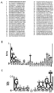
Fig. 4. DNA sequencing. (A) DNA sequences of oligonucleotides isolated after 4 rounds of binding-site selection. The 60 bp oligonucleotide population (with the random 20 bp sequences located at the centre) from round 4 of selection with E–S products of Trichinella pseudospiralis was cloned into pGEM-T. Clones were chosen at random for DNA sequence analysis. The 20 bp sequences were numbered 1–45. Selected sequences were analysed by the sequence logo technique. (B) The predicted pattern of selected sequences by aligning the 20 bp sequences. (C). The predicted pattern of selected sequences by aligning the 10 bp sequences obtained by splitting the selected sequences.

Fig. 5. Results of experiments showing that the DNA-binding protein is an authentic E–S product of Trichinella pseudospiralis (TpES). (A) Activity of DNA-binding protein was neutralized by anti-E–S antibodies in immunoprecipitation. E–S products were pre-incubated with various antibodies before being applied to electrophoretic mobility shift assay (EMSA). Pre-cleared E–S products were incubated with radio-isotope labelled oligonucleotide pool obtained from the 4th round of site selection in binding buffer. DNA–protein complexes were resolved by 8% polyacrylamide gel electrophoresis and autoradiography. Lane 1, probe alone; lane 2, TpES+control antibodies; lane 3, TpES+anti-TpES antibodies; lane 4, TpES without treatment. (B) Treatment with antibody led to supershift effect. A supershifted band was shown in EMSA when the E–S product was incubated with anti-E–S antibodies. Lane 1, probe alone; lane 2, TpES+control IgG; lane 3, TpES+anti-TpES IgG.

Fig. 6. Determination of molecular mass of DNA-binding peptide of Trichinella pseudospiralis by UV cross-linking. A 30 kDa band (arrow) was identified. E–S product was incubated with radio-isotope labelled 4th round-selected oligonucleotide (containing 5-bromo-2-deoxyuridine-5′-triphosphate). DNA–protein complexes were resolved by 8% polyacrylamide gel electrophoresis and then UV irradiated. The products were detected by autoradiography. DNA–protein complexes A1 and A3 were isolated and the products were resolved by 10% SDS–PAGE and detected by autoradiography. Lane 1, probe alone; lane 2, A3 complexes; lane 3, A1 complexes.
 You have
Access
- 11
- Cited by