Introduction
Cognitive dysfunction is increasingly recognized as a common side effect of breast cancer (BC) chemotherapy (Ahles, Reference Ahles2012; Janelsins et al., Reference Janelsins, Kohli, Mohile, Usuki, Ahles and Morrow2011; Rodin & Ahles, Reference Rodin and Ahles2012; Wefel, Saleeba, Buzdar, & Meyers, Reference Wefel, Saleeba, Buzdar and Meyers2010; Wefel & Schagen, Reference Wefel and Schagen2012). Neuroimaging studies of BC chemotherapy have significantly increased our understanding regarding these cognitive difficulties by indicating alterations of prefrontal cortex and hippocampus as well as distributed functional and structural networks (de Ruiter et al., Reference de Ruiter, Reneman, Boogerd, Veltman, Caan, Douaud and Schagen2012; Deprez, QJ;Billiet, Sunaert, & Leemans, Reference Deprez, Billiet, Sunaert and Leemans2013; Ferguson, McDonald, Saykin, & Ahles, Reference Ferguson, McDonald, Saykin and Ahles2007; Hosseini, Koovakkattu, & Kesler, Reference Hosseini, Koovakkattu and Kesler2012; Kesler, Janelsins, et al., Reference Kesler, Janelsins, Koovakkattu, Palesh, Mustian, Morrow and Dhabhar2013; Kesler, Bennett, Mahaffey, & Spiegel, Reference Kesler, Bennett, Mahaffey and Spiegel2009; Kesler, Kent, & O'Hara, Reference Kesler, Kent and O'Hara2011; McDonald, Conroy, Ahles, West, & Saykin, Reference McDonald, Conroy, Ahles, West and Saykin2010; McDonald, Conroy, Ahles, West, & Saykin, Reference McDonald, Conroy, Ahles, West and Saykin2012; Silverman et al., Reference Silverman, Dy, Castellon, Lai, Pio, Abraham and Ganz2007) [see (Reuter-Lorenz & Cimprich, Reference Reuter-Lorenz and Cimprich2013) for a review of functional studies].
A majority of previous neuroimaging studies used mass univariate analyses. We aimed to extend and advance this literature by using multivariate pattern analysis (MVPA). MVPA more closely matches the covarying nature of neural systems providing greater power for separating groups than univariate or behavioral methods (Hoeft et al., Reference Hoeft, McCandliss, Black, Gantman, Zakerani, Hulme and Gabrieli2011). This increased power stems from MVPA's ability to use subtle signals across voxels that tend to be undetectable by univariate analyses (Kamitani & Tong, Reference Kamitani and Tong2005). This increased sensitivity is especially important for the study of chemotherapy-related cognitive dysfunction, which tends to be subtle or difficult to detect with objective measures (Vardy, Reference Vardy2009).
MVPA applies machine learning algorithms to identify patterns in data that accurately differentiate between groups or conditions (Haynes & Rees, Reference Haynes and Rees2006; O'Toole et al., Reference O'Toole, Jiang, Abdi, Penard, Dunlop and Parent2007; Orru, Pettersson-Yeo, Marquand, Sartori, & Mechelli, Reference Orru, Pettersson-Yeo, Marquand, Sartori and Mechelli2012; Pereira, Mitchell, & Botvinick, Reference Pereira, Mitchell and Botvinick2009). MVPA is increasingly used to classify individuals to develop diagnostic and prognostic models based on neuroimaging biomarkers. For example, neuroimaging MVPA has been shown to accurately distinguish mild cognitive impairment from healthy aging and dementia (Aksu, Miller, Kesidis, Bigler, & Yang, Reference Aksu, Miller, Kesidis, Bigler and Yang2011; O'Dwyer, Lamberton, Bokde, et al., Reference O'Dwyer, Lamberton, Bokde, Ewers, Faluyi, Tanner and Hampel2012; Zhang & Shen, Reference Zhang and Shen2012; Zhang, Wang, Zhou, Yuan, & Shen, Reference Zhang, Wang, Zhou, Yuan and Shen2011), young APOE4 carriers from non-carriers (O'Dwyer, Lamberton, Matura, et al., Reference O'Dwyer, Lamberton, Matura, Scheibe, Miller, Rujescu and Hampel2012), and depressed from non-depressed subjects (Craddock, Holtzheimer, Hu, & Mayberg, Reference Craddock, Holtzheimer, Hu and Mayberg2009; Zeng et al., Reference Zeng, Shen, Liu, Wang, Li, Fang and Hu2012), among others [see (Orru et al., Reference Orru, Pettersson-Yeo, Marquand, Sartori and Mechelli2012) for a review].
In our recent study, we showed that MVPA of default mode network connectivity accurately distinguishes chemotherapy-treated (C+) from non-chemotherapy treated (C−) BC survivors and healthy controls with 90–91% accuracy (Kesler, Wefel, et al., Reference Kesler, Wefel, Hosseini, Cheung, Watson and Hoeft2013). Given that this previous study was the first to apply MVPA to chemotherapy-related cognitive dysfunction, it is useful to evaluate alternate potential classifiers. Therefore, in the present study, we applied MVPA to functional magnetic resonance imaging (fMRI) of prefrontal-executive connectivity. We used a common fMRI paradigm, the Go/Nogo task to measure prefrontal-executive neurocircuit function (Menon, Adleman, White, Glover, & Reiss, Reference Menon, Adleman, White, Glover and Reiss2001; Simmonds, Pekar, & Mostofsky, Reference Simmonds, Pekar and Mostofsky2008).
We focused on evaluation of prefrontal-executive neurocircuitry for several reasons. First, prefrontal dysfunction is thus far the most consistent finding among previous neuroimaging studies irrespective of imaging modality and even when whole-brain methods are used (Bruno, Hosseini, & Kesler, Reference Bruno, Hosseini and Kesler2012; de Ruiter et al., Reference de Ruiter, Reneman, Boogerd, Veltman, van Dam, Nederveen and Schagen2011, Reference de Ruiter, Reneman, Boogerd, Veltman, Caan, Douaud and Schagen2012; Ferguson et al., Reference Ferguson, McDonald, Saykin and Ahles2007; Kesler et al., Reference Kesler, Bennett, Mahaffey and Spiegel2009, Reference Kesler, Kent and O'Hara2011; McDonald et al., Reference McDonald, Conroy, Ahles, West and Saykin2010, Reference McDonald, Conroy, Ahles, West and Saykin2012; Silverman et al., Reference Silverman, Dy, Castellon, Lai, Pio, Abraham and Ganz2007). Second, executive function and memory deficits are the most common cognitive impairments in BC chemotherapy (Vardy, Reference Vardy2009; Wefel & Schagen, Reference Wefel and Schagen2012) and these skills rely on distributed prefrontal cortex networks (Eichenbaum, Reference Eichenbaum2000; Leh, Petrides, & Strafella, Reference Leh, Petrides and Strafella2010).
Third, the Go/Nogo task has been previously used in BC patients to evaluate pre-treatment neurobiologic status (Scherling, Collins, Mackenzie, Bielajew, & Smith, Reference Scherling, Collins, Mackenzie, Bielajew and Smith2012). Specifically, Scherling and colleagues demonstrated reduced activation in several prefrontal-executive network regions before adjuvant chemotherapy in patients compared to controls (Scherling et al., Reference Scherling, Collins, Mackenzie, Bielajew and Smith2012). Our study expands upon that of Scherling et al. by distinguishing between BC women with and without history of chemotherapy treatment and examining long-term survivors, thus emphasizing potential persistent cognitive dysfunction. We also focused on connectivity in prefrontal circuits rather than activation.
Fourth, prefrontal cortex abnormalities have been associated with self-report of executive function impairments (Kesler et al., Reference Kesler, Kent and O'Hara2011; McDonald et al., Reference McDonald, Conroy, Ahles, West and Saykin2012). These findings suggest that prefrontal status may be a sensitive biomarker for patients’ cognitive complaints. Finally, executive-prefrontal deficit is the best predictor of medication adherence in BC patients (Stilley, Bender, Dunbar-Jacob, Sereika, & Ryan, Reference Stilley, Bender, Dunbar-Jacob, Sereika and Ryan2010) and thus may be indirectly associated with health status. Therefore, we expected that a MVPA algorithm could be reliably trained to distinguish C+ from C− and healthy female controls (HC) based on connectivity within prefrontal-executive neurocircuitry. We hypothesized that the algorithm would have significant accuracy as defined by the proportion of correct classifications (positive and negative) as well as significant specificity (true negatives) and sensitivity (true positives).
Materials and Methods
Participants
We enrolled 56 primary (stages I–IIIA) female BC survivors in this study, 27 C+ and 29 C−, as well as 30 HC (see Table 1). This sample was included in previous studies by our group (Bruno et al., Reference Bruno, Hosseini and Kesler2012; Hosseini et al., Reference Hosseini, Koovakkattu and Kesler2012; Kesler, Janelsins, et al., Reference Kesler, Janelsins, Koovakkattu, Palesh, Mustian, Morrow and Dhabhar2013; Kesler et al., Reference Kesler, Kent and O'Hara2011; Kesler, Wefel, et al., Reference Kesler, Wefel, Hosseini, Cheung, Watson and Hoeft2013). BC survivors were recruited via the Army of Women (http://www.armyofwomen.org/), community-based BC support groups and local media advertisements. Healthy controls were recruited via the Army of Women and local media advertisements.
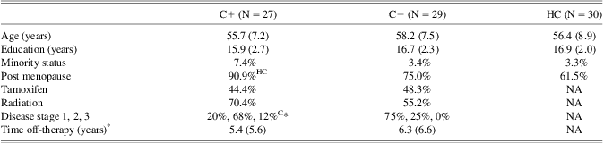
Demographic data for the C+, C−, and HC groups

Note. Data are shown as mean (standard deviation) except where noted.
C+ = chemotherapy-treated; C− = non-chemotherapy treated; HC = healthy control.
HC Significantly different from HC group (p < 0.05).
C− Significantly different from C− group (p < 0.01).
*Time off-therapy refers to chemotherapy and/or radiation.
There were no significant differences between the groups in age (F = 0.74; p = .48), education (F = 1.43; p = .24) or minority status (χ2 p > .23). The C− and C+ groups did not differ in postmenopausal status (p = .27), time off-therapy (p = .6), radiation (p = .40), or tamoxifen (p = .79). There were significantly more women in the C+ group who were postmenopausal compared to the HC group, which was expected given that chemotherapy can induce early menopause (Mar Fan et al., Reference Mar Fan, Houédé-Tchen, Chemerynsky, Yi, Xu and Harvey2010). The number of postmenopausal women in the C− compared with HC group was non-significantly larger (p = .22). As expected, the disease stage at the time of diagnosis was significantly higher in the C+ group compared with C−.
BC survivors were excluded for history of relapse or prior chemotherapy treatment. Individual chemotherapy regimens included adriamycin/cytoxan/taxol or taxotere = 21, cytoxan/methotrexate/5-fluorouracil = 4 and adriamycin/cytoxan+cytoxan/methotrexate/5-fluorouracil = 2. Additionally, 26 women were treated with tamoxifen and 35 received radiation therapy. Four participants in the C+ group and six in the C− group were still taking tamoxifen at the time of assessment. No patients had received other anti-estrogen treatments. All participants were excluded for diagnosed psychiatric, neurologic or comorbid medical conditions that are known to affect cognitive function as well as MRI contraindications or major sensory deficits (e.g., blindness). This research was completed in accordance with the Helsinki Declaration. The study was approved by the Stanford University Institutional Review Board and all participants provided informed consent.
Go/Nogo Paradigm
The details of the Go/Nogo experiment are described in a previous study (Menon et al., Reference Menon, Adleman, White, Glover and Reiss2001). Briefly, the task consisted of a 30-s rest block, alternating 26-s blocks of Go and Nogo conditions, followed by another 30-s rest block. During the Go/Nogo blocks, participants viewed a series of letters once every 2 s (stimulus presentation: 500 ms, inter-stimulus interval: 1500 ms). In the Go blocks, participants were asked to respond to every stimulus by pressing a button. The Go condition measures sustained attention (Menon et al., Reference Menon, Adleman, White, Glover and Reiss2001). In the Nogo blocks, participants were asked to press the button for all the letters except for the letter X. The letter X was not part of the presented stimuli during the Go blocks. The Nogo condition measures both sustained attention and response inhibition (Menon et al., Reference Menon, Adleman, White, Glover and Reiss2001).
fMRI Data Acquisition
MRI scanning was performed on a GE Discovery MR750 3.0 Tesla whole body scanner (GE Medical Systems, Milwaukee, WI). fMRI scans were acquired while participants were lying supine in the scanner. Stimuli were presented using E-Prime software (Psychology Software Tools). Behavioral responses (accuracy, reaction time) were recorded using a four-button fiber optic finger switch system.
fMRI data were acquired using a T2* weighted gradient echo spiral pulse sequence (repetition time = 2000 ms; echo time = 30 ms, flip angle = 80° and 1 interleave; field of view = 200 × 200 mm2, matrix size = 64 × 64, in-plane spatial resolution = 3.125). Thirty-two axial slices (3-mm thick, 1 mm skip) covering the whole brain were acquired. Number of data frames collected was 160, thus the total scan time was 5:20. An automated high-order shimming method based on spiral acquisitions was used to reduce field heterogeneity (Glover & Lai, Reference Glover and Lai1998). To coregister and normalize functional images with a standardized template, a high-resolution T1-weighted fast spoiled gradient echo anatomical scan was acquired for each individual (relaxation time: minimum, echo time: minimum, flip: 11 degrees, inversion time: 300 ms, bandwidth: ±31.25 kHz, field of view: 24 cm, phase field of view: 0.75, slice thickness: 1.5 mm, 125 slices, 256 × 256 at 1 excitation, scan time: 4:26 min).
Two other task-based fMRI scans, a resting state fMRI scan and a diffusion-weighted scan were also acquired for some participants during the MRI session (total duration = 1 h). Participants were also administered a battery of neuropsychological measures (approximately 1.5 h) on the day of the MRI as described in our previous studies (Bruno et al., Reference Bruno, Hosseini and Kesler2012; Hosseini et al., Reference Hosseini, Koovakkattu and Kesler2012; Kesler, Janelsins, et al., Reference Kesler, Janelsins, Koovakkattu, Palesh, Mustian, Morrow and Dhabhar2013; Kesler et al., Reference Kesler, Kent and O'Hara2011). These data are not reported here.
fMRI Data Analyses
Image preprocessing was performed using Statistical Parametric Mapping 8 (SPM8; Wellcome Department of Cognitive Neurology, London, UK; http://www.fil.ion.ucl.ac.uk/spm/), as described in our previous publications (Kesler et al., Reference Kesler, Bennett, Mahaffey and Spiegel2009, Reference Kesler, Kent and O'Hara2011). First, effects of small head movements were corrected by realigning functional volumes using least square minimization. Realigned functional volumes were then coregistered to individuals’ anatomical images. Anatomical volumes were then segmented and normalized to a standard MNI template and the transformation parameters were applied to the functional volumes. To reduce the effect of noise, the normalized functional volumes were spatially smoothed with an 8-mm full-width at half-maximum (FWHM) Gaussian filter. Images were visually assessed for correct spatial normalization.
Accuracy (percent correct) and response time in milliseconds were collected during the Go/Nogo task. To determine the effects of group and task condition on performance, we conducted a two-way analysis of variance (Nogo vs. Go × C+ vs. C− vs. HC) in SPSS 19.0 (www.spss.com). The threshold for statistical significance was set at p < .05.
MVPA Feature Selection and Extraction
We used 12 cortical regions of interest (ROIs) from the Automated Anatomical Labeling (AAL) atlas using the WFU PickAtlas Toolbox (Tzourio-Mazoyer et al., Reference Tzourio-Mazoyer, Landeau, Papathanassiou, Crivello, Etard, Delcroix and Joliot2002). ROIs included the bilateral inferior frontal (triangular part) (IFTr), middle frontal (MFG), superior frontal (SFG), medial frontal (MedSF), inferior parietal (IPL), and supplementary motor area (SMA). These regions were consistently reported in previous neuroimaging studies of the Go/Nogo task (Mostofsky et al., Reference Mostofsky, Schafer, Abrams, Goldberg, Flower, Boyce and Pekar2003; Simmonds et al., Reference Simmonds, Pekar and Mostofsky2008), including one study of women with BC (Scherling et al., Reference Scherling, Collins, Mackenzie, Bielajew and Smith2012). These regions also are known to subserve executive function (Leh et al., Reference Leh, Petrides and Strafella2010). The ROIs were resliced to the same dimension as that of the functional images.
Functional connectivity data were extracted using the Functional Connectivity Toolbox (http://www.nitrc.org/projects/conn) (Whitfield-Gabrieli & Nieto-Castanon, Reference Whitfield-Gabrieli and Nieto-Castanon2012) with CompCor noise correction (Behzadi, Restom, Liau, & Liu, Reference Behzadi, Restom, Liau and Liu2007). The individual motion parameters were also used as covariates. Finally, ROI to ROI temporal correlations based on corrected BOLD (blood oxygen level dependent) signal were computed for the Nogo blocks resulting in a 12×12 correlation matrix, containing normalized Z-scores, for each individual. We analyzed only the Nogo blocks to include all cognitive aspects of this task (response inhibition, sustained attention). We did not compare the Nogo blocks with rest since this was a condition-dependent functional connectivity analysis (Whitfield-Gabrieli & Nieto-Castanon, Reference Whitfield-Gabrieli and Nieto-Castanon2012). Subtracting the Go images from Nogo images would eliminate group differences in sustained attention, which is an important component of executive functions.
MVPA Training and Testing
A linear support vector machine (SVM), as implemented by our in-house MVPA Toolbox (Hoeft et al., Reference Hoeft, McCandliss, Black, Gantman, Zakerani, Hulme and Gabrieli2011; Kesler, Wefel, et al., Reference Kesler, Wefel, Hosseini, Cheung, Watson and Hoeft2013; Marzelli, Hoeft, Hong, & Reiss, Reference Marzelli, Hoeft, Hong and Reiss2011) (http://ncnl.stanford.edu/tools), was used for pattern classification. The unique characteristic of linear SVM, compared with other linear classifiers, is that it attempts to maximize the distance between the data points that are closest to the boundary (support vectors) between classes (groups). This characteristic makes the classifier work quite efficiently when the number of samples (and, therefore, the training set) is small (Noble, Reference Noble2006; Orru et al., Reference Orru, Pettersson-Yeo, Marquand, Sartori and Mechelli2012).
Separate binary linear SVM classifiers were built for discriminating C+ and HC, C+ and C−, and C− and HC individuals using the pattern of connectivity in the Nogo blocks. For C+ versus C− or HC, a class vector was constructed comprising either “+1”s (C+) or “−1”s (C− or HC) depending on which group the participant belonged to. For HC versus C− classification, a class vector was constructed comprising “+1”s for HC and “−1”s for C−. Next, the connectivity matrices for each individual were converted to a feature vector containing 66 unique ROI to ROI connection strengths (12 × 11/2). The SVM uses the training data that have been previously categorized into groups (i.e., C+, C−, and HC) to build a model (classifier) that optimally separates the groups. In this way, the SVM learns by example how to classify the individual subjects. This process involves finding a weight vector (assigning a weight to each connection) that maximizes the margin of separation between groups (Orru et al., Reference Orru, Pettersson-Yeo, Marquand, Sartori and Mechelli2012). Connections with high weights are considered important contributors to classification. The farther the participant's data are from the separation margin (hyperplane), the more different the participant's functional connectivity pattern is from that of the comparison group.
The validity of the model (classifier) was tested using leave-one out cross-validation to avoid overfitting and allow generalization of the model (Hoeft et al., Reference Hoeft, McCandliss, Black, Gantman, Zakerani, Hulme and Gabrieli2011; Pereira et al., Reference Pereira, Mitchell and Botvinick2009). In each repetition, one subject's data were left out as a test case, and the remaining subjects’ data were used to train the classifier. This procedure was repeated such that each subject was left out once and the accuracy of the model was then estimated as the proportion of correct predictions. This process allows the training and test cases to remain independent (Orru et al., Reference Orru, Pettersson-Yeo, Marquand, Sartori and Mechelli2012). Finally, permutation analysis (5000 times) was performed to empirically determine whether the obtained classification accuracy was significantly greater than chance (Marzelli et al., Reference Marzelli, Hoeft, Hong and Reiss2011). We adjusted the alpha level for the permutation analysis to p < .017 (Bonferroni corrected) to account for multiple comparisons.
Connectivity maps were constructed using the mean absolute weights of each feature in the classifier. The connections were thresholded for visualization purposes to indicate the connections that carried the greatest weight (>1 SD) in the classifier (Hoeft et al., Reference Hoeft, McCandliss, Black, Gantman, Zakerani, Hulme and Gabrieli2011). These connections are referred to as “connections with high classification weights”, hereafter. As noted above, the SVM weights indicate the contribution of each connection in discriminating between groups. However, they do not indicate whether the corresponding functional connectivity is decreased or increased. Thus, for connections with high discriminative weights, we also quantified their difference in functional connectivity strength between groups using the ROI-to-ROI normalized Z scores (Greicius et al., Reference Greicius, Flores, Menon, Glover, Solvason, Reiss and Schatzberg2007).
Effect size of the classifier was quantified using area under the receiver operating characteristic (ROC) curve, which examines the relationship between true positives and false positives (Rice & Harris, Reference Rice and Harris2005). Brain maps were created using BrainNet Viewer software (http://www.nitrc.org/projects/bnv).
Finally, for connections with high classification weight, we performed an exploratory correlation analysis to identify the association between their connectivity strengths and behavioral performance in the Nogo task within each group, separately. In addition, we explored the relationship between clinical and treatment factors in the C+ group with task performance and distance from the hyperplane.
Results
Behavioral Results
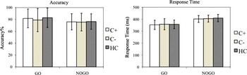
Mean Go/Nogo task accuracy and response time for each group are shown in Figure 1. There was a significant effect of condition (F 1,166 = 5.4; p < .05). However, the main effect of group (F 1,166 = 0.19; p = .83) and group by condition interaction (F 1,166 = 0.24; p = .79) were not significant. The response time data also showed a significant effect of condition (F 1,166 = 82.9; p < .001) but no significant effect of group (F 1,166 = 0.06; p = .97) or group by condition interaction (F 1,166 = 0.11; p = .89).
Behavioral results. Mean accuracy (percent correct) and response time in Go and Nogo blocks for C+, C−, and HC groups. For accuracy data, there was a significant main effect of condition (p < .05) but no significant main effect of group (p = .83) and group by condition interaction (p = .79). For response time data, there was a significant main effect of condition (p < .001) but no significant main effect of group (p = .97) and group by condition interaction (p = .89). The error bars indicate the standard deviation from the mean.

MVPA Results
For C+ versus HC classification, the SVM classifier achieved significant classification accuracy (72.0%; p = .006), sensitivity (70.0%; p = .007), and specificity (73.0%; p = .007). The area under the ROC curve was 0.72. Similarly, for C+ versus C− classification, the SVM classifier achieved significant classification accuracy (71.4%; p = .012), sensitivity (70.4%; p = .013), and specificity (72.4%; p = .017). The area under the ROC curve was 0.71. The classification accuracy for C− versus HC did not reach significance (50.9%; p = .46). The area under the ROC curve was 0.51.
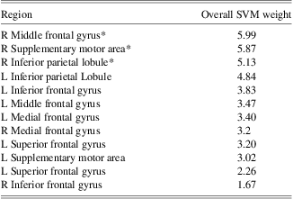
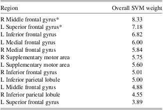
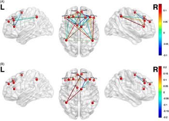
The contribution of each functional connection (absolute SVM weights) to the classifier is shown in Figure 2, Table 2, and Table 3. Connections involving right middle frontal gyrus, right supplementary motor area, and right inferior parietal lobule had the greatest weights (>1 SD) for discriminating C+ and HC, and those involving right middle frontal gyrus and left superior frontal gyrus had the greatest weight for discriminating C+ and C− groups.
Multivariate pattern classification of Nogo functional connectivity in (A) C+ compared to HC, and (B) C+ versus C−. For visualization purposes, only the connections with high classification weights (>1 SD) are shown. Connectivity pattern in the right middle frontal gyrus, right supplementary motor area and right inferior parietal lobule showed the highest overall contribution to C+ versus HC classification, and connectivity pattern in the right middle frontal and left superior frontal gyri showed the highest overall contribution to C+ versus C− classification. The colorbar represents the absolute weight of each connection in classification.

Overall contribution of each ROI in discrimination of C+ and HC, aggregating the weights of connections involving each ROI

* Indicates regions with overall contribution of 1 SD greater than mean.
ROI = region of interest; C+ = chemotherapy-treated; treated; HC = healthy control SVM, support vector machine.
Overall contribution of each ROI in discrimination of C+ and C−, aggregating the weights of connections involving each ROI

* Indicates regions with overall contribution of 1 SD greater than mean.
ROI = region of interest; C+ = chemotherapy-treated; C− = non-chemotherapy treated; SVM = support vector machine.
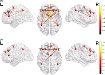
The between group differences (C+ vs. HC and C+ vs. C−) in functional connectivity strengths for regions with high SVM weights are shown in Figure 3. Of 45 connections with high classification weights, 23 connections showed decreased (cool colors) and 22 connections showed increased connectivity (warm colors) in C+ versus HC comparison. For C+ versus C− classification, six of nine connections showed increased connectivity.
Difference in functional connectivity between (A) C+ and HC and (B) C+ and C− for connections with high discriminant weights. Positive (negative) values indicate connections that are higher (lower) in C+ than in HC (or C−).

Exploratory Correlation Results
Within each group, an exploratory correlation analysis was performed to identify the relationship between participant's behavioral performance in the Nogo task and the strength of connections with high classification weights.
C+ versus HC Classification
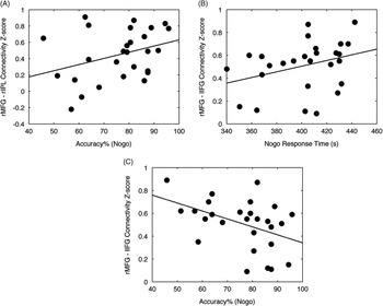
In the C+ group, Nogo accuracy was significantly correlated with the connectivity strength between right middle frontal gyrus and right inferior parietal lobule (r = 0.43; p < .05) (Figure 4A). The corresponding connectivity strength for these regions was higher in the C+ than in HC group as noted in Figure 3A. No significant correlations were observed in the HC group. Specifically, the correlation between connectivity strength of right middle frontal gyrus and right inferior parietal lobule and accuracy in the Nogo task was not significant within the HC group (r = 0.27; p = .14). The difference in correlations was also not significant between groups (z = 0.65; p = .26).
Correlation between performance and functional connectivity. (A) C+ participants with higher Nogo accuracy showed higher connectivity between right middle frontal gyrus and right inferior parietal lobule (r = 0.43; p < .05). (B) C+ participants with higher response time in Nogo task showed higher connectivity between right middle frontal gyrus and left inferior frontal gyrus (r = 0.39; p < .05). (C) C+ participants with lower Nogo accuracy showed higher connectivity between right middle frontal gyrus and left inferior frontal gyrus (r = −0.42; p < .05).

C+ versus C− Classification
Within the C+ group, the strength of connectivity between right middle frontal gyrus and left inferior frontal gyrus was significantly correlated with Nogo response time (r = 0.39; p < .05) and significantly negatively correlated with Nogo accuracy (r = −0.42; p < .05) (Figures 4B and 4C). The corresponding connectivity strength for these regions was higher in the C+ than in C− group as noted in Figure 3B. The correlation between connectivity strength of right middle frontal gyrus and left inferior frontal gyrus and Nogo response time and accuracy was not significant within the C− group (r = −0.04; p = .84 for response time; r = 0.15; p = .44 for accuracy). The between-group differences in correlations were significant for accuracy (z = 1.99; p < .05).
We did not find any significant correlation between C+ clinical/treatment status and their performance in the task. However, a significant correlation was found between C+'s distance from the hyperplane and their cancer stage (Spearman's r = 0.4; p < .05).
Discussion
We investigated whether the multivariate pattern of neural connectivity during an executive function task (Go/Nogo) could distinguish chemotherapy-treated from non-chemotherapy treated BC survivors and healthy women. While task performance of the C+ group was comparable to that of C− and HC, the SVM classifier achieved significant accuracy, sensitivity, and specificity in discriminating C+ from C− and HC groups. Classification performance resulted in ROCs of 0.71–0.72, which are considered large effects (Rice & Harris, Reference Rice and Harris2005). These findings suggest that altered functional connectivity during an executive function task may represent a sensitive indicator of cognitive dysfunction in C+. In prospective studies, MVPA classifiers could be used to predict individuals who will have persistent cognitive impairment using baseline neuroimaging data. This method has been used successfully in other conditions, such as the prediction of conversion from mild cognitive impairment to dementia (Aksu et al., Reference Aksu, Miller, Kesidis, Bigler and Yang2011).
The Nogo condition was more difficult than the Go condition for both groups, as expected. However, no significant between-group differences were observed in accuracy or response time. However, the absence of group difference in task performance or neuropsychological tests does not necessarily suggest that the underlying neurocircuitry is normal (Reuter-Lorenz & Cimprich, Reference Reuter-Lorenz and Cimprich2013). Neuroimaging evidence suggests that performance may be preserved in some women with BC through compensatory neural processes (Cimprich et al., Reference Cimprich, Reuter-Lorenz, Nelson, Clark, Therrien, Normolle and Welsh2010; Ferguson et al., Reference Ferguson, McDonald, Saykin and Ahles2007; Kesler et al., Reference Kesler, Bennett, Mahaffey and Spiegel2009) that cannot be detected by behavioral measures (McDonald et al., Reference McDonald, Conroy, Ahles, West and Saykin2012; Reuter-Lorenz & Cimprich, Reference Reuter-Lorenz and Cimprich2013).
Despite the absence of group difference in task performance, the MVPA classifier could accurately differentiate between C+ and C− or HC groups based on the pattern of prefrontal-executive network connectivity. The regions that showed the highest contribution to the C+ versus HC classification were the right middle frontal gyrus, right supplementary motor area and right inferior parietal lobule, and those that highly contributed to distinguishing C+ and C− groups were the right middle frontal gyrus and the left superior frontal gyrus. Multimodal neuroimaging evidence suggests that the supplementary motor area plays a role in conflict detection/resolution within the response generation network (Aron, Behrens, Smith, Frank, & Poldrack, Reference Aron, Behrens, Smith, Frank and Poldrack2007). Patients with lesions to the superior frontal regions showed increased incorrect responses to the Nogo stimulus (Picton et al., Reference Picton, Stuss, Alexander, Shallice, Binns and Gillingham2007). The frontoparietal network is critical for executive function and attention orientation as well as manipulating and monitoring information in working memory (Leh et al., Reference Leh, Petrides and Strafella2010). Alterations in frontal and parietal structural connectivity have been reported for C+ in previous studies (Deprez et al., Reference Deprez, Amant, Yigit, Porke, Verhoeven, Van den Stock and Sunaert2011, Reference Deprez, Amant, Smeets, Peeters, Leemans, Van Hecke and Sunaert2012). Additionally, our previous data showed changes in topological properties of prefrontal regions in both resting-state functional and structural association networks in C+ (Bruno et al., Reference Bruno, Hosseini and Kesler2012; Hosseini et al., Reference Hosseini, Koovakkattu and Kesler2012).
Examination of functional connectivity strengths revealed a profile of both hypo-and hyper-connectivity in the C+ group compared to HC and C−. Increased connectivity between the middle frontal gyrus and inferior parietal lobule was significantly associated with increased Nogo performance in the C+ group. This observation suggests a potential compensatory mechanism that helps C+ survivors preserve their performance during certain tasks. Consistent with these findings, previous fMRI studies reported hyper-activation during executive task performance in C+ survivors (Ferguson et al., Reference Ferguson, McDonald, Saykin and Ahles2007; McDonald et al., Reference McDonald, Conroy, Ahles, West and Saykin2012). For example, Ferguson and colleagues (2007) compared twins with and without BC during an n-back working memory performance. Compared with the healthy twin, the C+ twin recruited much broader brain regions during task performance, even at very low load conditions (Ferguson et al., Reference Ferguson, McDonald, Saykin and Ahles2007).
However, studies have also shown hypo-activation during other executive tasks (de Ruiter et al., Reference de Ruiter, Reneman, Boogerd, Veltman, van Dam, Nederveen and Schagen2011; Kesler et al., Reference Kesler, Kent and O'Hara2011). These differences in findings could reflect several factors including the nature and difficulty level of the task and/or specific sample characteristics. A recent review of fMRI studies in BC demonstrated a pattern of hyper-activation, or recruitment of additional neural resources, at low task difficulty (Reuter-Lorenz & Cimprich, Reference Reuter-Lorenz and Cimprich2013). As task difficulty increases, women with BC may be unable to maintain this compensatory response (Reuter-Lorenz & Cimprich, Reference Reuter-Lorenz and Cimprich2013), resulting in decreased activation or connectivity. The authors also suggest that neural compensation may mask behavioral performance effects (Reuter-Lorenz & Cimprich, Reference Reuter-Lorenz and Cimprich2013), which is consistent with our findings.
It should also be noted that studies showing hypo-activation tend to involve very long-term survivors (5 or more years off-therapy) (de Ruiter et al., Reference de Ruiter, Reneman, Boogerd, Veltman, van Dam, Nederveen and Schagen2011; Kesler et al., Reference Kesler, Kent and O'Hara2011). However, progressive decline is supported by neuropsychological data (Wefel et al., Reference Wefel, Saleeba, Buzdar and Meyers2010) and is observed in other conditions that affect cognitive function. For example, increased functional connectivity is demonstrated during early phases of amnestic mild cognitive impairment followed by decreased connectivity later on (Bai et al., Reference Bai, Watson, Shi, Wang, Yue, Yuhuan and Zhang2011; Jones et al., Reference Jones, Machulda, Vemuri, McDade, Zeng, Senjem and Jack2011). Our previous studies of network topology in long-term C+ survivors demonstrate decreased brain network efficiency, which may reduce the ability to coordinate neural responses to cognitive demands and compensate for neurologic dysfunction (Bruno et al., Reference Bruno, Hosseini and Kesler2012; Hosseini et al., Reference Hosseini, Koovakkattu and Kesler2012). Longitudinal studies of BC that include long-term follow up assessments are necessary to examine changes in neurocircuitry response over time and how these changes may impact cognitive status.
Among features that distinguished between C+ and C−, the connectivity strength between right middle frontal gyrus and left inferior frontal gyrus showed a positive correlation with Nogo response time and a negative correlation with accuracy in the C+ group. Multiple studies have shown the involvement of inferior frontal cortex in response inhibition (Aron et al., Reference Aron, Behrens, Smith, Frank and Poldrack2007; Picton et al., Reference Picton, Stuss, Alexander, Shallice, Binns and Gillingham2007; Swick, Ashley, & Turken, Reference Swick, Ashley and Turken2008). This observation may reflect over-inhibition of responding in chemotherapy-treated BC survivors during tasks with low difficulty that results in higher response time and lower accuracy in Nogo blocks.
Within the C+ group only, we found a positive correlation between distance from the hyperplane and cancer stage. Data that are more distant from the hyperplane are those that are more distinguishable from the comparison group. Therefore, the results suggest an association between disease severity and alterations in connectivity pattern in C+. Consistently, our previous data showed a significant correlation between disease severity and hypo-activation in the left prefrontal regions in C+ but not C− (Kesler et al., Reference Kesler, Kent and O'Hara2011). Disease severity tends to be highly interrelated with treatment intensity; patients with high stage at diagnosis are more likely to receive chemotherapy (Du & Goodwin, Reference Du and Goodwin2001).
We did not find a pattern of prefrontal connectivity that could distinguish between C− and HC despite previous research suggesting that cancer pathology as well as other adjuvant treatments (radiation, tamoxifen) may contribute to cognitive changes in BC independently from chemotherapy (Eberling, Wu, Tong-Turnbeaugh, & Jagust, Reference Eberling, Wu, Tong-Turnbeaugh and Jagust2004; Kesler et al., Reference Kesler, Kent and O'Hara2011; McDonald et al., Reference McDonald, Conroy, Ahles, West and Saykin2012; Phillips et al., Reference Phillips, Jim, Small, Laronga, Andrykowski and Jacobsen2012; Scherling, Collins, Mackenzie, Bielajew, & Smith, Reference Scherling, Collins, Mackenzie, Bielajew and Smith2011; Scherling et al., Reference Scherling, Collins, Mackenzie, Bielajew and Smith2012). Compared to previous studies, the present findings were based on examination of functional connectivity rather than functional activation. Our results could suggest that cancer pathology, radiation, and/or hormonal blockade treatments may affect regional activation but are not sufficient to impact the connectivity of larger-scale brain networks. Additionally, MVPA involves a different inferential question compared to univariate methods. MVPA is concerned with the reliability of the difference between two groups whereas univariate analysis is concerned with whether or not there is a between-group difference (O'Toole et al., Reference O'Toole, Jiang, Abdi, Penard, Dunlop and Parent2007). The Go/Nogo task may not be the best for distinguishing C− from HC. Studies demonstrating differences between C− and HC with respect to prefrontal function used tasks measuring other executive functions (working memory, cognitive flexibility) (Kesler et al., Reference Kesler, Kent and O'Hara2011; McDonald et al., Reference McDonald, Conroy, Ahles, West and Saykin2012). Further research is required to identify the different profiles of brain changes that may be associated with individual and combined candidate mechanisms of cognitive difficulties in BC.
While our current results were associated with strong effect sizes, our previous MVPA results suggest that default mode network (DMN) connectivity may provide a better classifier of chemotherapy-related cognitive difficulty. DMN connectivity distinguished C+ from both C− survivors and HC with 90–91% accuracy with ROCs of 0.97–0.98 (Kesler, Wefel, et al., Reference Kesler, Wefel, Hosseini, Cheung, Watson and Hoeft2013). This is consistent with previous research suggesting that resting state DMN connectivity is highly sensitive to disease states (Sheline et al., Reference Sheline, Raichle, Snyder, Morris, Head, Wang and Mintun2010). In future studies, evaluation of various neuroimaging classifiers against other models (e.g., neuropsychological or treatment variables) and combined models (neuroimaging + neuropsychological variables) could reveal the most precise classifiers.
Our study is limited by its cross-sectional design. Prospective, longitudinal designs are required to determine individual differences in neural connectivity that may contribute to individual cognitive outcome. Our subject recruitment materials specifically mentioned cognitive dysfunction and therefore the sample may have been biased toward survivors who were concerned about their cognitive function. The Go/Nogo task is a well-established, commonly used fMRI paradigm that is brief and easy to implement. However, other fMRI paradigms may yield alternate results. Our MVPA feature selection was based on previous studies of the Go/Nogo task as well as literature regarding prefrontal-executive neurocircuitry but alternate features might provide alternate results.
In summary, our findings demonstrate that MVPA is a very promising tool for examining BC chemotherapy-related cognitive dysfunction. Methods like MVPA with increased sensitivity are especially important for this field given that cognitive deficits may be difficult to detect and/or masked by compensatory responses. If used in prospective studies, MVPA could potentially identify individuals at the highest risk for persistent cognitive dysfunction. These patients could then be prioritized for regular neuropsychological surveillance and possible early or preventative intervention. Our results also increase our understanding of the neurobiologic mechanisms underlying cognitive impairment in C+. Specifically, we demonstrated an altered pattern of functional connectivity during an executive function task. Consistent with previous studies, our findings suggest that performance during certain tasks may be preserved in some C+ survivors via recruitment of additional frontoparietal neurocircuitry. The identification of demographic, neurobiologic, medical, and/or other factors that enhance or maintain this compensation will be essential for the development of interventions.
Acknowledgments
This work was supported by the National Institutes of Health Director's New Innovator Award (DP2 OD004445-01 to SK). The authors report no conflict of interest.