Chest pain frequently affects adolescents, but is rarely cardiac in origin. Although uncommon, cardiac aetiologies of chest pain include myocarditis, pericarditis, arrhythmia, cardiomyopathy, or even myocardial infarction.Reference Kane, Fulton, Saleeb, Zhou, Lock and Geggel 1 In the adult, an acute episode of chest pain directs physicians to check cardiac biomarkers, particularly troponin I and T, because of the likelihood that the chest pain is due to ischaemic coronary artery disease.
Troponin levels are sometimes sent in the evaluation of adolescents with chest pain, partly because of the widespread use of these assays in adults. Rather than myocardial infarction, myocarditis or pericarditis are more likely to lead to chest pain with an elevated troponin in an adolescent who has no history of cardiac disease. In some patients, differentiating between myocardial infarction and myocarditis or pericarditis can be challenging.Reference Desai, Patel and Book 2
Despite the increasing use of the troponin assay in adolescents with chest pain, no guidelines and few reports exist to assist physicians in the management of these patients.Reference Desai, Patel and Book 2 – Reference Brown, Hirsh and Mahle 4 We aim to describe the evaluation, treatment, and follow-up of a group of adolescents without history of significant cardiac abnormality who presented to our institution with chest pain and an elevated troponin level. Patients with a typical presentation of myocarditis or pericarditis were not included in the analysis, as significant literature exists describing the management of such patients.Reference Cooper 5 , Reference Lange and Hillis 6
Materials and methods
After obtaining approval from the Institutional Review Board at the Children's Hospital of Philadelphia, a retrospective chart review was performed. Patients aged 10–18 years who were admitted to the Children's Hospital of Philadelphia between 2000 and 2010 with chest pain and an abnormal troponin I assay were reviewed. At our institution, troponin I values greater than 0.4 nanograms per millilitre are considered elevated. Patients with a known history of coronary artery anomaly or haemodynamically significant cardiac disease were excluded. Patients with a typical presentation of myocarditis or pericarditis were also excluded. A diagnosis of typical myocarditis was made in those patients who presented with a viral prodrome and globally decreased left ventricular shortening on echocardigram.Reference Cooper 5 Pericarditis was considered typical in patients who had pleuritic chest pain that was worse when supine, a history of viral prodrome, an electrocardiogram showing diffuse ST-segment elevation, and pericardial effusion on echocardiogram.Reference Lange and Hillis 6
The hospital admission for each patient was reviewed and relevant information extracted, including patient demographic and historical characteristics, laboratory values, patient treatment, and clinical outcome. The presenting electrocardiogram of each patient and any diagnostic imaging including echocardiogram, catheterisation, cardiac computed tomography angiogram, or cardiac magnetic resonance imaging were also reviewed. The discharge diagnosis for the patient's chest pain as determined by the treating physicians was recorded. Patient medical records were further reviewed to record any recurrent presentations due to chest pain and most recent clinical follow-up.
Results
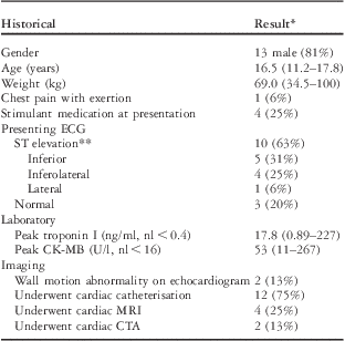
A total of 16 patients were included and their baseline characteristics are described in Table 1. Most patients (81%) were male with a median age of 16.5 years (range 11.2–17.8 years). Only one patient presented with chest pain on exertion. There were 10 patients (63%) who showed ST elevation greater than 1 millimetre in two consecutive leads in a coronary distribution on their presenting electrocardiogram. There were three (20%) patients who had a normal electrocardiogram at presentation. All patients had normal electrolytes and lipid panel. All patients were screened for drug use; one patient had a positive screen for marijuana and was later found to have myopericarditis. All patients underwent echocardiography and only two (13%) showed a left ventricular focal wall motion abnormality: one patient showed posterior wall hypokinesis after presenting with inferolateral ST elevation on electrocardiogram and another showed inferior wall hypokinesis after presenting with inferior ST elevation. In all, 15 (94%) patients underwent catheterisation, cardiac magnetic resonance imaging, and/or cardiac computed tomography angiography to definitively identify the coronary artery origins; one patient thought to have a myocardial infarction from prolonged (greater than 48 hours) supraventricular tachycardia did not undergo further imaging after echocardiogram suggested normal coronary origins.
Baseline characteristics in 16 adolescents with chest pain and elevated troponin I level.

ECG = electrocardiogram; nl = normal; MRI = magnetic resonance imaging; CTA = computed tomography angiography
*Median (range) or number (%)
**ST elevation >1 mm in at least two consecutive leads in a coronary distribution
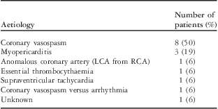
Table 2 shows the discharge diagnosis as determined by treating physicians. There were eight (50%) patients who suffered myocardial infarction from suspected coronary vasospasm and three (20%) were thought to have atypical myopericarditis. The one patient presenting with chest pain on exertion had an infarction from an anomalous left coronary artery from the right sinus of Valsalva. Other suspected aetiologies included essential thrombocythaemia and prolonged supraventricular tachycardia.
Discharge diagnoses in 16 patients presenting with chest pain and elevated troponin I level.

LCA = left coronary artery; RCA = right coronary artery
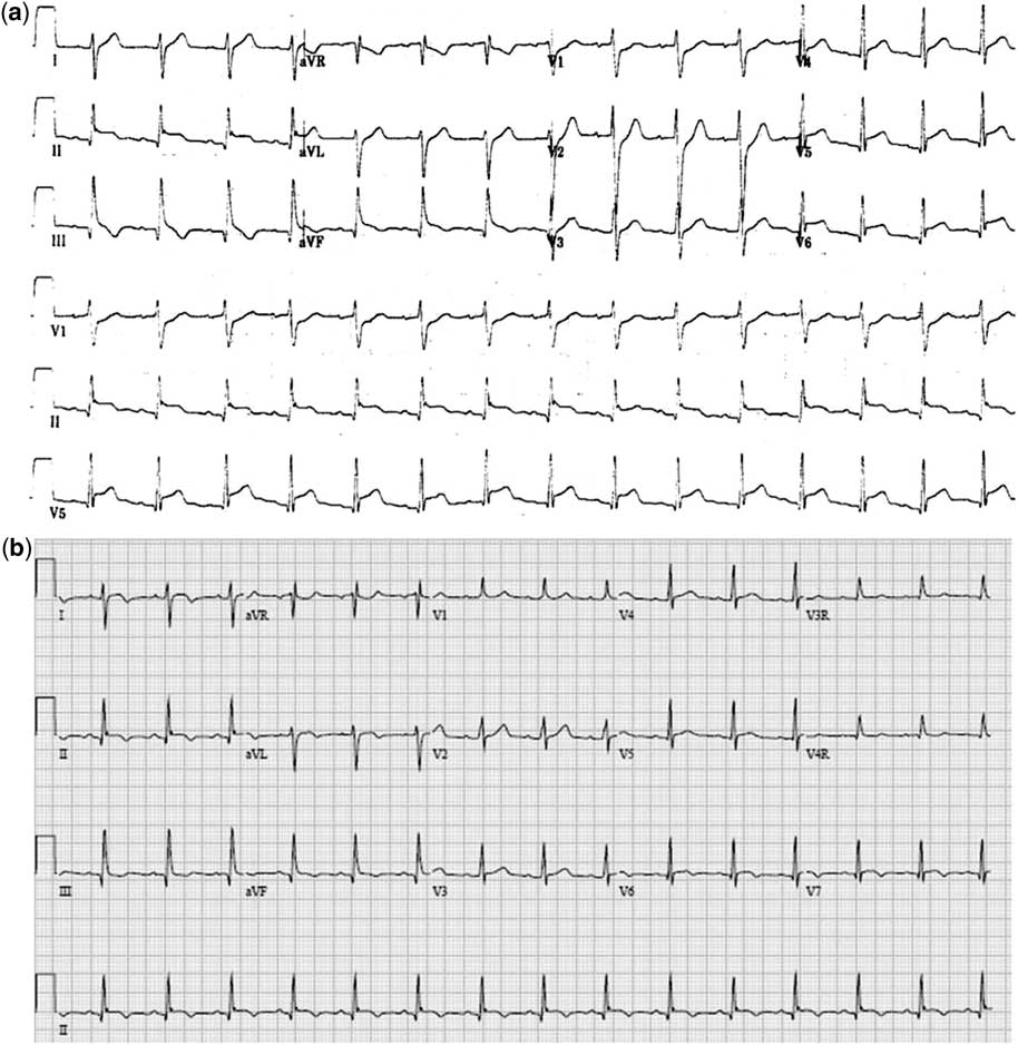
Chest pain with elevated troponin was attributed to coronary vasospasm in eight patients, all males. On review, each patient suspected to have vasospasm by treating physicians met commonly used criteria for the diagnosis of coronary vasospasm including (1) a presenting electrocardiogram showing dynamic ST elevation greater than 1 millimetre in at least two consecutive leads in a coronary distribution and (2) no evidence of coronary anomaly or obstruction on diagnostic imaging. 7 The median peak troponin I in this subgroup was 17.6 nanograms per millilitre (range 12.9–66). In all, four patients presented with inferolateral ST elevation, three with inferior ST elevation, and one with lateral ST elevation. The presenting electrocardiogram in a 15-year-old boy showing inferolateral ST elevation is shown in Figure 1. The four patients taking stimulant medication to treat attention deficit disorder at the time of presentation were all diagnosed with coronary vasospasm. Of the eight patients diagnosed with coronary vasospasm, two were awoken by pain early in the morning. No patient had a history of migraine headaches and one patient was being treated for Crohn's disease with mesalamine and azathioprine. Only one patient had focal left ventricular wall motion abnormality (posterior wall) on echocardiogram. There were five patients who were treated with intravenous nitrates at the time of presentation. All eight patients underwent coronary artery evaluation, five with cardiac catheterisation, two with cardiac computed tomography angiography, and one patient with cardiac magnetic resonance imaging. There were no coronary anomalies or stenoses detected. Provocative testing for vasospasm was not performed in any patient. All eight patients were discharged on calcium channel blocker therapy – three nifedipine, two diltiazem, two verapamil, and one amlodipine. In the four patients taking stimulant therapy at presentation, three were permitted to continue therapy at the time of discharge. At a median follow-up of 9.3 months (0–29.5 months), no patient had experienced a recurrent event.
( a ) Presenting electrocardiogram in a 15.5-year-old boy with chest pain and elevated troponin I level due to coronary vasospasm showing inferolateral ST elevation and inferior t-wave inversions. The patient underwent urgent catheterisation showing normal coronary arteries. ( b ) Electrocardiogram obtained 72 hours after presentation in the same patient showing the evolution of his myocardial infarction with resolved ST segment elevation and inferior repolarisation abnormalities (t-wave inversions).

Atypical myopericarditis was diagnosed in three patients (20%). No patient had a clear viral prodrome. There was one patient who had a normal electrocardiogram, another had non-specific t-wave changes, and the third patient had inferior ST elevations. All three patients underwent catheterisation showing normal coronary arteries. There were two patients who subsequently underwent cardiac magnetic resonance imaging, which confirmed the diagnosis of myopericarditis; one of these patients had findings localised to the free wall of the right ventricle with mildly decreased right ventricle function and received intravenous immunoglobulin therapy. The third patient presented with pleuritic chest pain, but a normal electrocardiogram and no viral prodrome. The patient's pain was promptly relieved with ibuprofen. After normal findings on catheterisation, treating physicians concluded that his pain was secondary to myopericarditis. After 5 months of discharge, this patient experienced recurrence of pain and elevation in troponin with no electrocardiogram changes and was successfully treated with ibuprofen for presumed recurrent inflammatory disease.
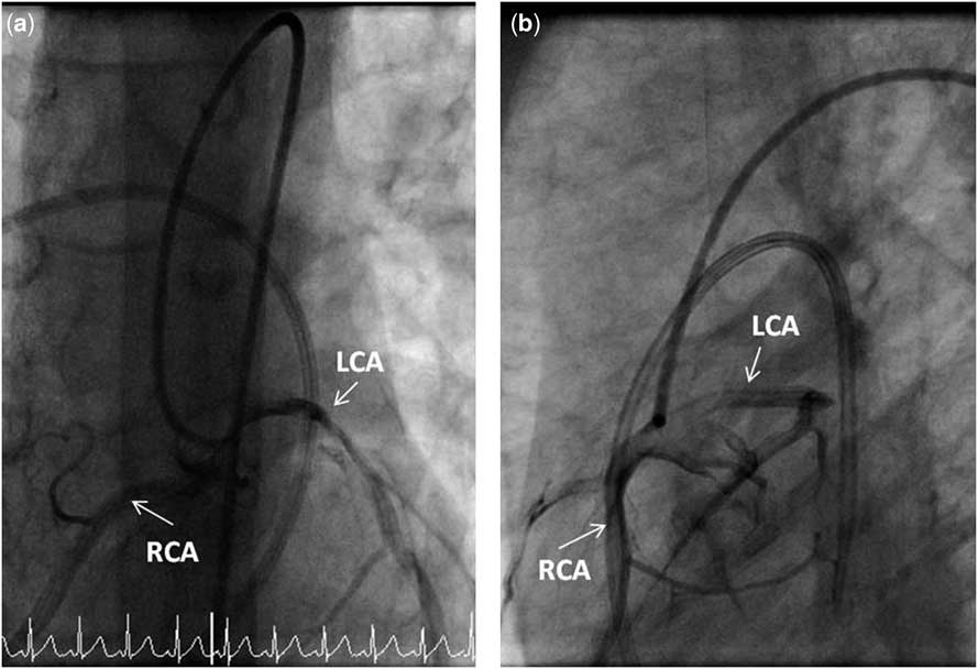
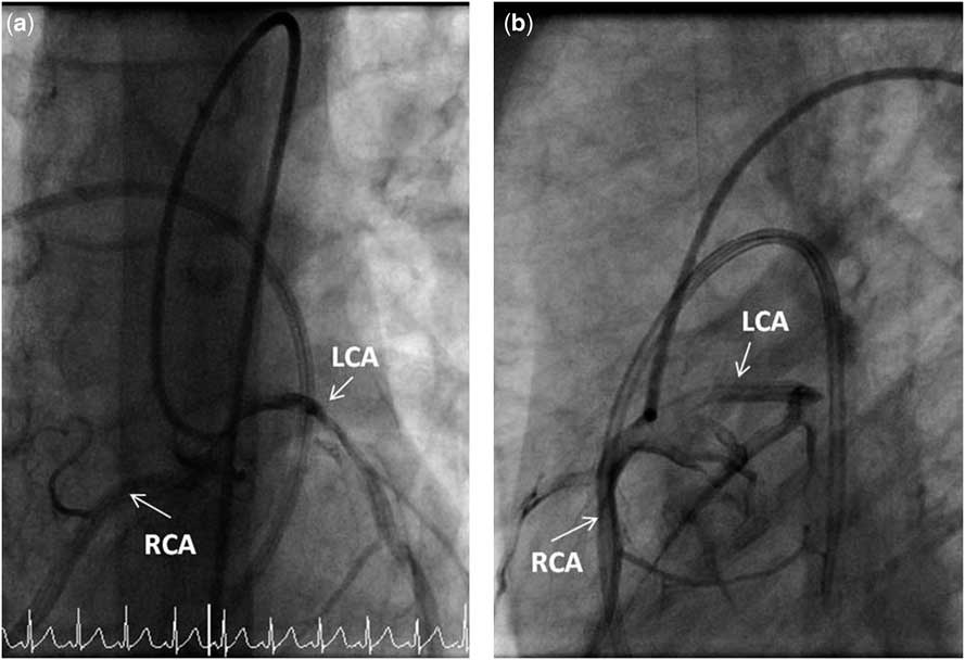
Anomalous left coronary artery from the right sinus of valsalva was diagnosed in one patient. He was an 11-year-old boy who presented with chest pain and light-headedness while “playing”, which progressed to cardiac arrest and ventricular tachycardia. He was the only patient who presented with chest pain associated with activity. The patient was resuscitated and urgent catheterisation demonstrated left coronary artery arising from the right sinus of Valsalva (Fig 2). The patient went directly to the operating room where unroofing of the left coronary artery was performed. He is doing well 3.6 years after surgery. A 15-year-old girl presented with chest pain and palpitations for 2 days and electrocardiogram showed supraventricular tachycardia. Her rhythm responded to adenosine and she was discharged on atenolol. A 17-year-old girl was diagnosed with essential thrombocythaemia after presenting with chest pain, vomiting, and confusion. Her platelet count was 857,000 per microlitre and electrocardiogram showed inferior ST elevations. Echocardiogram showed left ventricular inferior wall hypokinesis and cardiac catheterisation showed normal coronary arteries. She was suspected to have suffered an inferior wall myocardial infarction due to sluggish coronary flow and was treated with aspirin and heparin. Her platelet count rose to over 1,500,000 per microlitre and bone marrow biopsy showed findings consistent with essential thrombocythaemia and hydroxyurea was started.
( a and b ) Right anterior oblique and left anterior oblique views of retrograde catheter in the aortic root showing that the right coronary artery (RCA) and left coronary artery (LCA) both arise from the right sinus of Valsalva (anomalous LCA from the right sinus of Valsalva).
In two patients, no definitive diagnosis was made. The first patient was suspected to have suffered myocardial infarction due to coronary vasospasm, but did not meet criteria for the diagnosis because of a lack of electrocardiogram abnormalities. This patient was a 14-year-old boy with a history of Wolff–Parkinson–White syndrome who presented with the sudden onset of substernal chest pain. His electrocardiogram was normal, but troponin I mildly elevated, peaking at 1.0 nanogram per millilitre. He had normal echocardiogram, cardiac catheterisation, and cardiac magnetic resonance imaging. Although intermittent supraventricular tachycardia was suspected as an aetiology for his presentation, a diagnosis of coronary vasospasm was also entertained given the normal coronary arteries and lack of evidence for myopericarditis. He was discharged on calcium channel blocker therapy and has had no further events 1.5 years later. The second patient, a 17-year-old boy with a history of mitral valve prolapse, but no mitral regurgitation, presented with chest pain soon after eating and was found to have an elevated troponin I. He had a normal electrocardiogram and echocardiogram and no evidence of pulmonary embolus on chest computed tomography angiography. He then underwent cardiac catheterisation during which a dissection of the right coronary artery was appreciated. The dissection likely occurred during catheter engagement of the right coronary os and was not related to his original presentation. The patient had urgent coronary stent placement to treat the dissection. He eventually had an extensive evaluation for connective tissue disorder that was negative and the aetiology of his presentation was not clear. He was doing well without further events at a follow-up of 3.2 years.
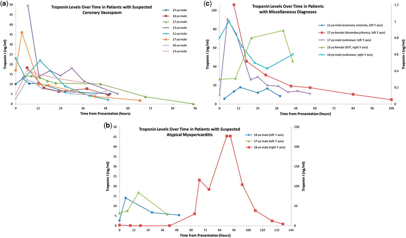
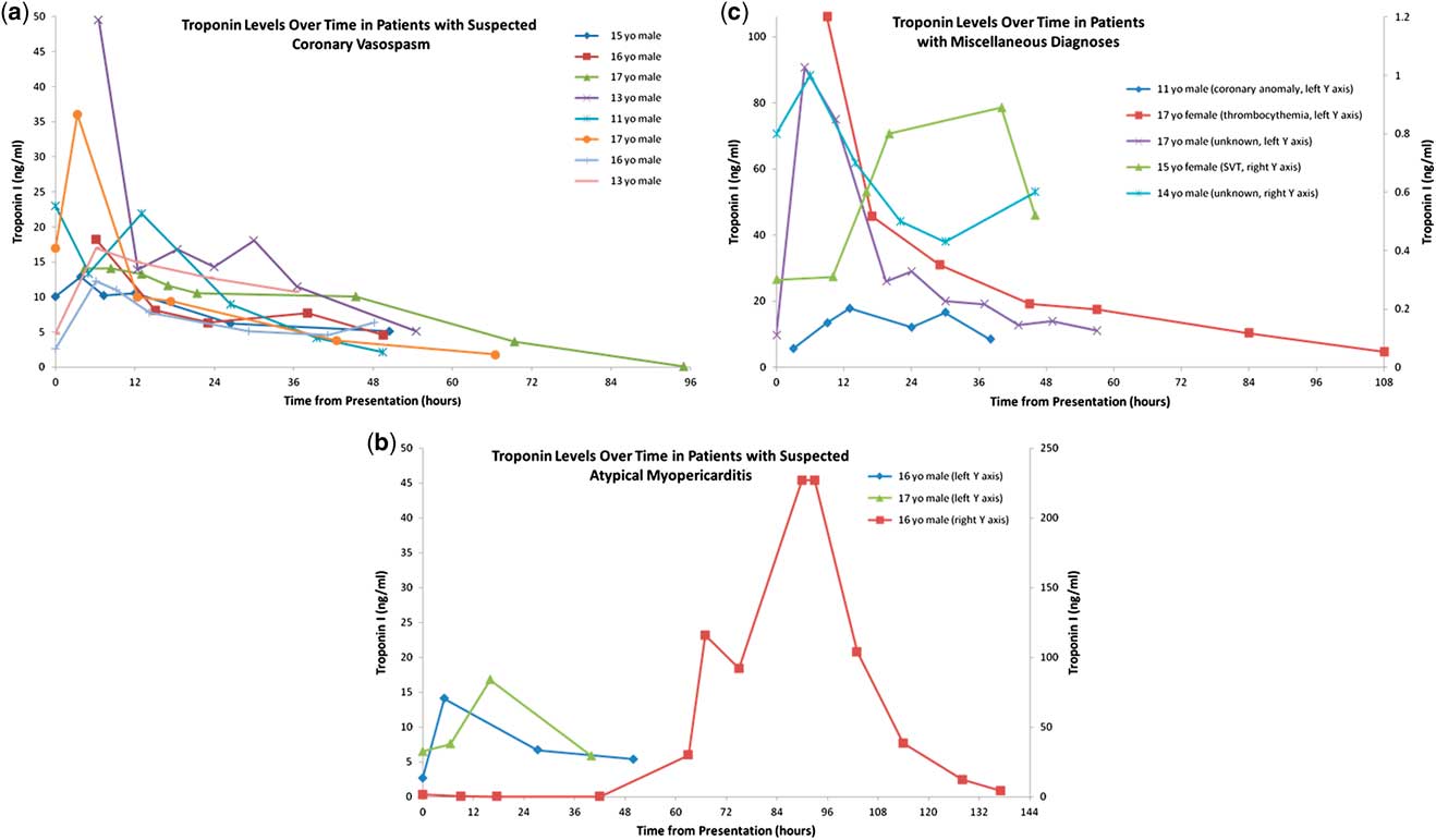
Figure 3 shows the troponin I level over time for each of the patients: 3a shows patients presumed to have coronary vasospasm, 3b shows those suspected to have atypical myopericarditis, and 3c shows the remaining assigned diagnoses. The troponin level peaked at 6 hours or less in all patients with vasospasm and, then, gradually declined (Fig 3a). In these patients, the median time to peak troponin value from presentation was 4.1 hours (range 0.5–6.1 hours) and the median time after presentation at which the troponin value decreased to 50% of the peak value was 19.8 hours (range 12.4–69.5 hours). The three patients with atypical myopericarditis are heterogeneous; two patients show an early peaking troponin with slow decline, whereas the third patient has a prolonged troponin leak with late peak value.
( a , b , and c ) Graphic display of troponin I levels over time in 16 adolescents with no history of significant cardiac disease who presented with chest pain. SVT = supraventricular tachycardia.
Discussion
Adolescents who present with chest pain and are found to have elevated cardiac biomarkers represent a challenge for treating physicians because there is little information to guide appropriate evaluation and management.Reference Desai, Patel and Book 2 , Reference Lane and Ben-Shachar 3 We describe 16 adolescents with no history of significant cardiac disease who were admitted to a single centre with chest pain and an elevated troponin I assay. Patients with a typical presentation of myocarditis or pericarditis were excluded from our series. In all, 15 patients (94%) had diagnostic imaging in addition to echocardiography to define the coronary anatomy, most having catheterisation. Most of our patients were male and the most common discharge diagnosis in our patient cohort was myocardial infarction from presumed coronary vasospasm. Coronary anomalies were rare, but the only patient who had chest pain with exertion suffered myocardial infarction because of the anomalous left coronary artery arising from the right sinus of Valsalva.
Cardiac biomarkers, particularly troponin I and troponin T, are commonly used in the evaluation of chest pain in adults owing to the high frequency of ischaemic heart disease in this population. Troponin is an extremely sensitive marker of myocardial necrosis and is an important part of the diagnosis of myocardial infarction.Reference Thygesen, Alpert and White 8 Myocardial infarction in otherwise healthy children is particularly rare, but can occur in the setting of drug use, anomalous coronary arteries, Kawasaki disease with coronary aneurysms, familial hypercholesterolaemia, and coronary vasospasm.Reference Desai, Patel and Book 2 , Reference Lane and Ben-Shachar 3 , Reference Holt, Singh, Rhee, Billadello and Ludomirsky 9 – Reference Towbin, Bricker and Garson 13
Few other reports exist that describe adolescents with chest pain and elevated cardiac biomarkers. Lane et al described nine adolescents, mostly boys, who presented in this manner. All had normal coronary arteries at catheterisation and were thought to have experienced myocardial infarction from coronary vasospasm.Reference Lane and Ben-Shachar 3 Desai et al described nine patients, also mostly boys, who presented with chest pain, elevated cardiac enzymes, and ischaemic electrocardiogram changes. In all, three patients were thought to have coronary vasospasm, two patients myocarditis, and one patient a coronary anomaly. The diagnosis was unclear in three patients.Reference Desai, Patel and Book 2 Brown et alReference Brown, Hirsh and Mahle 4 described 37 patients less than 22 years of age with chest pain and elevated troponin; the most common diagnosis was myocarditis, but patients were also diagnosed with cardiac contusion, acute myocardial infarction, pericarditis, and sepsis.
Coronary vasospasm leads to myocardial ischaemia, but does not typically cause infarction. However, if the vasospasm persists or if coronary collateral circulation is incomplete, infarction can occur. The diagnosis of coronary vasospasm can be challenging. In our cohort, patients who were diagnosed with vasospasm by treating physicians all experienced chest pain in the setting of an electrocardiogram showing ST elevation in a coronary distribution and were found to have normal, unobstructed coronary arteries. 7 Vasospasm can also be diagnosed at catheterisation using provocative testing, although no patients in our series underwent this testing.Reference Desai, Patel and Book 2 , 7 All patients with vasospasm in our series were boys, a predisposition that has previously been reported.Reference Lane and Ben-Shachar 3 In addition, nearly all of the patients presented with ST elevation on electrocardiogram that involved the inferior leads, suggesting spasm of the right coronary artery. In adult series, the epicardial right coronary has been reported to be the most susceptible vessel to vasospasm. 7 In our cohort, all patients with vasospasm were discharged on calcium channel blocker therapy and there were no recurrent admissions for chest pain; although follow-up is modest, ours is the only study to report follow-up in adolescents with presumed coronary vasospasm.
Although it is striking that four of the eight patients diagnosed with vasospasm were taking stimulants at presentation, our cohort is small and it would be inappropriate to suggest a causal role for these medications from this retrospective report. Nonetheless, the stimulant methylphenidate has been associated with myocardial infarction from presumed coronary vasospasm.Reference George, Kunwar and Awasthi 14 , Reference Thompson and Thompson 15 One report describes the case of a 27-year-old man who suffered infarction from likely vasospasm while taking methylphenidate and pseudoephedrine. The patient took several extra doses of methylphenidate the night before presentation; the authors hypothesised that the combined sympathetic effects of both drugs may have led to coronary spasm.Reference Thompson and Thompson 15 Another report describes the case of a 16-year-old boy who suffered infarction from vasospasm presumably because of a medication interaction between methylphenidate, bupropion, and erythromycin.Reference George, Kunwar and Awasthi 14 Further study of the association between stimulant therapy and vasospasm is warranted.
Both myocarditis and pericarditis can lead to chest pain and an elevated troponin value.Reference Cooper 5 , Reference Lange and Hillis 6 Typically, inflammation leads to myocyte cell membrane degradation and resultant leakage of troponin into the blood. When the presentation of myocarditis or pericarditis is typical, the diagnosis is readily made and appropriate treatment given. However, both can present atypically, making diagnosis challenging.Reference Bruce and Spodick 16 In our study, three patients were diagnosed with atypical myopericarditis, two based on magnetic resonance imaging findings. Magnetic resonance imaging can be very useful in the evaluation of chest pain and positive biomarkers, as delayed enhancement imaging can be used to diagnose myocarditis.Reference Gerbaud, Harcaut and Coste 17 , Reference Friedrich, Sechtem and Schulz-Menger 18
Coronary anomalies have been reported to cause myocardial infarction in adolescents, particularly an anomalous left coronary artery from the right sinus of Valsalva or from the pulmonary artery.Reference Desai, Patel and Book 2 , Reference Towbin, Bricker and Garson 13 Although coronary anomalies are rare, clearly defining the origins of coronary arteries with diagnostic imaging is important in adolescents who present with chest pain and abnormal cardiac biomarkers.
Coronary atherosclerotic disease is particularly rare in adolescents and there were no acute coronary occlusions associated with atherosclerosis in our cohort. Premature atherosclerosis can occur in the setting of familial hypercholesterolaemia and all patients in our series had normal lipid panels. Although defining the origins of coronary arteries is important in order to rule out a congenital coronary anomaly, routine diagnostic angiography for coronary atherosclerosis is likely not warranted in adolescents with chest pain and biomarker elevation in the absence of severe hypercholesterolaemia.
In our cohort, a patient presented with supraventricular tachycardia that was associated with chest pain and elevated troponin, likely because of increased cardiac oxygen demand. Another patient was diagnosed with essential thrombocythaemia. The elevated troponin level in this patient was likely due to sluggish coronary flow as no acute coronary clot was identified. Pulmonary embolus is another diagnosis that can cause chest pain and biomarker elevation, presumably because of an increase in right ventricular demand. This diagnosis was not represented in our cohort; pulmonary embolus is quite rare in otherwise healthy children.
In our cohort, the pattern of troponin elevation over time was not specific for any diagnosis. Most patients, regardless of diagnosis, showed an early peak troponin with gradual decline. The patients with suspected coronary vasospasm all followed this pattern. An acute mismatch in cardiac oxygen supply and demand from any cause leads to infarction and troponin release. After the mismatch resolves and oxygen delivery is restored, troponin release stops; however, troponin clearance is gradual, typically requiring 7 days for complete resolution.Reference Kehl, Iqbal, Fard, Kipper, De La Parra Landa and Maisel 19 We hypothesise that atypical myopericarditis is associated with a prolonged leak of troponin due to ongoing inflammation, but this could not be clearly demonstrated in our small cohort.
Limitations
Although unlikely, it is possible that eligible patients may have been missed given the retrospective nature of the study. All 16 patients in our study presented to community hospital emergency departments and were subsequently transferred to our institution for further management. In all cases, biomarker evaluation was initiated at the referring institution. The frequency with which adult practitioners check biomarkers in patients with chest pain may be different from practitioners in a children's hospital emergency department. This study cannot comment on indications for biomarker testing in this population. In addition, the echocardiographic assessment of wall motion abnormalities in our cohort was subjective, and thus some cases of wall motion abnormality may have been missed.
Recommendations
No treatment algorithm exists to guide physicians who are caring for adolescents with chest pain in the setting of an elevated troponin level. In this series from a single institution where all 16 patients were admitted to an inpatient cardiology unit, management varied. Nevertheless, a general management algorithm can be considered based on our experience and previous reports in the literature (Fig 4). All such patients should undergo electrocardiogram evaluation for evidence of localised ischaemia or changes suggestive of pericarditis or myocarditis. We also recommend prompt echocardiogram to determine whether there are signs of focal or global decrease in ventricular shortening or pericardial effusion. Lipid panel screening and drug screening are also warranted in each patient. Patients with a classic presentation of myocarditis or pericarditis should be treated accordingly. Likewise, patients with suspected pulmonary embolus should be evaluated with chest computed tomography angiography and, possibly, lower extremity Doppler ultrasound. Otherwise, we recommend that coronary artery anatomy be clearly defined in all patients to rule out congenital coronary anomaly. Coronary anatomy can be determined using echocardiography, but is more reliably done using cardiac computed tomography angiography, cardiac magnetic resonance imaging, or cardiac catheterisation. In institutions where the expertise exists, cardiac magnetic resonance imaging is particularly attractive; there is no exposure to ionising radiation, and delayed enhancement imaging can be useful in evaluating the patient for myocarditis.Reference Gerbaud, Harcaut and Coste 17 , Reference Friedrich, Sechtem and Schulz-Menger 18 Diagnostic coronary angiography is not without risk and should be reserved for cases where non-invasive imaging studies are inconclusive or where significant concern exists for an atherosclerotic acute coronary syndrome. Empiric systemic anticoagulation is likely not warranted because atherosclerotic coronary disease is very rare. If concern exists for coronary embolus or pulmonary embolus, then anticoagulation can be considered. Anticoagulation can be risky as it may lead to a haemorrhagic effusion in those with pericarditis. Otherwise, aetiologies are heterogeneous and treatment should be tailored to the particular cause of the patient's symptoms.
Algorithm to evaluate adolescents who present with chest pain and are found to have elevated troponin level. CTA = computed tomography angiography; ECG = electrocardiogram; LE = lower extremity; MRI = magnetic resonance imaging; PE = pulmonary embolus; STE = ST-segment elevation.

Conclusion
The adolescent with no history of cardiac disease who presents with chest pain and elevated troponin can pose a diagnostic challenge to treating physicians. Myocarditis and pericarditis should always be considered, although patients with these diagnoses may not present in a typical manner. In our series, coronary vasospasm was the most common attributed cause of chest pain and elevated cardiac biomarker. Non-invasive imaging should be performed to confirm coronary anatomy. Coronary angiography should be reserved for instances where non-invasive studies are non-diagnostic.
Acknowledgement
The authors would like to thank John W. Hirshfeld, Jr, MD for his helpful contributions to the production of the manuscript. Dr. Hirshfeld is a Professor of Medicine in the Department of Cardiology at the University of Pennsylvania.