INTRODUCTION
The liver fluke, Fasciola hepatica, is a parasitic trematode that infects ruminants and humans in temperate regions of the world. The benzimidazole anthelmintic, triclabendazole (TCBZ) is currently the treatment of choice for the control of fascioliasis in livestock. However, an over-reliance on this fasciolicide has lead to the development of TCBZ-resistant F. hepatica infections. An experimental fasciolicide has been developed by The National Autonomous University of Mexico as a possible alternative to TCBZ for the treatment of fascioliasis. Compound alpha [5-chloro-2-methylthio-6-(1-naphthyloxy)-1H-benzimidazole] is a derivative of TCBZ (Hernández-Campos et al. Reference Hernández-Campos, Ibarra-Velarde, Vera-Montenegro, Rivera-Fernandez and Castillo2002) and the preliminary trial data gathered against a Mexican field isolate showed that compound alpha is highly efficacious against F. hepatica, with a spectrum of activity similar to that of TCBZ. Several studies in sheep and cattle have shown that compound alpha is capable of killing flukes from 3 days old up to adult flukes at least 12 weeks old (Ibarra et al. Reference Ibarra, García, Fernández, Vera, Castillo and Hernández1997a, Reference Ibarra, Vera, Hernández and Castillob, Reference Ibarra, Montenegro, Flores, Hernández and Castillo2000; Rivera et al. Reference Rivera, Ibarra, Olazaràn, Vera, Castillo and Hernández2002).
A trial carried out in our laboratory has confirmed the activity of compound alpha against an adult TCBZ-susceptible fluke infection, although not against a TCBZ-resistant isolate (McConville et al. Reference McConville, Brennan, Flanagan, Edgar, Hanna, McCoy, Gordon, Castillo, Hernández-Campos and Fairweather2009). Fluke material from that trial has been processed for electron microscopical and histological analysis. It was collected at 3 time-points post-treatment (p.t.): 24, 48, 72 h. The present paper is concerned with fine-structural changes to the tegument, somatic musculature and gut of F. hepatica. The tegument plays major roles in fluke biology and acts as a protective barrier between the fluke and its environment (Hanna, Reference Hanna1980; Fairweather et al. Reference Fairweather, Threadgold, Hanna and Dalton1999). Maintenance of the tegument, especially the apical plasma membrane, is essential for the survival of the fluke in the face of stressful situations, such as drug action. Previous ultrastructural studies have shown that it is sensitive to the action of several fasciolicides, including TCBZ and compound alpha, both in vivo and in vitro (see review by Fairweather and Boray, Reference Fairweather and Boray1999; also, Robinson et al. Reference Robinson, Trudgett, Hoey and Fairweather2002; McConville et al. Reference McConville, Brennan, McCoy, Castillo, Hernandez-Campos, Ibarra and Fairweather2006, Reference McConville, Brennan, McCoy, Castillo, Hernandez-Campos, Ibarra and Fairweather2007, Reference McConville, Brennan, Flanagan, Edgar, McCoy, Castillo, Hernández-Campos and Fairweather2008). The tegument has also been shown to be the principal route of entry of TCBZ and its metabolites into the fluke (Mottier et al. Reference Mottier, Alverez, Ceballos and Lanusse2006; Toner et al. Reference Toner, McConvery, Meaney, Brennan and Fairweather2009). This may be true of compound alpha as well. In contrast, the gut is the main route of uptake for other compounds such as clorsulon and closantel (Verheyen et al. Reference Verheyen, Vanparijs, Lauwers, Thienpont and Van den Bossche1980; Meaney et al. Reference Meaney, Fairweather, Brennan and Forbes2004, Reference Meaney, Haughey, Brennan and Fairweather2005a, Reference Meaney, Haughey, Brennan and Fairweatherb). So, the relative changes in the two tissues will indicate which is more severely affected and more likely to be involved in drug uptake. The somatic muscle layers are essential for locomotory, feeding and reproductive movements of the fluke (Fairweather et al. Reference Fairweather, Threadgold, Hanna and Dalton1999).
The study had 3 aims: to determine the time-scale of drug action, by monitoring changes to these tissues over a 3-day period; to shed light on the route of entry of compound alpha into the fluke (whether oral uptake or trans-tegumental): and to obtain information on the possible mode of action of the drug. These aspects of drug action have received little attention in the past and this is the first detailed ultrastructural study on adult flukes following treatment with compound alpha in the natural sheep host.
MATERIALS AND METHODS
The trial was conducted in 2007 under a project licence, held by Dr R. E. B. Hanna, and was carried out at the Agri-Food Biosciences Institute (AFBI) in Stormont, Belfast. The results have been published separately (McConville et al. Reference McConville, Brennan, Flanagan, Edgar, Hanna, McCoy, Gordon, Castillo, Hernández-Campos and Fairweather2009).
Animals and infective material
Six indoor-reared sheep (aged between 9 and 10 months) of similar breed type (Dorset×Suffolk and Dorset×Cheviot), evenly matched for sex and weight (31 kg – 49 kg), with no pre-exposure to F. hepatica were selected for this trial. Five sheep were assigned to Group A and 1 sheep was allocated to Group B as an untreated control sheep. Each sheep was identified by an individual ear-tag number and housed indoors throughout the course of the trial. To confirm no prior exposure to F. hepatica, faecal samples were collected from all sheep at 1 week and at 2 days prior to artificial infection and examined for fluke eggs. A coproantigen F. hepatica ELISA (Bio K201; Bio-X Diagnostics, Jemelle, Belgium) was also performed 1 week prior to artificial infection to test for infection with F. hepatica.
The sheep in Groups A and B were infected by oral gavage with 200 and 250 cysts of the TCBZ-susceptible Cullompton isolate, respectively. The Cullompton isolate was originally derived from an abattoir in Cullompton (Co. Devon, England), and has been shown to be susceptible to the action of TCBZ in vivo and in vitro (Robinson et al. Reference Robinson, Trudgett, Hoey and Fairweather2002; McCoy et al. Reference McCoy, Fairweather, Brennan, Kenny, Ellison and Forbes2005; McConville et al. Reference McConville, Brennan, Flanagan, Edgar, Hanna, McCoy, Gordon, Castillo, Hernández-Campos and Fairweather2009).
Treatment and necropsy
For the 5 sheep in the treatment group (Group A), anthelmintic dosing occurred at 12 weeks p.i. using 15 mg/kg compound alpha. This concentration has been established as the most suitable one to use in sheep (Ibarra et al. Reference Ibarra, Vera, Hernández and Castillo1997b) and it has been shown to be effective against different stages of the liver fluke's life cycle (Ibarra et al. Reference Ibarra, Montenegro, Flores, Hernández and Castillo2000; Rivera et al. Reference Rivera, Ibarra, Olazaràn, Vera, Castillo and Hernández2002). One treated sheep was taken at 24 h p.t. with compound alpha, and 2 sheep were taken at 48 h and 72 h p.t.; they were euthanized by captive bolt stunning followed by exsanguination and their liver removed. The sheep allocated to Group B received no treatment and was euthanized at the same time as the 24 h compound alpha-treated sheep.
The first 6 (living) flukes retrieved from the bile ducts of each sheep were placed directly in warm (37°C) RPMI-1640 medium (Sigma-Aldrich Co. Ltd, Poole, Dorset, UK). The flukes were washed twice in fresh RPMI-1640 medium before being processed for electron microscopical examination. The time between the removal of flukes from the liver until the beginning of fixation was no longer than 3 min.
Tissue preparation for TEM
The 6 flukes from each sheep were lightly flat-fixed for 1 h at room temperature in 4% (w/v) glutaraldehyde in 0·1 m sodium cacodylate buffer (pH 7·4) containing 3% (w/v) sucrose. The specimens were dissected into oral cone (including ventral sucker), midbody and tail regions. For the purpose of this study, all TEM analysis was carried out on the midbody regions of the flukes. The midbody was further divided into transverse sections of approximately 2 mm in width. The sections of fluke were then free-fixed for a further 3 h in fresh fixative, after which they were washed in 0·1 m sodium cacodylate buffer (pH 7·4) containing 3% (w/v) sucrose and left overnight at 4°C. Following post-fixation in 1% (w/v) osmium tetroxide for 1 h, the tissue was washed in fresh buffer, dehydrated in an ascending series of ethanol and infiltrated and embedded in Agar 100 resin. Ultrathin sections, 55–70 nm in thickness, were cut on a Reichert Ultracut E ultramicrotome, mounted on bare 200-mesh copper grids and double-stained with uranyl acetate (5 min) and lead citrate (3 min); all staining was carried out in a 60°C incubator. The specimens were examined in a FEI CM 100 transmission electron microscope operating at 100 keV.
RESULTS
The tegumental syncytium, its underlying tegumental cells and connections, and the somatic musculature
Untreated control flukes
The ultrastructure of the tegumental syncytium and its underlying tegumental cells and connections, and of the somatic musculature were examined and seen to retain a normal morphology as described in Kumar et al. (Reference Kumar, McGeown, Reynoso-Ducoing, Ambrosio and Fairweather2003) and Fairweather et al. (Reference Fairweather, Threadgold, Hanna and Dalton1999).
Twenty-four hours post-treatment
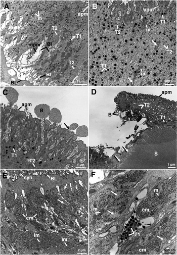
A number of changes were observed in the tegumental syncytium. Swelling of the mucopolysaccharide masses (Threadgold and Brennan, Reference Threadgold and Brennan1978) occurred in patches along the length of the syncytium (Fig. 1A) and, in the vast majority of sections examined, an increase in the number of T1 and T2 secretory bodies was observed within the tegumental syncytium (Fig. 1A, B). The T1 secretory bodies increased in number throughout the depth of the syncytial layer (Fig. 1A), sometimes accumulating at the apex of the syncytium (Fig. 1B). The T2 secretory bodies were typically more abundant in the upper half of the syncytium (Fig. 1B). Changes were commonly observed in the condition of the mitochondria: in some instances, the cristae were slightly swollen (Fig. 1B) and, in approximately one-third of the sections examined, the mitochondria had an electron-lucent appearance. Other forms of disruption included blebbing of the apical membrane (Fig. 1C) and loss of tegument from the tips of the spines (Fig. 1D). Where tegument was lost from the spines, the remaining syncytium contained very few T1 and T2 secretory bodies (Fig. 1D). The subtegumental musculature typically appeared undisrupted, although the circular muscle bundles were occasionally loosely-packed, with a reduced number of muscle fibres (Fig. 1E, 1F). Running between the muscle layers, the T1 cytoplasmic processes typically held more T1 secretory bodies compared to the number of T2 secretory bodies observed within the T2 cytoplasmic processes (Fig. 1F).
Transmission electron micrographs (TEMs) of the tegumental syncytium, cytoplasmic connections and the underlying musculature of 12-week-old, adult Fasciola hepatica 24 h p.t. with 15 mg/kg compound alpha. (A) TEM showing the full depth of the tegumental syncytium from the apical plasma membrane (apm) to the basal lamina (BL). A large number of T1 secretory bodies (T1) are distributed evenly throughout the syncytium. Clusters of T2 secretory bodies (T2) are present throughout the syncytium. At the base of the syncytium are swollen mucopolysaccharide masses (mpm), which surround the basal infolds (BI). m, mitochondria. (B) Transverse section of the tegumental syncytium showing increased numbers of T1 (T1) and T2 (T2) secretory bodies throughout the upper half of the syncytial layer. The mitochondria (m) contain swollen cristae (arrows). apm, apical plasma membrane. (C) High-power TEM of the apex of the tegumental syncytium, showing blebs (B) budding off (arrow) from the apical plasma membrane (apm). T1, T1 secretory body; T2, T2 secretory body. (D) TEM showing loss of the tegumental syncytium covering the tip of a spine (S). The tip of the spine is disrupted (arrows) and, where the syncytium remains, the number of T1 (T1) and T2 (T2) secretory bodies are greatly reduced. apm, apical plasma membrane; B, blebs. (E) Micrograph of the outer circular (cm) and the inner longitudinal (lm) muscle layers situated beneath the tegumental syncytium (syn). The circular muscle bundles appear loosely-packed with few muscle fibres (arrows). The longitudinal muscle retains a normal morphology. BL, basal lamina. (F) High-power TEM, showing T1 secretory bodies (T1) within a cytoplasmic connection situated between the circular muscle bundles (cm), which appear loosely-packed with few muscle fibres (arrows). m, mitochondria.

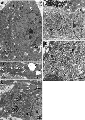
The T1 tegumental cells contained a high number of T1 secretory bodies (Fig. 2A). Though the mitochondria within the T1 tegumental cells generally retained a normal morphology (Fig. 2A), a number of cells contained mitochondria that were swollen and rounded and occasionally appeared electron-lucent (Fig. 2B, C). Although vacuoles were present in the T1 tegumental cells, they were not a common feature (Fig. 2B). The Golgi complexes in the T1 tegumental cells were sometimes reduced in size with indistinct cisternae (Fig. 2C). T2 secretory bodies were observed in all of the T2 tegumental cells examined and their numbers were similar to those observed in untreated control T2 tegumental cells (Fig. 2D, E). The mitochondria in the T2 tegumental cells were sometimes swollen and, within the cell's cytoplasm, small electron-lucent areas were observed (Fig. 2E); the T2 Golgi complexes did not appear to have been disrupted (Fig. 2E).
Transmission electron micrographs (TEMs) of the tegumental cells of 12-week-old, adult Fasciola hepatica 24 h p.t. with 15 mg/kg compound alpha. (A) TEM of two T1 tegumental cells, showing an increased number of T1 secretory bodies (T1) within the cells. The mitochondria (m) retain a normal morphology. G, Golgi complex; N, nucleus. (B) A micrograph of a portion of a T1 tegumental cell containing swollen mitochondria (m), some of which are electron-lucent (white arrows). Vacuoles (black arrows) are also present within the cell. pm, plasma membrane; T2, T2 secretory bodies. (C) High-power TEM, showing Golgi complexes (G) within a T1 tegumental cell. There is an accumulation of T1 secretory bodies (T1) in the vicinity of the Golgi complexes, which are small with diffuse cisternae. Some of the mitochondria (m) are swollen and have assumed a rounded shape. N, nucleus. (D) TEM of a T2 tegumental cell containing a large number of T2 secretory bodies (T2) and some of the mitochondria (m) appear slightly swollen. N, nucleus. (E) Micrograph of a T2 tegumental cell showing a Golgi complex (G) and small electron-lucent areas within the cytoplasm (arrows). The mitochondria (m) retain a normal morphology. T2, T2 secretory bodies.

Forty-eight hours post-treatment
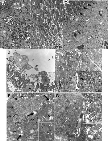
The number of T1 secretory bodies throughout the depth of the tegumental syncytium was substantially less than normal (Fig. 3A–C). The mitochondria were disrupted in the majority of sections examined, occasionally appearing swollen and electron-lucent (Fig. 3A), whilst in other areas of the syncytium the mitochondria were smaller than normal and some contained swollen cristae. The T1 secretory bodies that remained were frequently accumulated in the apical region of the syncytium and the basal infolds were slightly swollen in some areas (Fig. 3B). T1 secretory bodies in what appeared to be the process of breaking down were present in areas of the syncytium where the number of T1 secretory bodies was low (Fig. 3C). The number of T2 secretory bodies was typically normal, though their numbers had reduced in discrete areas along the length of the syncytium (Fig. 3C). Where blebbing occurred on the surface of the tegument, autophagic vacuoles and ‘open’ bodies were typically present in the apex of the tegumental syncytium (Fig. 3D).
Transmission electron micrographs (TEMs) of the tegumental syncytium, cytoplasmic connections and the underlying musculature of 12-week-old, adult Fasciola hepatica 48 h post-treatment with 15 mg/kg compound alpha. (A) TEM showing swollen, electron-lucent mitochondria (m) within the tegumental syncytium. Numerous T2 secretory bodies (T2) are present at the apex of the tegument. apm, apical plasma membrane; BI, basal infolds. (B) TEM of the tegumental syncytium. There is minor swelling of the basal infolds (BI). The mitochondria do not appear to be disrupted. BL, basal lamina; T1, T1 secretory body. (C) High-power TEM, showing possible degraded T1 secretory bodies (white arrows). The number of T2 secretory bodies (T2) is fewer than normal and the mitochondria (m) are electron-dense. T1, T1 secretory bodies. (D) High-power TEM of the apex of the tegumental syncytium, showing blebs (B) on the surface of the tegument and ‘open’ bodies (arrows) can be seen just below the apical plasma membrane (apm). (E) TEM showing disrupted circular muscle (cm) bundles below the basal lamina (BL). The longitudinal muscle (lm) retains a normal morphology. Inset shows a high-power micrograph of the area marked by a dashed box in the main micrograph, showing circular muscle bundles (cm) which are loosely-packed and contain few muscle fibres. One of the mitochondria appears to be breaking down (arrow). (F) TEM of a T1 tegumental cell containing numerous small, inactive Golgi complexes (G). The mitochondria (m) are swollen and electron-lucent (white arrow). A number of mitochondria have begun to break down (small black arrows) and areas of cytoplasm appear to be degenerating (large black arrows). A singular T1 secretory body (T1) is also visible. N, nucleus. Inset is a high-power micrograph of a small Golgi complex. T1, T1 secretory body. (G) TEM of a T2 tegumental cell, showing a few T2 secretory bodies (T2) which are present in small clusters. A mitochondrion (m) is breaking down. N, nucleus. Inset shows the disrupted cytoplasm of a T2 tegumental cell. Areas of the cytoplasm (arrows) are electron-lucent and a small collection of T2 secretory bodies (T2) is visible.

Below the tegumental syncytium, although the circular and longitudinal muscle layers generally retained a normal morphology, at times, the circular muscle was disrupted (Fig. 3E). Specifically, the circular muscle bundles sometimes appeared to contain fewer muscle fibres than normal and the mitochondria within these muscle bundles appeared to be breaking down (Fig. 3E). A very low number of T1 secretory bodies were present in the T1 tegumental cells and the Golgi complexes, though numerous, were small and possessed few discernible cisternae (Fig. 3F). The mitochondria were disrupted in the majority of the T1 tegumental cells. The mitochondria were typically swollen and electron-lucent and some were beginning to break down (Fig. 3F). In the cells containing disrupted mitochondria, the cell cytoplasm often contained small areas that were electron-lucent in appearance (Fig. 3F). In approximately half of the T2 tegumental cells examined, the number of T2 secretory bodies remained at a level similar to those observed in the untreated control flukes. In the remaining T2 tegumental cells, the T2 secretory bodies were few in number and occurred in small clusters throughout the cell (Fig. 3G). The mitochondria within the T2 tegumental cells were often swollen and electron-lucent (Fig. 3G) and the cytoplasm of some cells contained small, electron-lucent areas (inset, Fig. 3G).
Seventy-two hours post-treatment
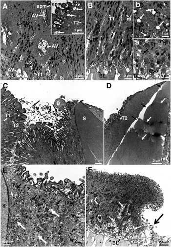
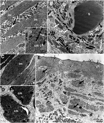
Changes to the tegumental syncytium, 72 h after treatment were severe, with large areas of tegumental sloughing and substantial degeneration of the underlying cell bodies. Large autophagic vacuoles occurred throughout the syncytium (Fig. 4A) and ‘open’ bodies were occasionally present just below the apical plasma membrane (Fig. 4A inset). Within the syncytium, the numbers of T1 and T2 secretory bodies were considerably reduced in the majority of the sections examined (Fig. 4A) and what secretory bodies remained were generally significantly smaller than normal (Fig. 4B); some T1 secretory bodies appeared to have broken down (Fig. 4B inset a). The mitochondria were frequently swollen and had assumed a rounded appearance, no longer retaining the typical chain-like formation within the syncytium (Fig. 4B inset a). This disruption was more severe in isolated areas along the length of the syncytium, and the mitochondria were seen to be breaking down; where this occurred, secretory bodies were largely absent (Fig. 4B inset b). Disruption associated with the spines was observed, generally taking the form of swelling and blebbing of the tegument covering them (Fig. 4C, D). Fibrous projections extending from the surface of the tegument frequently occurred in close proximity to the spines, either on the tegument covering the spines or from the surface of the inter-spinal tegument (Fig. 4C). In some instances, the spines were breaking down and appeared to be fractured along their length and the surrounding tegument had separated from them (Fig. 4D).
Transmission electron micrographs (TEMs) of the tegumental syncytium, cytoplasmic connections and the underlying musculature of 12-week-old, adult Fasciola hepatica 72 h p.t. with 15 mg/kg compound alpha. (A) TEM of the tegumental syncytium containing very few secretory bodies and large autophagic vacuoles (AV). BI, basal infolds. Inset shows the apex of the tegumental syncytium which contains very few T2 secretory bodies (T2) and numerous ‘open’ bodies (arrows) are present below the apical plasma membrane (apm) of the tegument. T1 secretory bodies are not visible. (B) Micrograph of the tegumental syncytium, showing very few secretory bodies. Where T1 secretory bodies (T1) are visible, they are very small. The basal infolds (BI) are slightly swollen. m, mitochondria. Inset a shows the disorganized arrangement of swollen, rounded mitochondria at the base of the tegumental syncytium. The T1 secretory bodies (T1) are very small, and some appear to be breaking down (arrows). BL, basal lamina. Inset b shows a number of swollen, rounded mitochondria (m); other mitochondria appear to be breaking down (arrows). No secretory bodies are visible. (C) TEM of swollen tegument surrounding a spine (S). A bleb (B) is visible on the surface of the tegument covering the tip of the spine. The numbers of T1 (T1) and T2 (T2) secretory bodies are greatly reduced. Where present, the T1 secretory bodies are smaller than normal. Extending from the surface of the tegument is a collection of fibrous projections (arrows). (D) Micrograph showing a spine with multiple fractures (white arrows) along its length. The tegument covering the top of the spine is swollen and has separated (black arrows) from the spine. Only a small number of T2 secretory bodies (T2) are present in the syncytium. (E) High-power TEM of the apex of the tegumental syncytium. The majority of T1 secretory bodies appear to be breaking down (black arrows). The T2 secretory bodies (T2) are few in number and the syncytium is breaking down (white arrows). Swollen and electron-lucent mitochondria (m) are visible. S, spine. (F) Micrograph showing the interface between where the tegument remains, and where the entire syncytial layer has sloughed off. The remaining tegumental syncytium contains small and swollen mitochondria (m), and the mucopolysaccharide masses (small arrows) surrounding the basal infolds (BI) are swollen. BL, basal lamina.

Sections of the tegument taken from areas close to where the syncytium had sloughed off revealed disrupted cytoplasm which contained a significantly reduced number of secretory bodies and almost completely degraded mitochondria (Fig. 4E). The syncytium at the interface between intact tegument and where the syncytial layer had sloughed off contained areas of swollen mucopolysaccharide masses and, where present, the secretory bodies and mitochondria were severely disrupted (Fig. 4F). Below the syncytium, though the circular and longitudinal muscle bundles appeared to contain a normal number of tightly-packed muscle fibres (Fig. 5A and inset), the layers were often widely-spaced due to flooding of the intercellular spaces between the cells separating the layers (Fig. 5A, B). In areas where the syncytium was more severely disrupted, the musculature was also severely affected. Both circular and longitudinal muscle bundles were electron-dense and seemed smaller than normal (Fig. 5B–D) and, between the muscle bundles, the cytoplasmic processes were distended and the parenchymal tissues were loosely-packed (Fig. 5B). The muscle fibres within the muscle bundles frequently appeared disorganized and fewer in number (Fig. 5C) and, within the circular muscle bundles, the mitochondria were sometimes swollen and rounded (Fig. 5D). Where the basal lamina was exposed following sloughing of the syncytial layer, the underlying musculature and parenchymal tissue appeared to be breaking down (Fig. 5E).
Transmission electron micrographs (TEMs) of the underlying musculature and cytoplasmic processes of 12-week-old, adult Fasciola hepatica 72 h p.t. with 15 mg/kg compound alpha. (A) TEM of the longitudinal muscle (lm) layer, showing flooding of the interstitial tissue (arrows) between the muscle bundles. The longitudinal muscle layers retain a normal morphology. Inset shows undisrupted bundles of circular muscle (cm). m, mitochondria. (B) Low-power TEM of the musculature below the tegumental syncytium. The organization of the circular (cm) and longitudinal (lm) muscle layers is irregular. The circular and longitudinal muscle bundles are small and electron-dense, there is distension of the cytoplasmic processes (black arrows) and the parenchymal tissue is more loosely-packed than normal (white arrows). BL, basal lamina; S, spine. (C) High-power micrograph of a longitudinal muscle (lm) bundle, which contains a low number of muscle fibres and appears disorganized. (D) High-power micrograph of small circular muscle (cm) bundles. Within the muscle bundles, the mitochondria (m) are swollen and electron-dense. (E) TEM showing the subtegumental tissues and muscle bundles (arrows) which are breaking down. The basal lamina (BL) remains, but the entire syncytial layer has been sloughed off.

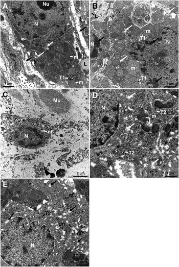
The T1 tegumental cells were in various states of disruption. The condition of T1 cells ranged from those with swollen, and sometimes electron-lucent, mitochondria, minimal numbers of T1 secretory bodies and no Golgi complexes (Fig. 6A), to inactive tegumental cells with degrading cytoplasm, containing electron-lucent areas and severely swollen mitochondria (Fig. 6B). No Golgi complexes were evident in these cells and, when present, T1 secretory bodies were smaller than normal (Fig. 6B). Flooding of the parenchymal tissues surrounding the tegumental cells was common (Fig. 6A) and, in the most severely-affected areas, the T1 tegumental cells and the surrounding parenchyma and musculature were disintegrating (Fig. 6C). A similar range of disruption was observed in the T2 tegumental cells, which typically contained swollen, electron-lucent mitochondria and reduced numbers of T2 secretory bodies (Fig. 6D). The cytoplasm of T2 tegumental cells became progressively more disrupted and mitochondria within these cells were completely broken down (Fig. 6E).
Transmission electron micrographs (TEMs) of tegumental cells of 12-week-old, adult Fasciola hepatica 72 h p.t. with 15 mg/kg compound alpha. (A) TEM of a T1 tegumental cell containing a small number of T1 secretory bodies (T1). Some of the mitochondria appear electron-lucent (white arrows) and others are swollen and rounded (m). Flooding can also be seen outside the cell (black arrows) and a lipid droplet (L) is present in a cytoplasmic process. N, nucleus; Nu, nucleolus. (B) TEM of two T1 cells that are beginning to break down. The mitochondria (m) are severely swollen and beginning to degenerate. The cell's nucleus (N) has lost its typical oval shape and there is clumping of heterochromatin around its periphery (black arrows). The cell cytoplasm (white arrows) is degrading and the T1 secretory bodies (T1) are very small; also, a lipid droplet (L) is visible. (C) TEM of a disintegrating T1 tegumental cell. m, mitochondrion; Mu, muscle; N, nucleus. (D) TEM of a T2 tegumental cell, showing swollen mitochondria (m) that are beginning to break down. N, nucleus; T2, T2 secretory bodies. (E) T2 tegumental cell containing clusters of T2 secretory bodies (T2). The mitochondria (m) have broken down and electron-lucent patches (arrows) are present throughout the cytoplasm. N, nucleus.

Gastrodermal cells
Untreated control flukes
The gastrodermal cells of the control specimens retained a normal morphology similar to that described by Robinson and Threadgold (Reference Robinson and Threadgold1975).
Twenty-four hours post-treatment
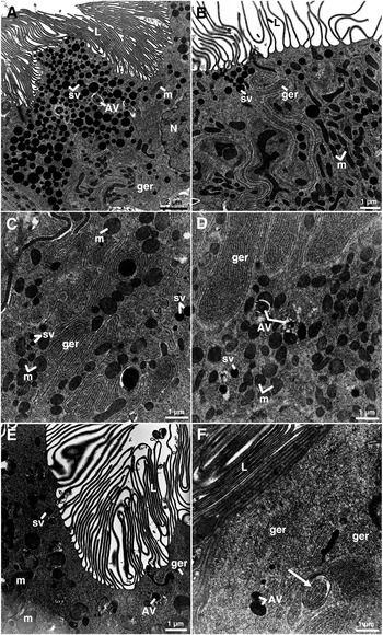
No changes to the morphology of gastrodermal cells were apparent 24 h after treatment with compound alpha (15 mg/kg) (Fig. 7A, B).
Transmission electron micrographs (TEMs) of the gastrodermal cells of 12-week-old, adult Fasciola hepatica 24 h (A and B), 48 h (C and D) and 72 h (E and F) p.t. with 15 mg/kg compound alpha. (A) TEM showing numerous secretory vesicles (sv), cisternae of granular endoplasmic reticulum (ger) and lamellae (L). An autophagic vacuole (AV) can also be seen. m, mitochondrion; N, nucleus. (B) TEM showing normal mitochondria (m) and secretory vesicles (sv). ger, cisternae of granular endoplasmic reticulum; L, lamellae. (C) TEM of a gut cell 48 h following treatment with compound alpha, showing few secretory vesicles (sv) and the mitochondria (m) are swollen. ger, cisternae of granular endoplasmic reticulum. (D) The granular endoplasmic reticulum is extensive and the mitochondria (m) are numerous, retaining a normal morphology. There are few secretory vesicles (sv) and a number of autophagic vacuoles (AV) are present in the cell. (E) TEM of a gut cell 72 h following treatment with compound alpha, showing a small number of secretory vesicles (sv). Some of the mitochondria (m) are swollen and autophagic vacuoles (AV) are present. The lamellae (L) remain unaffected. ger, cisternae of granular endoplasmic reticulum. (F) TEM of a gut cell showing the cisternae of granular endoplasmic reticulum (ger) and autophagic vacuoles (AV). Cisternae of granular endoplasmic reticulum (ger) can be seen within an autophagic vacuole (arrow). L, lamellae.

Forty-eight hours post-treatment
After 48 h treatment with compound alpha (15 mg/kg), the number of secretory vesicles in the gastrodermal cells was reduced (Fig. 7C) and, although the cisternae of the granular endoplasmic reticulum retained a normal morphology, there was an increase in the amount of granular endoplasmic reticulum, particularly at the apex of the cells (Fig. 7C, D). The mitochondria were occasionally swollen and autophagic vacuoles were present in most of the gut cells (Fig. 7D). The apical lamellae remained unchanged.
Seventy-two hours post-treatment
There were very few secretory vesicles remaining in the gastrodermal cells following 72 h treatment with compound alpha (15 mg/kg), and there was an increase in the number of autophagic vacuoles (Fig. 7E). The mitochondria and apical lamellae generally retained a normal morphology, although occasionally the mitochondria were swollen (Fig. 7E). Often, there was a large amount of granular endoplasmic reticulum present throughout the gut cells, and cisternae were seen within autophagic vacuoles (Fig. 7F).
DISCUSSION
The tegumental system, the underlying musculature and the gastrodermal cells of the gut were examined for the purpose of investigating ultrastructural changes in adult TCBZ-susceptible Cullompton flukes in response to treatment with compound alpha. The disruption observed in the tegumental system and in the somatic musculature became increasingly severe with time p.t.. By 72 h p.t., extensive tegumental loss and degeneration of the tegumental cell bodies and muscle bundles had occurred. In contrast, disruption to the gut was relatively mild, even after 72 h treatment. The morphological changes observed will be discussed in terms of the 3 aims: the time-course of drug action (concentrating on the tegument), the route of uptake of compound alpha and its mechanism of drug action.
Dealing with the time sequence of drug action first, surface blebbing and apical accumulations of secretory bodies were observed at 24 h p.t.. These features are typical of a stress response and have been seen in studies with other fasciolicides (Fairweather et al. Reference Fairweather, Anderson and Baldwin1987; Stitt et al. Reference Stitt and Fairweather1993a; Buchanan et al. Reference Buchanan, Fairweather, Brennan, Trudgett and Hoey2003; McKinstry et al. Reference McKinstry, Fairweather, Brennan and Forbes2003; Meaney et al. Reference Meaney, Fairweather, Brennan, McDowell and Forbes2003, Reference Meaney, Fairweather, Brennan and Forbes2004, Reference Meaney, Haughey, Brennan and Fairweather2005b; Halferty et al. Reference Halferty, Brennan, Hanna, Edgar, Meaney, McConville, Trudgett, Hoey and Fairweather2008). The response represents an attempt by the fluke to replace ‘damaged’ tegument, and is dependent on a continuous supply and release of secretory bodies. The tegumental cells were still synthetically active at 24 h, although disruption of the Golgi complexes was observed in some T1 tegumental cells: their reduced size and diffuse cisternae were accompanied by accumulations of T1 secretory bodies at the trans face of the complexes. Microtubules are known to be involved in the dispersal of vesicles away from the Golgi complex (Kelly, Reference Kelly1990), and a build-up of tegumental secretory bodies at their point of synthesis has been observed in liver flukes after treatment with the microtubule inhibitors tubulozole-C and colchicine (Stitt et al. Reference Stitt and Fairweather1993b; Robinson et al. Reference Robinson, Trudgett, Hoey and Fairweather2003).
Following the initial stress response, the number of secretory bodies, particularly T1 bodies, fell dramatically within the tegumental system by 48 h p.t. and (by 72 h) T1 secretory bodies were scarce. The T1 secretory bodies were also smaller in size than normal, suggesting that they were being malformed by the disrupted Golgi complexes. T2 secretory bodies, though reduced in number, were always more frequent than the T1 secretory bodies. This was most notable in the tegumental cells, where they remained even as the cell cytoplasm appeared to be breaking down, suggesting that they were no longer being transported out of the cell. By 72 h p.t., there was an absence of recognizable Golgi complexes in both T1 and T2 cells and this would have a direct bearing on the production and packaging of secretory bodies. Similar changes have been seen in flukes treated in vitro with the microtubule inhibitor tubulozole-C (Stitt and Fairweather, Reference Stitt and Fairweather1993b; Robinson et al. Reference Robinson, Trudgett, Hoey and Fairweather2003) and compound alpha.SO (McConville et al. Reference McConville, Brennan, McCoy, Castillo, Hernandez-Campos, Ibarra and Fairweather2006, Reference McConville, Brennan, McCoy, Castillo, Hernandez-Campos, Ibarra and Fairweather2007) and in vivo with compound alpha (McConville et al. Reference McConville, Brennan, Flanagan, Edgar, McCoy, Castillo, Hernández-Campos and Fairweather2008). It is not unexpected that the reductions in the number of secretory bodies preceded the loss of the tegumental syncytium, as the secretory bodies are vital components in the maintenance of the syncytial layer (Fairweather et al. Reference Fairweather, Threadgold, Hanna and Dalton1999).
Other time-related changes were noted in this study. For example, the mitochondria became progressively more disrupted with time p.t., beginning at 24 h with a swollen and electron-lucent appearance. Breakdown of the mitochondria was observed at 48 h and was more extensive at 72 h p.t.. At 72 h, lipid droplets were present in the tegumental cells and surrounding parenchymal cells. Disruption of the mitochondria within the tegumental syncytium and the tegumental cells has been a feature of compound alpha treatment in previous studies (McConville et al. Reference McConville, Brennan, McCoy, Castillo, Hernandez-Campos, Ibarra and Fairweather2007, Reference McConville, Brennan, Flanagan, Edgar, McCoy, Castillo, Hernández-Campos and Fairweather2008). Autophagic vacuoles were present in the syncytium at 48 h p.t. and more evident at 72 h p.t. and are indicative of cellular breakdown.
Fracturing of the spines was occasionally observed in sections at 72 h p.t.. This has been observed in previous in vitro studies involving compound alpha.SO and TCBZ-resistant flukes: treatment resulted in the loss of spines, whilst the inter-spinal tegument surrounding them remained in place (McConville et al. Reference McConville, Brennan, McCoy, Castillo, Hernandez-Campos, Ibarra and Fairweather2006). More commonly, the tegument covering the spines was covered in blebs and small patches of tegument had sloughed off, exposing the underlying surface of the spines. This would allow the drug to penetrate further into the tissues of the fluke, causing more widespread disruption. The spines generally remained attached to the basal lamina until the tegumental syncytium was lost.
Disruption of the somatic muscle was evident, but not extensive, at 24 h and 48 h in the present study. By 72 h, the circular and longitudinal muscle bundles appeared electron-dense and disorganized, were sometimes smaller than usual and frequently contained fewer muscle fibres. The internal flooding observed, coupled with the limited swelling of the mucopolysaccharide masses and basal infolds in the syncytium, could be due to osmotic changes in the fluke. Similar changes previously have been attributed to an impairment of energy-dependent ion pumps on the tegumental membranes. However, such forms of disruption have been reported following treatment with fasciolicides not known for their effects on energy metabolism (Fairweather et al. Reference Fairweather, Anderson and Threadgold1986; Stitt and Fairweather, Reference Stitt and Fairweather1994; Anderson and Fairweather, Reference Anderson and Fairweather1995; Buchanan et al. Reference Buchanan, Fairweather, Brennan, Trudgett and Hoey2003) and so could be the result of a number of mechanisms. Certainly, the reduction in secretory body production and transport to, and release from, the apical plasma membrane would impair the integrity of the membrane and potentially lead to the osmotic effects observed.
With regard to the route of uptake, previous EM studies with compound alpha have focussed on the tegument, to the neglect of the gut (Rivera et al. Reference Rivera, Ibarra, Zepeda, Fortoul, Hernández, Castillo and Cantó2004, Reference Rivera, Ibarra, Zepeda, Fortoul, Cantó, Hernández and Castillo2005; McConville et al. Reference McConville, Brennan, McCoy, Castillo, Hernandez-Campos, Ibarra and Fairweather2006, Reference McConville, Brennan, McCoy, Castillo, Hernandez-Campos, Ibarra and Fairweather2007, Reference McConville, Brennan, Flanagan, Edgar, McCoy, Castillo, Hernández-Campos and Fairweather2008). In the present study, the tegumental system was more severely affected than the gastrodermis at all time-points p.t. and disruption became increasingly severe with longer treatment times, as described above. Changes to the gastrodermal cells were relatively minor and, at 24 h p.t., the condition of the gastrodermal cells could not be differentiated from control specimens. The number of secretory vesicles showed a progressive decrease at 48 and 72 h p.t., while the number of autophagic vacuoles increased, indicating a fall in secretory activity. The changes observed in the gastrodermal cells may not necessarily be the direct result of drug action, but instead be due to a cessation of feeding that would likely have accompanied the reduction, and later loss, of motility of the flukes observed from 48 h onwards (McConville, unpublished Ph.D. thesis, The Queen's University of Belfast, 2008). By 72 h p.t., a significant reduction in gut content was observed, in conjunction with low levels of motility in those flukes recovered that were not totally inactive. Once flukes cease movement, they stop feeding and enter a state of starvation. Comparing the tegumental and gut changes, then, the present results indicate that trans-tegumental diffusion is the more likely route of entry than oral uptake. It is possible that the different tegumental and gut responses are due to differences in the tissue expression of whatever is the target molecule for compound alpha, but this alternative explanation is considered unlikely.
A similar conclusion for the route of entry has been reached for TCBZ. Thus, despite the strong plasma protein binding shown by TCBZ, experiments have shown that entry of this compound is predominantly across the tegument (Mottier et al. Reference Mottier, Alverez, Ceballos and Lanusse2006; Toner et al. Reference Toner, McConvery, Meaney, Brennan and Fairweather2009). For compound alpha, it is most likely the sulphoxide and sulphone metabolites that penetrate the tegument, as they are present in the blood plasma of sheep, whereas compound alpha itself has not been detected (Rivero et al. Reference Rivero, Jung, Castillo and Hernández Campos1998; Ramírez et al. Reference Ramírez, Mayet, Del Rivero, Ibarra-Velarde, Castillo, Hernández-Campos and Jung-Cook2008). In contrast to the results with compound alpha and TCBZ, disruption of the gastrodermal cells following treatment with the fasciolicides nitroxynil and clorsulon was consistently more severe than that of the tegument, pointing to oral uptake as the most important route of entry (McKinstry et al. Reference McKinstry, Brennan, Halferty, Forbes and Fairweather2007; Meaney et al. Reference Meaney, Haughey, Brennan and Fairweather2005a, Reference Meaney, Haughey, Brennan and Fairweatherb, Reference Meaney, Allister, McKinstry, McLaughlin, Brennan, Forbes and Fairweather2007). Clorsulon, for example, binds to red blood cells and is ingested by the fluke (Meaney et al. Reference Meaney, Haughey, Brennan and Fairweather2005a, Reference Meaney, Haughey, Brennan and Fairweatherb). If compound alpha was entering the flukes during ingestion of host blood, a similar degree of gut disruption would have been expected.
In relation to clarification of its mechanism of action, compound alpha is a benzimidazole compound and so it may act by inhibiting microtubule-based processes. The block in secretory body synthesis (via disruption of the Golgi complex) and transport in the tegumental system supports this idea and the results confirm those of previous in vitro studies (McConville et al. Reference McConville, Brennan, McCoy, Castillo, Hernandez-Campos, Ibarra and Fairweather2006, Reference McConville, Brennan, McCoy, Castillo, Hernandez-Campos, Ibarra and Fairweather2007) and an in vivo study on juvenile flukes (McConville et al. Reference McConville, Brennan, Flanagan, Edgar, McCoy, Castillo, Hernández-Campos and Fairweather2008). Similar changes have been seen in TCBZ.SO-, albendazole sulphoxide- and microtubule inhibitor-treated liver flukes (Stitt and Fairweather, Reference Stitt and Fairweather1993a, Reference Stitt and Fairweatherb, Reference Stitt and Fairweather1994; Buchanan et al. Reference Buchanan, Fairweather, Brennan, Trudgett and Hoey2003; Robinson et al. Reference Robinson, Trudgett, Hoey and Fairweather2003). The immunocytochemical results in McConville et al. (Reference McConville, Brennan, McCoy, Castillo, Hernandez-Campos, Ibarra and Fairweather2006) suggest a depolymerization of microtubules (as a result of loss of tubulin-immunoreactivity) in TCBZ-susceptible Cullompton flukes following in vitro incubations with compound alpha.SO. However, the results were equivocal for TCBZ-resistant Sligo flukes, as treatment with compound alpha.SO did not significantly disrupt tubulin-immunoreactivity, although the EM results revealed significant changes to the tegumental system that could be interpreted in this way (McConville et al. Reference McConville, Brennan, McCoy, Castillo, Hernandez-Campos, Ibarra and Fairweather2006). There was indication of metabolic disruption, through changes to the mitochondria, presence of lipid deposits and autophagic vacuoles, along with the internal flooding observed. However, the changes have been features of treatment with fasciolicides not believed to target energy metabolism, such as TCBZ and diamphenethide (Fairweather et al. Reference Fairweather, Anderson and Threadgold1986; Stitt and Fairweather, Reference Stitt and Fairweather1994; Anderson and Fairweather, Reference Anderson and Fairweather1995; Robinson et al. Reference Robinson, Trudgett, Hoey and Fairweather2002; Buchanan et al. Reference Buchanan, Fairweather, Brennan, Trudgett and Hoey2003). The possibility that compound alpha results in an uncoupling of oxidative phosphorylation within mitochondria, as has been proposed for TCBZ (Carr et al. Reference Carr, McCracken and Stillwell1993), was deemed unlikely as disruption of the mitochondria failed to occur at all time-points p.t. in a separate study on juvenile flukes (McConville et al. 2008 a). Morphological changes in mitochondria are known to accompany changes in their respiratory state (Heytler, Reference Heytler1979), but could also be due to osmotic effects or broader metabolic disruption accompanying cellular breakdown. This was not the case in the present study on adult flukes, as mitochondrial disruption was a feature of all times p.t.. However, the inconsistency of these results means that uncoupling is an unlikely action for compound alpha. It is possible that another cytoskeletal element, actin, may be a target of drug action rather than tubulin, because of the disruption of the spines (McConville et al. Reference McConville, Brennan, McCoy, Castillo, Hernandez-Campos, Ibarra and Fairweather2006; present study) and of the muscle fibres in the subtegumental muscle bundles (McConville et al. Reference McConville, Brennan, McCoy, Castillo, Hernandez-Campos, Ibarra and Fairweather2007, Reference McConville, Brennan, Flanagan, Edgar, McCoy, Castillo, Hernández-Campos and Fairweather2008; present study). Clearly, further research is required before a mechanism(s) of action can be confidently proposed for compound alpha.
The present study has shown that compound alpha is a relatively slow-acting fasciolicide. While peak blood levels occur fairly quickly (10 h p.t.: Rivero et al. Reference Rivero, Jung, Castillo and Hernández Campos1998; Ramírez et al. Reference Ramírez, Mayet, Del Rivero, Ibarra-Velarde, Castillo, Hernández-Campos and Jung-Cook2009), morphological changes take considerably longer to become apparent. They are not marked until 48 h p.t., becoming very severe by 72 h. This observation resembles the time-sequence observed with juvenile flukes (McConville et al. Reference McConville, Brennan, Flanagan, Edgar, McCoy, Castillo, Hernández-Campos and Fairweather2008). The severity of the changes will inevitably lead to the elimination of the flukes and the high efficacy reported for the drug (Ibarra et al. Reference Ibarra, García, Fernández, Vera, Castillo and Hernández1997a, Reference Ibarra, Vera, Hernández and Castillob; Rivera et al. Reference Rivera, Ibarra, Olazaràn, Vera, Castillo and Hernández2002; McConville et al. Reference McConville, Brennan, Flanagan, Edgar, Hanna, McCoy, Gordon, Castillo, Hernández-Campos and Fairweather2009). The observations will contribute to an understanding of why the fluke is eliminated. Thus, the changes to the tegument and the internal spread of disruption will lead to the destruction of the flukes. This process will be exacerbated by changes to the muscle, which will reduce the fluke's motility and trigger a state of starvation. However, precise clarification of drug targets and action awaits further study.
This work was supported by a post-graduate studentship given by the Department of Agriculture and Rural Development Northern Ireland (DARDNI) to Maeve McConville. The authors would like to thank the technical staff at the Agri-Food Biosciences Institute (AFBI), Veterinary Sciences Division (VSD), Stormont, Belfast, for the provision and care of the animals involved in this study.