Hypoplasia of the left ventricle exists on a spectrum, from the severely diminutive left ventricle with the atretic mitral and aortic valves, to the slightly undersized left ventricle with a small mitral valve annulus. Although an objective definition does not exist, borderline left ventricle is a term used to describe a left ventricle that may potentially be inadequate to provide systemic cardiac output.Reference Corno 1 This is often couched in the context of a neonate in the preoperative planning for a single- or two-ventricle repair, when attempting to predict whether the left ventricle can perform as the systemic pumping chamber throughout life. Although these decisions have traditionally been based on the neonatal echocardiograms, the advent of foetal echocardiography and foetal cardiac intervention has brought the question of borderline left ventricle to the forefront earlier in gestation. Counselling families regarding surgical options and expected outcomes can be very difficult in the setting of borderline left ventricle, as reliable predictors of whether a single-ventricle palliation or a two-ventricle repair will be performed have not been identified.
Multiple scoring systems have been developed to guide surgical planning in critical aortic stenosis based on postnatal echocardiography.Reference Hickey, Caldarone and Blackstone 2 – Reference Colan, McElhinney, Crawford, Keane and Lock 5 However, these scoring systems have been invalidated in patients with borderline left ventricle without valvar aortic stenosis.Reference Tani, Minich, Pagotto, Shaddy, McGough and Hawkins 6 , Reference Alboliras, Mavroudis, Pahl, Gidding, Backer and Rocchini 7 From a prenatal perspective, there are several studies that have examined the foetal predictive features of a borderline ventricle in the context of other cardiac lesions, such as pulmonary atresia with intact ventricular septum,Reference Peterson, Levi, Williams, Lai, Sklansky and Drant 8 – Reference Salvin, McElhinney and Colan 10 critical aortic stenosis,Reference Makikallio, McElhinney and Levine 11 , Reference McElhinney, Marshall and Wilkins-Haug 12 and atrioventricular septal defect or double outlet right ventricle.Reference Pitkanen, Hornberger and Miner 13 The foetal echocardiographic features that predict the need for any form of neonatal intervention in the borderline left ventricle was evaluated by Weber et al;Reference Weber, Ayala-Arnez, Atiyah and Etoom 14 however, the small number of patients who required single-ventricle palliation (n=2) likely precluded any prognostication regarding the type of intervention. No study to date has utilised foetal echocardiography to discriminate the type of surgical intervention in foetuses with borderline left ventricle without double outlet right ventricle or atrioventricular septal defect.
The aim of this study was to identify foetal echocardiographic features that help discriminate which infants with borderline left ventricle in utero will undergo univentricular palliation versus biventricular repair, to aid in prenatal counselling for the anticipated postnatal surgery.
Material and methods
This was a retrospective cohort study approved by the University of Michigan Institutional Review Board before initiation. The foetal cardiology database at our institution, created from a review of medical records and cataloguing of performed foetal echocardiograms, was searched to identify foetuses with borderline left ventricle, as determined subjectively by a foetal cardiologist, from 2000 through 2012. Search terms used were “coarctation”, “coarctation?”, “hypoplastic left heart syndrome”, “hypoplastic left heart syndrome?”, and “right ventricle/left ventricle size discrepancy” to capture all foetuses that might have a borderline left ventricle as part of the diagnosis. Medical records were examined to verify the concerns for borderline left ventricle as documented by the attending physician counselling the patient. Exclusion criteria included initial foetal echocardiogram after 36 weeks’ gestation, as measurements too close to delivery were felt to not be useful for prediction earlier in gestation; severe left ventricular hypoplasia requiring single-ventricle palliation without doubt; atrioventricular septal defect; double outlet right ventricle; or the absence of a postnatal echocardiogram. Follow-up foetal echocardiograms after 36 weeks’ gestation were included in the longitudinal data. Medical records were then reviewed for demographic data, including maternal age, gestational age of initial foetal echocardiogram, gestational age at birth, birth weight, the presence of extracardiac anomalies, postnatal cardiac diagnosis, postnatal death at any point following surgical decision, and surgical approach – univentricular palliation or biventricular repair. The presence of extracardiac anomalies or medical risk factors diagnosed at birth were recorded, including defects of the brain, lungs, kidneys, intestines, or extremities, as well as prematurity. The type or intended type of surgical approach was obtained from review of the medical records, and the patients were divided into two cohorts depending on whether they underwent a univentricular or biventricular repair.
Foetal echocardiographic data
Foetal echocardiograms were performed using a Philips iE33 (Philips Healthcare USA, Andover, Maryland, United States of America) or ACUSON Sequoia (Siemens Healthcare USA, Malvern, Pennsylvania, United States of America) ultrasound machine, and retrospective offline analysis of these images was performed on a Siemens syngo Dynamics (Siemens Healthcare USA, Malvern, Pennsylvania, United States of America) work station by a single investigator blinded to the surgical approach. Gestational age at the time of the foetal echocardiogram was calculated by maternal last menstrual period. Initial and follow-up echocardiograms were all analysed independently using the same methods of measurement. Measurements obtained on foetal echocardiogram included: mitral and tricuspid valve annulus dimensions, from the hinge points in late diastole; right ventricular and left ventricular end-diastolic dimensions in short axis; right ventricular and left ventricular end-diastolic length in long axis, measured at end-diastole immediately before atrioventricular valve closure in the four-chamber view; pulmonary valve and aortic valve annulus dimensions, measured in systole at the hinge points in separate short-axis and long-axis views, respectively; ascending aorta diameter; and transverse aorta diameter. Z-scores for gestational age were calculated on all appropriate measurements using an unpublished normal measurement database from Boston Children’s Hospital. Ratios between corresponding right- and left-sided structures were then calculated. To evaluate interobserver reproducibility, a second investigator recorded independent measurements of the morphologic variables as described above. Colour Doppler flow direction was evaluated across the foramen ovale, ascending aorta, and transverse aorta. A monophasic versus biphasic mitral inflow pattern was documented, in addition to mitral valve abnormalities. Mitral valve abnormalities were defined as those with indistinct papillary muscles, close papillary muscles, a single papillary muscle, abnormal chordal attachments, or leaflet abnormalities.
For patients with serial foetal echocardiograms, changes in dimensional sizes and ratios over time were calculated as a rate of growth per week by using the following calculation:
Growth rate of an echo measurement=(echo measurement Z-score or ratio at the last follow-up−echo measurement Z-score or ratio at baseline)/(gestational age of foetus at the last echo follow-up measurement−gestational age of foetus at baseline echo)
Statistical analysis
Data are presented as frequency (percentage) for categorical variables and median (interquartile range or range) or mean±standard deviation, as appropriate, for continuous variables. Univariate comparison of patient and echocardiographic characteristics were made between the two surgical outcomes using χ2 test or Fisher’s exact test for categorical variables and Wilcoxon rank sum test or t-test, as appropriate, for continuous variables. For continuous echocardiographic variables found to be significantly associated with two surgical outcomes in univariate comparisons and their ratios, the cut-off values optimising sensitivity and specificity from receiver operating characteristic curves were determined for significant discrimination between two surgical approaches. The area under the curve, sensitivity, specificity, positive predictive value, and negative predictive value for each cut-off value were reported. Interobserver variability was evaluated using limits of agreement, coefficient of variability, and intraclass correlation coefficient. Finally, growth rates of the echocardiographic measurement were also compared between types of repairs using Wilcoxon rank sum test. All analyses were performed with SAS Version 9.3 (SAS Institute Inc., Cary, North Carolina, United States of America) with statistical significance set at p-values <0.05 using two-sided tests.
Results
A total of 62 patients were identified as having borderline left ventricle using the foetal database. Of these patients, six died in utero, four were delivered and received subsequent care at outside institutions, four patients were excluded owing to obvious hypoplastic left heart syndrome on initial foetal echocardiogram, and nine had initial foetal echocardiograms after 36 weeks’ gestation. Thus, a total of 39 patients were included in the analysis. Postnatally, 15 patients were planned for a single-ventricular palliation, and 24 patients were planned for a biventricular repair. One patient, diagnosed after delivery with a small muscular ventricular septal defect and a tortuous aortic arch without coarctation, did not require any surgery and was included in the biventricular outcome group.
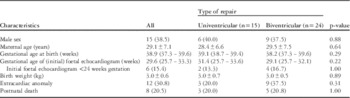
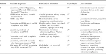
There were no significant differences in patient characteristics between the two surgical groups (Table 1). Of the patients, eight died after the determination of the intended surgery: three from the univentricular group (20%), and five from the biventricular group (20.8%) (Table 2). In both the univentricular and biventricular pathway groups, one patient died before the intended operation. Of the three univentricular patients who died, two (67%) had extracardiac anomalies, whereas four (80%) of the five biventricular patients who died had extracardiac manifestations. A moderate-to-large ventricular septal defect was present in six (75%) of the eight patients who died, including two univentricular patients and four biventricular patients.
Characteristics of patients with foetal identification of borderline left ventricle:univentricular palliation versus biventricular repair (n=39).

Data are presented as n (%) for categorical variables and median (interquartile range) or mean±SD, as appropriate, for continuous variables
Patients with borderline LV who died following determination of surgery.

1V=univentricular palliation; 2V=biventricular repair; HLHS=hypoplastic left heart syndrome; IVC=inferior vena cava; LV=left ventricle; MV=mitral valve; VSD=ventricular septal defect
* Patients 1 and 8 died before intended surgery
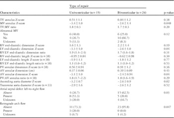
The foetal echocardiographic results for the two outcome groups are shown in Table 3. Variables with significant differences between the groups in univariate comparisons were as follows: mitral valve annulus dimension Z-score, left ventricular end-diastolic dimension Z-score, aortic valve annulus dimension Z-score, ascending aorta diameter Z-score, left-to-right flow across the foramen ovale, and retrograde aortic arch flow. There was no significant difference in tricuspid valve size, mitral valve abnormalities, tricuspid:mitral valve ratio, right ventricular end-diastolic dimension, right:left ventricular end-diastolic dimension ratio, right ventricular or left ventricular end-diastolic length, pulmonary valve annulus dimension, or transverse aorta diameter between the two outcome groups. Of the 34 patients with available mitral inflow data, only one patient, who ultimately underwent single-ventricle palliation, had monophasic mitral inflow. Although the tricuspid:mitral valve ratio and right:left ventricular end-diastolic dimension ratio were not significantly different between groups in the univariate comparisons (both p-value >0.05), their components, such as mitral valve annulus dimension Z-score and left ventricular end-diastolic dimension Z-score, were significantly different; thus, they were included in the receiver operating characteristic curve analysis.
Foetal echocardiographic characteristics in patients with foetal identification of borderline left ventricle:univentricular palliation versus biventricular repair (n=39).

AV=aortic valve; LV=left ventricle; MV=mitral valve; PV=pulmonary valve; RV=right ventricle; TV=tricuspid valve
(n=X) denotes number of patients with available on the measurement. Data are presented as n(%) for categorical variables and median (interquartile range) or mean±SD, as appropriate, for continuous variables
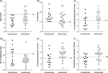
The area under the curve and cut-off values determined using receiver operating characteristic curves are listed in Table 4. Mitral valve annulus dimension Z-score ⩽−1.9 and tricuspid:mitral valve ratio ⩾1.5 were extremely sensitive for discriminating univentricular palliation (sensitivity 100% and negative predictive value 100%), but with low specificity (33%) and positive predictive value (48%). Right:left ventricular end-diastolic dimension ratio ⩾2.1 was very specific for discriminating a single-ventricle palliation (specificity 95.8%), while having low sensitivity (40%). Figure 1 demonstrates the distribution of data between the two groups around the receiver operating characteristic curve-determined cut-off point for the selected variables. On the basis of the distributions, it appears that the mitral valve Z-score and tricuspid:mitral valve ratio cut-off values can define a subset of foetuses with very low probability of single-ventricle palliation, whereas the right:left ventricular end-diastolic dimension cut-off value can define a subset of infants with a high probability of single-ventricle palliation. Interobserver variability testing of the morphologic variables demonstrated good reproducibility, as limits of agreement were all within −0.34–0.31, coefficient of variability ranged from 10.4 to 17.8%, and intraclass correlation coefficients ranged from 0.73 to 0.88.
Distributions of foetal echocardiographic variables in the two repair groups, univentricular and biventricular, with cut-off values determined from the receiver operating characteristics curves to discriminate between two surgical outcomes. ( a ) Mitral valve (MV) annulus Z-score, ( b ) tricuspid valve (TV):MV ratio, ( c ) left ventricular (LV) end-diastolic dimension Z-score, ( d ) right ventricular (RV):LV end-diastolic dimension ratio, ( e ) aortic valve (AV) annular dimension Z-score, and ( f ) ascending aorta diameter Z-score.

Diagnostic characteristics of selected foetal echocardiographic variables for discriminating univentricular palliation (n=39).

AUC=area under the receiver operating characteristic (ROC) curve; AV=aortic valve; LV=left ventricle; MV=mitral valve; NPV=negative predictive value; PPV=positive predictive value; RV=right ventricle; TV=tricuspid valve
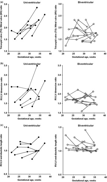
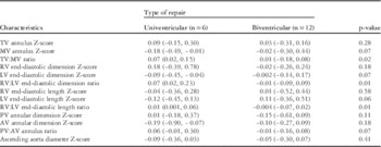
A total of 27 follow-up foetal echocardiograms were performed on 18 patients, 6 univentricular and 12 biventricular. Although mitral valve annulus, left ventricular end-diastolic dimension, and left ventricular end-diastolic length showed trends towards significance in differences in growth rate between the univentricular and biventricular groups, none were considered statistically significant (p=0.07, 0.07, and 0.06, respectively) (Table 5). More discriminative of the eventual choice of a univentricular or biventricular repair was the relative growth of the right and left heart structures. The tricuspid:mitral valve, right:left ventricular end-diastolic dimension, and right:left ventricular end-diastolic length growth ratios were significantly greater in those foetuses ultimately selected for univentricular palliation. Figure 2 illustrates the differential growth of right- and left-sided structures between foetuses in the univentricular and biventricular groups. Ratios between right and left heart dimensions in the univentricular group show a steady increase throughout gestation, whereas in the biventricular group there was a comparable growth of the right- and left-sided structures.
Follow-up data for ( a ) TV:MV ratio, ( b ) RV:LV end-diastolic dimension ratio, and ( c ) RV:LV length ratio:univentricular palliation versus biventricular repair. LV=left ventricle; MV=mitral valve; RV=right ventricle; TV=tricuspid valve.

Growth rates (per week) of foetal echocardiographic characteristics in patients with foetal identification of borderline left ventricle:univentricular versus biventricular repair (n=18).

AV=aortic valve; LV=left ventricle; MV=mitral valve; PV=pulmonary valve; RV=right ventricle; TV=tricuspid valve
Data are presented as median (range)
Discussion
A major aim of foetal echocardiography is to provide an accurate cardiac diagnosis to the family and the care team to develop appropriate plans and foster realistic expectations for postnatal care. Given the important differences in the treatment and outcomes of patients requiring single-ventricle palliative surgeries compared with those who are candidates for biventricular repair, the ambiguity of the “borderline left ventricle” diagnosis can render counselling unhelpful or even anxiety-provoking. Therefore, identifying echocardiographic measures that reliably predict which foetuses will ultimately undergo univentricular palliative surgery will allow the cardiologist to provide supportive and informative counselling in terms of the different surgical and non-surgical options following delivery in patients, with the not uncommon diagnosis of “borderline left ventricle”.
Previous studies have assessed the ability of various scoring criteria to assist in surgical planning for the borderline left ventricle in aortic stenosis. Rhodes et alReference Rhodes, Colan, Perry, Jonas and Sanders 4 published a scoring system based on retrospective analysis, which found that body surface area, indexed aortic root dimension, left ventricular long-axis dimension, and indexed mitral valve area predicted in-hospital mortality in patients with critical aortic stenosis, who underwent an attempted “biventricular” repair, typically a balloon valvuloplasty. On the basis of the equation that was formed using multivariate analysis, a cut-off was determined to help predict a survival advantage with a univentricular palliation. However, Rhodes criteria factors for borderline left ventricle in valvar aortic stenosis are not applicable to patients without valvar abnormalities; infants without valvar aortic stenosis have better biventricular outcomes than would be predicted by traditional scoring methods.Reference Tani, Minich, Pagotto, Shaddy, McGough and Hawkins 6 , Reference Alboliras, Mavroudis, Pahl, Gidding, Backer and Rocchini 7 , Reference Tchervenkov, Tahta, Jutras and Beland 15 The reason for this difference could be explained by the associated myocardial and endocardial abnormalities in critical aortic stenosis, which trigger endocardial fibroelastosis, poor diastolic function, elevated left atrial pressures, and pulmonary hypertension. In fact, the presence of endocardial fibroelastosis is now recognised as an important factor in the decision-making process in critical aortic stenosis, as noted by its appearance in more recent scoring calculators.Reference Hickey, Caldarone and Blackstone 2 , Reference Lofland, McCrindle and Williams 3 , Reference Colan, McElhinney, Crawford, Keane and Lock 5 In infants with borderline left ventricle without valvar aortic stenosis, these myocardial factors are typically not present. With delivery and the subsequent increase in pulmonary blood flow, left heart blood flow, and correction of any arch abnormalities, there is a much higher probability of appropriate left ventricle growth, volume, and function.
Previous literature has demonstrated the importance of mitral valve annulus size and ratios between right- and left-sided structures in predicting outcomes.Reference Schwartz, Gauvreau and Geva 16 , Reference Quartermain, Cohen, Dominguez, Tian, Donaghue and Rychik 17 In our study, we found that the mitral valve annulus size, tricuspid:mitral valve ratio, and right:left ventricular end-diastolic dimension ratios could be used to discriminate surgical preference in borderline left ventricle, with mitral valve size and tricuspid valve:mitral valve ratio being the most sensitive, and right:left ventricular end-diastolic dimension ratio being the most specific. With these three parameters, there were clear, clinically useful cut-offs that can be used to counsel regarding probable surgical outcome. A family can be told with a degree of certainty that, if the foetal mitral valve annulus Z-score is >−1.9 (in the normal range), or the tricuspid:mitral valve ratio is <1.5, their child will likely undergo a biventricular repair. However, if the foetal right:left ventricular end-diastolic dimension is >2.1, then the family can be told that their infant will likely undergo a single-ventricle palliation.
With serial foetal echocardiographic data, there were significant differences in the right/left ventricular size discrepancy parameters – tricuspid:mitral valve ratio, right:left ventricular end-diastolic dimension ratio, and right:left ventricular end-diastolic length ratio – over time between the univentricular and biventricular outcomes. The right and left heart discrepancy remained constant in the patients who eventually had biventricular repair, whereas the discrepancy increased over time in those who underwent a single-ventricle palliation. This relative growth rate of foetal right and left heart structures appears to be an important factor that warrants follow-up during gestation. Determination of whether the right/left ventricular size discrepancy is worsening or staying stable may help separate out some of the overlap of the two groups that occurred with the measurements on a single foetal echocardiogram, but our small number of serial studies prevented this type of analysis.
Certain functional parameters, such as atrial septal defect left-to-right flow and the presence of retrograde arch flow, were also helpful, if present; however, the small numbers again precluded accurate statistical and clinical conclusions. Given that nearly all patients had biphasic mitral inflow, this parameter was not found to be a good discriminator in our study, although the presence of monophasic mitral inflow would likely signify the need for single-ventricle palliation. Finally, we expected structural mitral valve abnormalities to play a prominent role in the determination of single ventricle versus biventricular outcome, but our study did not bear out a significant difference between the two groups with respect to structural mitral valve findings. This may be partly because of small sample size.
Analysis of the postnatal deaths was limited owing to small numbers, but the presence of extracardiac anomalies certainly appeared to be a major factor (present in 75% of postnatal deaths versus 19% of survivors). Another interesting finding was that 75% of patients who died had a moderate-to-large ventricular septal defect, compared with the presence of a moderate-to-large ventricular septal defect in 8/31 (26%) survivors. This is consistent with the previous literature that had demonstrated a moderate-to-large ventricular septal defect as a predictor for failure of biventricular repair in patients with multiple left heart obstructive lesions.Reference Schwartz, Gauvreau and Geva 16 Ventricular septal defects and extracardiac manifestations may not be independent variables, and it is possible that this group represents an entirely different cohort of patients that should not be compared with those with an intact ventricular septum.
Severe diastolic dysfunction and resultant pulmonary hypertension from an inadequate sized left ventricle did not appear to be a theme in the patients who died following biventricular repair, although it may have been an unrecognised factor in these patients with multifactorial causes of death. Would these patients have had a different outcome if a single pathway was chosen? The antithesis of this question cannot be answered by our study but is still important to consider: Are there patients who underwent a univentricular palliation who could have done well with a biventricular repair? We chose to evaluate surgical preference (single ventricle versus two ventricle) rather than post-surgical outcome as an endpoint, as our goal was to find predictors that may improve prenatal counselling. Evaluation of actual post-surgical outcome, that is, whether the correct surgery was performed, would be better assessed using postnatal echocardiography.
Limitations of this study include the inherent problems with a retrospective study. Not all of the echocardiographic parameters could be measured or determined in every patient or every study. Although two measurements of interobserver variability, limits of agreement and intraclass correlation coefficient, showed excellent reproducibility of our echocardiographic measurements, our coefficient of variation was relatively high. This may affect our receiver operating characteristic-derived cut-offs with a larger sample size, and a prospective multicentre study would be appropriate for further investigation. Another major limitation is that it is a single institution study of surgical preference, to improve counselling. There is lack of defined criteria and likely significant institutional variance in the postnatal management and decision making of borderline left ventricle in the neonate. Some use strict cut-offs for mitral valve Z-score to define a borderline left ventricle, although we felt that using one of our measured variables – mitral valve annulus size – to define our patient selection could lead to bias. In addition to the decision between single-ventricle palliation and two-ventricle repair, novel approaches to management, including hybrid strategies and staged left ventricular recruitment, make this discussion even more complex, depending on the institutional experiences and preferences.Reference Davis, Pastuszko, Lamberti, Moore, Hanley and El Said 18 , Reference Emani, McElhinney and Tworetzky 19 For example, our institution may direct more borderline patients towards univentricular palliation, given our relative success with the single-ventricle approach. Although this single-centre data can provide a guide to foetal counselling at any centre, a multicentre study would again be the best way to determine more specific foetal predictors that are fully applicable to patients at all cardiac centres. Finally, the number of patients included in this study is relatively small, given the overall low incidence of any particular congenital heart defect in the general population, and less than half of the patients had follow-up on the foetal echocardiograms available. This likely contributed to our relatively low area under the curve values, making generalisation of conclusions less reliable. Nevertheless, this is the largest study investigating foetal borderline left ventricle to date.
Conclusions
Foetal echocardiography can be helpful in guiding counselling for surgical outcome in the foetus with borderline left ventricle. In this single-centre cohort study, the best predictors of biventricular repair were a mitral valve annulus Z-score ⩾−1.9 or tricuspid:mitral valve ratio ⩽1.5, whereas the best predictor for univentricular palliation was a right:left ventricular end-diastolic dimension ratio ⩾2.1. However, there is still a significant degree of overlap between groups, and longitudinal analysis, including change of right ventricle/left ventricle discrepancy over time, may provide additional insight.
Acknowledgements
None.
Financial Support
This research received no specific grant from any funding agency, commercial, or not-for-profit sectors.
Conflicts of Interest
None.
Ethical Standards
The authors assert that all procedures contributing to this work comply with the ethical standards of the relevant national guidelines on human experimentation and with the Helsinki Declaration of 1975, as revised in 2008, and has been approved by the Institutional Review Board of the University of Michigan Medical School.