INTRODUCTION
ATP-buffering systems based on phosphagens (i.e. phosphocreatine and phosphoarginine) maintain the ATP homeostasis during high and fluctuating cell energy demands. Phosphoarginine is the main reserve of high-energy phosphate compounds in a wide variety of invertebrates, whereas in vertebrates only phosphocreatine was found. Arginine kinase (ATP:arginine phosphotransferase; EC 2.7.3.3) catalyses the reversible transphosphorylation between N-phospho-L-arginine and ATP (Ellington, Reference Ellington2001). We have previously demonstrated the presence of an arginine kinase-mediated phosphagen system in Trypanosoma cruzi and Trypanosoma brucei, the aetiological agents of Chagas' disease and human sleeping sickness, respectively (Pereira et al. Reference Pereira, Alonso, Paveto, Flawia and Torres1999, Reference Pereira, Alonso, Paveto, Iribarren, Cabanas, Torres and Flawia2000, Reference Pereira, Alonso, Torres and Flawia2002). Over-expression of T. cruzi arginine kinase improves the capability of transfectant parasites to grow and survive under nutritional and pH stress conditions (Pereira et al. Reference Pereira, Alonso, Ivaldi, Bouvier, Torres and Flawiá2003b). These phenomena have also been observed when a heterologous arginine kinase was expressed in organisms lacking any kind of phosphagen kinase, such as yeast and bacteria (Canonaco et al. Reference Canonaco, Schlattner, Pruett, Wallimann and Sauer2002, Reference Canonaco, Schlattner, Wallimann and Sauer2003). In addition, T. cruzi epimastigotes treated with hydrogen peroxide showed a time-dependent increase in arginine kinase expression, suggesting the participation of this phosphagen system in oxidative stress responses (Miranda et al. Reference Miranda, Canepa, Bouvier and Pereira2006). The finding in these parasites of a phosphagen and its biosynthetic pathway, which are totally different from those in mammalian host tissues, points to arginine kinase as a possible chemotherapy target (Pereira et al. Reference Pereira, Alonso, Paveto, Iribarren, Cabanas, Torres and Flawia2000).
It was postulated that phosphagen kinases act together with other phosphotransferases such as adenylate kinases and nucleoside diphosphate kinases forming an enzymatic ‘phosphotransfer network’ that allows the communication between spatial separation of ATP consumption and production processes inside the cell (Dzeja and Terzic, Reference Dzeja and Terzic2003). The number and localization of the different phosphotransferases in the intracellular space is a critical parameter for the network efficiency. Consequently, in this work we studied the presence of different arginine kinase isoforms and their subcellular localization by multiple approaches including digitonin extraction, immunofluorescence and bioinformatic prediction.
MATERIALS AND METHODS
Parasite cultures and cell extracts
Epimastigotes of the clone CL Brener, were cultured at 28°C in 25 cm2 plastic flasks, containing 5 ml of LIT medium supplemented with 10% (v/v) fetal calf serum, 100 U/ml penicillin, and 100 mg/l streptomycin (Camargo, Reference Camargo1964). Cells were counted using a haemocytometric chamber, harvested by centrifugation at 1500 g for 10 min and washed 3 times with phosphate-buffered saline (PBS). Cell pellets were then re-suspended in 50 mm Tris-HCl buffer, pH 7·3, containing 10 μm of the protease inhibitor trans-Epoxysuccinyl-L-leucylamido(4-guanidino)butane (E64), and lysed by 5 cycles of freezing and thawing. Trypomastigote and amastigote samples were kindly provided by Dr Maria Júlia Manso Alves (University of São Paulo, Brazil).
Western blot analysis
Arginine kinase antiserum was obtained from BALB/c mice immunized with T. cruzi recombinant arginine kinase. Hexokinase, NADP+-linked glutamate dehydrogenase, cruzipain and mitochondrial aspartate amino transferase antibodies were kindly provided by Dr Juan Luis Concepción (Universidad de Los Andes, Venezuela), Dr Juan José Cazzulo (Universidad Nacional de General San Martín, Argentina), and Dr Cristina Nowicki (Universidad de Buenos Aires, Argentina), respectively. Samples from Trypanosoma cruzi epimastigotes were resolved by SDS polyacrylamide gel electrophoresis. Proteins were electrotransferred from polyacrylamide gels to PVDF membranes (Pierce). The transferred membranes were blocked with 5% (w/v) non-fat milk suspension for 60 min. T. cruzi arginine kinase, hexokinase, NADP+-linked glutamate dehydrogenase, cruzipain and aspartate amino transferase antibodies were added and incubated for 12 h at a dilution of 1:2000, 1:2000, 1:5000, 1:5000 and 1:2500 respectively. Antiserum detection was carried out by incubating with a 1:5000 dilution of anti-mouse or anti-rabbit IgG conjugated with horseradish peroxidase (Vector Laboratories). The latter was developed with Supersignal™ West Pico Chemiluminescent Substrate (Pierce).
Digitonin extractions
The extraction protocol was performed as previously described (Bouvier et al. Reference Bouvier, Miranda, Canepa, Alves and Pereira2006). Briefly, 5·2×109 epimastigote cells from a 200 ml culture were washed twice and re-suspended in 4 ml of 50 mm Tris-HCl buffer, pH 7·5, containing 0·25 m sucrose and 10 μm E64. Aliquots of 950 μl containing 6·5×108 parasites were mixed with 50 μl of the same buffer containing increasing amounts of digitonin (0–5 mg⋅ml−1). After incubation for 2·5 min at room temperature, tubes were centrifuged at 16 100 g for 2 min. Supernatant fractions were kept frozen for enzyme assays. Pellets were re-suspended in the same buffer, lysed by 5 cycles of freezing and thawing and kept frozen for further assays.
Enzyme assays
Arginine kinase activity was determined measuring the ADP formation using an enzyme-coupled assay that produces pyruvate by PK and lactate by LDH, the consequent oxidation of NADH is quantified by absorbance at 340 nm (Pereira et al. Reference Pereira, Alonso, Ivaldi, Bouvier, Torres and Flawiá2003a). Briefly, a 25 μl aliquot of protein fraction was added to the reaction mixture (100 mm Tris-HCl buffer, pH 8·2, 1·5 mm phosphoenolpyruvate, 1·5 mm MgCl2, 0·5 mm DTT, 0·3 mm NADH, 10 mm L-arginine, 5 units per ml pyruvate kinase and 5 units per ml lactate dehydrogenase) in a cuvette to give a final volume of 0·5 ml. After 5 min at 35°C the reaction was started by the addition of a small volume of ATP to a final concentration of 1·5 mm. Pyruvate kinase and hexokinase activities were determined using a lactic dehydrogenase and glucose-6-phosphate dehydrogenase coupled assay, respectively (Cazzulo et al. Reference Cazzulo, Cazzulo Franke and Franke de Cazzulo1989; Caceres et al. Reference Caceres, Portillo, Acosta, Rosales, Quinones, Avilan, Salazar, Dubourdieu, Michels and Concepcion2003). NADP+-dependent isocitrate dehydrogenase activity was determined as previously described (Denicola et al. Reference Denicola, Rubbo, Haden and Turrens2002).
Immunofluorescence
Indirect immunofluorescence was performed with freshly grown trypanosomes washed twice in PBS. After letting the cells settle for 30 min at room temperature onto poly-L-lysine coated cover-slips, they were fixed at room temperature for 15 min with 3% formaldehyde in PBS, followed by 10 min with 0·1% Triton X-100 in PBS. After blocking with 20% horse serum in PBS for 1 h. T. cruzi arginine kinase, hexokinase, cruzipain and NADP+-linked glutamate dehydrogenase antibodies were added and incubated for 1 h at a dilution of 1:50, 1:200, 1:200 and 1:500, respectively. Texas Red or FITC conjugated anti-mouse or anti-rabbit IgGs, were used as secondary antibodies. DAPI at a concentration of 1·5 μg/ml was used to visualize the DNA. For mitochondrial staining, cells were previously incubated for 30 min in complete LIT medium containing 500 nm MitoTracker (Red CMXRos, Molecular Probes), washed and incubated for 30 min in fresh complete LIT medium before fixation. Cells were observed in an Olympus BX60 fluorescence microscope. Images were recorded with an Olympus DP71 camera and processed using the Olympus DP software.
Bioinformatics
Sequences from the ‘Tritryps’ genome projects were obtained at GeneDB (http://www.genedb.org/) and TcruziDB (http://tcruzidb.org/). Assembly and analysis of the DNA sequence data, including prediction of open reading frames, were carried out using the software package Vector NTI v. 10.3.0 (Invitrogen) and the online version of BLAST at the NCBI (http://www.ncbi.nlm.nih.gov/BLAST/).
RESULTS
Arginine kinase isoform prediction in trypanosomatids
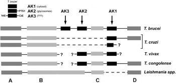
In order to determine the number of arginine kinase isoforms and the predicted subcellular localization, a database screening was performed using GeneDB followed by bioinformatics sequence analysis. The results showed that all analysed species of the genus Trypanosoma present putative arginine kinase genes, including T. brucei (3 isoforms), T. cruzi (2 isoforms), T. vivax (2 isoform) and T. congolense (2 isoforms). All of them have a ‘short’ cytosolic isoform (AK1), which has been previously characterized in T. cruzi (357 aa) and T. brucei (356 aa) (Pereira et al. Reference Pereira, Alonso, Paveto, Iribarren, Cabanas, Torres and Flawia2000, Reference Pereira, Alonso, Torres and Flawia2002). T. cruzi presents an identical but truncated, AK1 variant (237 aa) which was also found at the GeneDB (Tc00.1047053482369.29), such isoform was analysed below. On the other hand, T. brucei has 2 additional isoforms, one with a glycosomal localization signal (PTS-1, -SNL) called AK2 (370 aa) and a large AK3 isoform with unusual N- and C-terminal extensions (404 aa, upper inset Fig. 1). T. congolense and T. vivax present a similar arginine kinase genomic organization but, due to the low coverage of their genome projects, the existence of an AK3 isoforms has not been confirmed. The overall amino acid sequence identity between Trypanosoma spp. arginine kinases was very high, 64% (88% positives). Moreover, synteny analysis revealed a strong conservation of genomic organization around the arginine kinase locus in trypanosomatids (Fig. 1). In T. brucei, the best characterized genome from the ‘Tritryp project (Berriman et al. Reference Berriman, Ghedin, Hertz-Fowler, Blandin, Renauld, Bartholomeu, Lennard, Caler, Hamlin, Haas, Böhme, Hannick, Aslett, Shallom, Marcello, Hou, Wickstead, Alsmark, Arrowsmith, Atkin, Barron, Bringaud, Brooks, Carrington, Cherevach, Chillingworth, Churcher, Clark, Corton, Cronin, Davies, Doggett, Djikeng, Feldblyum, Field, Fraser, Goodhead, Hance, Harper, Harris, Hauser, Hostetler, Ivens, Jagels, Johnson, Johnson, Jones, Kerhornou, Koo, Larke, Landfear, Larkin, Leech, Line, Lord, Macleod, Mooney, Moule, Martin, Morgan, Mungall, Norbertczak, Ormond, Pai, Peacock, Peterson, Quail, Rabbinowitsch, Rajandream, Reitter, Salzberg, Sanders, Schobel, Sharp, Simmonds, Simpson, Tallon, Turner, Tait, Tivey, Van Aken, Walker, Wanless, Wang, White, White, Whitehead, Woodward, Wortman, Adams, Embley, Gull, Ullu, Barry, Fairlamb, Opperdoes, Barrell, Donelson, Hall, Fraser, Melville and El-Sayed2005) presents the following gene order: protein phosphatase 2A regulatory subunit gene (PP2A, Tb09.160.4540), hypothetical conserved protein gene 1 (HCP1, Tb09.160.4550), 2 arginine kinase genes (AK3 and AK2), hypothetical conserved protein gene 2 (HCP2, Tb09.160.4580), AK1 gene and ABC transporter gene (ABCT, Tb09.160.4600). A similar gene order was found in T. vivax and T. congolense; however, T. cruzi lost AK3, AK2 and HCP2 genes. Interestingly, Leishmania spp. lost only AK genes, which is consistent with previous biochemical data (Pereira et al. unpublished observations).
Genetic architecture of arginine kinase loci in trypanosomatids. Genes flanking arginine kinase loci from different trypanosomatids were predicted by database screening at the GeneDB (http://www.genedb.org/) and gene prediction using a BLAST based approach. Gene A: protein phosphatase 2A regulatory subunit (PP2A), B: hypothetical conserved protein 1 (HCP1), C: hypothetical conserved protein 2 (HCP2), and D: ABC transporter gene (ABCT). Dotted lines indicate intergenic regions out of scale, and question marks indicate unknown genome regions. Upper inset: A schematic representation of Trypanosoma brucei arginine kinase isoforms (AK1, AK2 and AK3), NtE: N-terminal extension, CtE: N-terminal extension and PTS1: peroxisomal (glycosomal) targeting signal 1.

Arginine kinase isoforms expression in different stages of the Trypanosoma cruzi life cycle
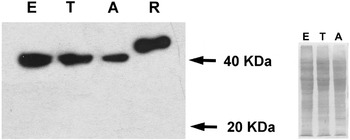
In order to test the expression of the putative truncated arginine kinase isoform, Western Blot analyses were performed using samples obtained from different T. cruzi stages and a polyclonal anti-AK antibody. As Fig. 2 shows, a 40 kDa arginine kinase was detected in epimastigotes, trypomastigotes and amastigotes samples; however, no evidence of the truncated 20 kDa isoform was found.
Arginine kinase expression in different stages of the Trypanosoma cruzi life cycle. Western blot analyses were performed using a mouse polyclonal anti-T. cruzi arginine kinase antibody and samples from T. cruzi epimastigotes (E), trypomastigotes (T), amastigotes (A) and a T. cruzi 6x his-tagged recombinant arginine kinase (45 kDa) as positive control (R). Samples were obtained from approximately 107 parasites. The Ponceau S-stained membrane was also included (right inset).

Determination of arginine kinase subcellular localization by digitonin extraction
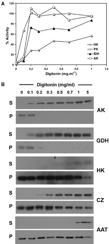
To determine the subcellular localization of T. cruzi arginine kinase, digitonin extraction experiments were carried out (Fig. 3A). The pattern of extraction of arginine kinase activity was compared with the following enzyme markers: hexokinase (HK), pyruvate kinase (PK) and NADP+-dependent isocitrate dehydrogenase (IDH). HK and PK are enzymes involved in glucose metabolism in T. cruzi. HK localizes inside the glycosomes and PK in the cytosol (Caceres et al. Reference Caceres, Portillo, Acosta, Rosales, Quinones, Avilan, Salazar, Dubourdieu, Michels and Concepcion2003; Cazzulo, Reference Cazzulo1994). IDH participates in the citric acid cycle in the mitochondrial matrix (Denicola et al. Reference Denicola, Rubbo, Haden and Turrens2002). At a digitonin concentration of 0·1 mg⋅ml−1, where 67% of the cytosolic marker (PK) has been extracted, about 16% of the arginine kinase activity was measured. In addition, at digitonin concentrations of 0·2 mg⋅m−1 and 0·5 mg⋅m−1, where the mitochondrial (IDH) and glycosomal (HK) markers were respectively released, all the arginine kinase activity was detected. About 16% of arginine kinase is soluble and the remaining 84% is extracted with the mitochondrial marker, suggesting the possibility of AK association with this organellum. Digitonin extraction samples were also analysed by Western blot. HK, NADP+-linked glutamate dehydrogenase (GDH), which in T. cruzi is located exclusively in the cytosol, cruzipain (CZ), the major cysteine proteinase of T. cruzi epimastigotes, located in reservosomes (Barderi et al. Reference Barderi, Campetella, Frasch, Santome, Hellman, Pettersson and Cazzulo1998; de Souza et al. Reference de Souza, Carreiro, Miranda and Silva2000) and the mitochondrial aspartate amino transferase (AAT, Marciano et al. Reference Marciano, Llorentea, Maugeri, Fuentea, Opperdoes, Cazzulo and Nowicki2008) were used as enzyme markers. The results obtained are in agreement with the biochemical enzyme measurements of the different digitonin samples (Fig. 3A and B). A similar digitonin extraction pattern was observed only between arginine kinase and the cytosolic marker (GDH).
Arginine kinase activity and Western blot analysis of digitonin-extracted samples. (A) The experiment was carried out as described in the Materials and Methods section using Trypanosoma cruzi epimastigotes samples. The measured activities represent the release of the indicated enzyme markers of intracellular compartments (HK, hexokinase; PK, pyruvate kinase; IDH, NADP+-dependent isocitrate dehydrogenase) and the arginine kinase activity (AK) by selective permeation with digitonin in the range 0–1 mg⋅ml−1. (B) Western blot analyses were performed using mouse polyclonal anti-T. cruzi arginine kinase (AK), NADP+-linked glutamate dehydrogenase (GDH), hexokinase (H), cruzipain (CZ) and mitochondrial aspartate amino transferase (AAT) antibodies. Samples were equal amounts of supernatants and pellets from a digitonin extraction in the range 0–5 mg⋅ml−1.

Determination of arginine kinase subcellular localization by immunofluorescence
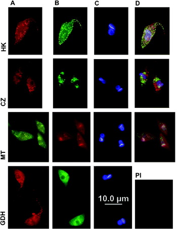
Digitonin extraction analysis suggests that arginine kinase activity is localized mainly in mitochondria, in addition to a minor cytosolic localization. However, Western blot analysis revealed no association with this organellum. In order to understand this observation, arginine kinase localization was also analysed by immunofluorescence. A predominant punctuated pattern has been observed and a minor proportion in the cytosol (details in Fig. 4 column A). The obtained pattern does not match with those observed by the cytosolic marker (NADP+-linked glutamate dehydrogenase, Fig. 4, GDH) or by the glycosomal marker (hexokinase, Fig. 4, HK) and the reservosome marker (cruzipain, Fig. 4, CZ). Furthermore, no co-localization was observed between arginine kinase and the mitochondrial reticulum when epimastigotes were stained with MitoTracker. In summary, arginine kinase does not localize in any of the tested organella and the identity of the observed punctuated pattern requires further investigation.
Immunofluorescence of Trypanosoma cruzi arginine kinase. T. cruzi epimastigotes were attached to multi-well slides, fixed, permeabilized and reacted with arginine kinase antibody (column A) and specific subcellular markers (column B): hexokinase antibody (HK), cruzipain antibody (CZ), MitoTracker staining (MT) and NADP+-linked glutamate dehydrogenase antibody (GDH). Arginine kinase pre-immune serum was also included (PI). All the slides were also stained with DAPI (column C). Merged images are shown in column D.

DISCUSSION
Energy communication and balance are essential processes in cellular homeostasis. Trypanosomatid organisms have an unusually large number of phosphotransferases involved in nucleotide recycling and interconversion. In T. cruzi 6 adenylate kinases probably with different subcellular localization were recently described and also 4 different nucleoside diphosphate kinases were identified by our group and the T. cruzi genome project (Bouvier et al. Reference Bouvier, Miranda, Canepa, Alves and Pereira2006; Miranda et al. Reference Miranda, Canepa, Bouvier and Pereira2008). In the specific case of arginine kinase, the evolutionary processes that determined the presence of 3 different isoforms in other trypanosomes different from T. cruzi and the complete absence of those genes in Leishmania remain to be elucidated. In mammalian cells the energy exchange between organella (i.e. mitochondria and nucleus) is sustained by spatial repositioning of organella and the existence of an integrated enzymatic phosphotransfer network constituted by guanidino kinases (creatine kinase), adenylate kinases and nucleoside diphosphate kinases (Dzeja et al. Reference Dzeja, Bortolon, Perez-Terzic, Holmuhamedov and Terzic2002). An efficient phosphotransfer network requires the correct subcellular localization of each enzyme. Previous studies from our group reported that T. cruzi arginine kinase remains mainly in soluble fractions obtained by freezing and thawing followed by centrifugation (Pereira et al. Reference Pereira, Alonso, Paveto, Iribarren, Cabanas, Torres and Flawia2000). In this work arginine kinase subcellular localization was studied by different approaches, i.e. in silico prediction, digitonin extraction and immunofluorescence. In contrast to the genomic data, the existence of a unique 40 kDa enzyme isoform was confirmed by Western blot analysis. These results could be attributable to mistakes during the preliminary contig assembly or the automatic gene annotation process. T. cruzi arginine kinase was localized mainly in unidentified structures, with similar shape and quantity to glycosomes (Fig. 4, HK), but with different localization and membrane properties. A similar digitonin extraction pattern and a partial co-localization were observed between arginine kinase and cytosolic markers. Punctuated patterns have been observed in trypanosomatids using different microscopic techniques for several cytosolic proteins. For example, the cytosolic isoforms of malate dehydrogenase from Leishmania and T. brucei, the farnesyl diphosphate synthase from T. cruzi and T. brucei, and also an actin-like protein called TcActin, present a similar pattern to that of arginine kinase (Aranda et al. Reference Aranda, Maugeri, Uttaro, Opperdoes, Cazzulo and Nowicki2006; Leroux et al. Reference Leroux, Fleming-Canepa, Aranda, Maugeri, Cazzulo, Sánchez and Nowicki2006; Ferella et al. Reference Ferella, Li, Andersson and Docampo2008; De Melo et al. Reference De Melo, Sant'Anna, Reis, Lourenço, De Souza, Lopes and Cunha-e-Silva2008). Recent studies from our laboratory showed that the localization of arginine kinase could be observed as a diffuse or a punctuate pattern (unpublished observation). It could be hypothesized that different patterns are a consequence of regulatory mechanisms acting upon the varying intra- extracellular conditions.
We are deeply grateful to Juan Jose Cazzulo, Wilfredo Quiñones, Juan Luis Concepción, Cristina Nowicki, Carlos Labriola, Ivan Schumacher, Carolina Carrillo, Pablo Gargantini, Wanderley de Souza, Milagros Camara and Esteban Erben. This study was supported by Consejo Nacional de Investigaciones Científicas y Técnicas (CONICET, PIP 5492), and Agencia Nacional de Promoción Científica y Tecnológica (FONCyT PICT 33431 and FONCYT-PICT REDES 2003-00300). C.A.P. is member of the career of Scientific Investigator of CONICET (Argentina), M.R.M. is a Research Fellow from Fundación YPF and L.A.B. and G.E.C. are Research Fellows from CONICET.