




No CrossRef data available.
Article contents
Acute pulmonary oedema following percutaneous balloon pulmonary valvoplasty in children
Published online by Cambridge University Press: 24 May 2005
Abstract
Pulmonary oedema complicating balloon dilation of the pulmonary valve is extremely rare. We describe two children who developed acute pulmonary oedema soon after successful dilation of the pulmonary valve. They were treated with diuretics, ionotropes, and ventilatory support. Despite the supportive therapy, the complication proved fatal in one.
Information
- Type
- Brief Report
- Information
- Copyright
- © 2003 Cambridge University Press
Percutaneous balloon pulmonary valvoplasty is accepted as a safe and effective alternative to surgical valvotomy, and has become the preferred method of therapy in children for isolated congenital pulmonary valvar stenosis.1, 2 This procedure has also been extended to treat patients with a combination of subvalvar and valvar pulmonary stenosis in the setting of cyanotic congenital heart disease.3 We describe two children who underwent successful percutaneous balloon valvoplasty complicated by life threatening pulmonary oedema, sadly resulting in the death of one child.
Case report
Clinical data of both babies is given in Table 1.
Table 1. Clinical data.

Case 1
Echocardiography revealed severe valvar pulmonary stenosis, severe tricuspid regurgitation, and suprasystemic right ventricular pressures. Cardiac catheterization and percutaneous balloon dilation of pulmonary valve was performed under general anesthesia. The right atrial pressure was elevated at 20 mmHg (mean), and the right ventricular pressure was suprasystemic, with a systolic pressure of 150 mmHg against the aortic systolic pressure of 110 mmHg. The size of the pulmonary valvar orifice was measured as 14 mm, so balloon dilation was performed sequentially, using balloons with diameters of 10, 15, and 18 mm. At the end of the procedure, the right ventricular systolic pressure had reduced to half the systemic pressure, then measured as 60 mmHg. The child was transferred to the intensive care unit while still ventilated. Within 15 min, the patient was seen to be frothing via the endotracheal tube. Chest auscultation revealed wheezes and crepitations. The chest radiograph was consistent with bilateral pulmonary oedema, so the child was continued on ventilatory support, and diuretics and ionotropes were started. He remained haemodynamically stable but, despite positive pressure ventilation and diuretic therapy, the pulmonary oedema did not resolve. Instead, the clinical condition deteriorated further, requiring a very high level of ventilatory support. Despite this, he developed frequent episodes of hypotension and bradycardia, and died 2 days after the procedure.
Case 2
Echocardiography revealed usual atrial arrangement, a common atrium with a common atrioventricular valve, albeit with atresia of the left-sided component, hypoplasia of the left ventricle, double outlet right ventricle, a large inlet ventricular septal defect, anterior deviation of outlet septum, and valvar pulmonary stenosis. The pulmonary arteries were confluent but hypoplastic. Balloon dilation of the pulmonary valve was planned as an alternative to constructing a systemic-to-pulmonary arterial shunt.
The valvotomy was performed uneventfully via the percutaneous route using ketamine intravenous infusion. The diameter of the pulmonary valvar orifice was measured as 7.8 mm, so dilation of the valve and outflow tract was performed using balloons having diameters of 8 and 10 mm. Arterial oxygen saturation after dilation rose to 92–96%, from a basal level of 54–56%. The baby was transferred to the intensive care unit in a stable state. Two hours later, metabolic acidosis was revealed by the blood gases, which could not be corrected by replacement of volume. Chest radiography, which had shown clear lung fields prior to the dilation (Fig. 1a), now revealed haziness of the right lung consistent with pulmonary oedema (Fig. 1b), so we commenced with diuretic therapy. The acidosis persisted, and the patient developed respiratory arrest, requiring intubation with positive pressure ventilation. Blood gases then improved dramatically, and the pulmonary oedema resolved rapidly over next 8–12 h (Fig. 1c). The ventilatory support was required for 48 h, subsequent to which he remained stable without oxygen, accepted oral feeds, and was discharged on decongestive therapy 6 days after the procedure. Echocardiographic examination then revealed a total gradient across the pulmonary outflow tract of 40 to 45 mmHg, most of the gradient being at the valvar level, with minimal turbulence at the sub-pulmonary area.

Figure 1. The chest radiograph of our second patient (a) is shown prior to dilation of the pulmonary valve. Subsequent to the dilation (b), there is right-sided pulmonary oedema. This cleared completely (c) after ventilatory support and diuretic therapy.
Discussion
Acute pulmonary oedema complicating percutaneous balloon pulmonary valvoplasty is extremely rare.1, 4 Walker et al.5 reported one instance in an adult patient aged 66 years, who underwent balloon dilation for long-standing pulmonary valvar stenosis. He developed acute pulmonary oedema soon after the procedure, which had been deemed successful, and the problem resolved rapidly with the institution of ventilation and diuretic therapy. It was thought to occur as a result of the sudden increase in flow of blood to the lungs after the dilation of the long-standing valvar stenosis, and rapid resolution of the pulmonary oedema reinforced this belief, rather than indicating that the complication was due to left ventricular dysfunction. The authors reasoned that, in chronic and severe pulmonary stenosis, the pulmonary arteriolar smooth muscle is diminished and susceptible to leakage when flow is suddenly increased. They presumed that such a situation is not seen in children because of the relatively immature cardiopulmonary vasculature, which rapidly adapts to the acute changes in pulmonary blood flow after such procedures. Our first case defies their presumption. In our patient, who unfortunately died, the factors contributing to pulmonary oedema could have been:
- Presence of prolonged severe tricuspid regurgitation, which could have contributed to chronic lung hypoperfusion as a result of decreased forward flow. The sudden opening of pulmonary valve could then have resulted in a torrential increase in pulmonary blood flow.
- Use of a large balloon, measured at 128% of the diameter of the pulmonary outflow tract. Although dilation has been reported using a balloon measuring 140% of the diameter of the pulmonary valvar orifice, it is possible that staged dilation with smaller balloons, with relatively incomplete opening of pulmonary valve during the first procedure, followed by repeat dilation at a later stage, could have prevented the complication.
The pulmonary oedema seen in our second patient is most likely due to the absence of a protective infundibular stenosis, thus acutely exposing the lungs to a large volume of blood in the already compromised situation of a functionally univentricular heart. We appreciated this fact only at our follow-up echocardiographic investigation, when we found no significant turbulence at infundibular level. Thus, in effect, the patient had valvar dilation in the setting of a large ventricular septal defect, thus exposing the lungs to torrential flow. The reason for right-sided pulmonary oedema could be attributed to the peculiar orientation of the outflow tract towards the right, thus preferentially increasing flow of blood to the right lung.
Pulmonary oedema, therefore, is a rare complication after successful percutaneous dilation of the stenotic pulmonary valve, and is presumed to be due to an acute increase in flow of blood to the lungs immediately subsequent to the dilation. Preexisting chronic severe tricuspid regurgitation, clinical signs of congestive heart failure, and use of large balloons, may all be predisposing factors. In patients with the physiology of tetralogy of Fallot, it is particularly important to search echocardiographically for subvalvar pulmonary stenosis. In its absence, attempts should not be made to dilate the outflow tract for fear of exposing the lungs to limited flow of blood.
References
Kan JS, White RI Jr, Mitchell SE, Gardner TJ. Percutaneous balloon valvuloplasty: a new method for treating congenital pulmonary valve stenosis. N Engl J Med 1982; 307: 540–542.Google Scholar
Chen CR, Cheng TO, Huang T, et al. Percutaneous balloon valvuloplasty for pulmonic stenosis in adolescents and adults. N Engl J Med 1996; 335: 21–25.Google Scholar
Rao PS. Transcatheter treatment of pulmonary outflow tract obstruction: a review. Prog Cardiovasc Dis 1992; 35: 119–158.Google Scholar
Matisonn RE, Mitha AS, Williams MA, Chesler E. Pulmonary oedema following pulmonary valvotomy. Ann Thor Surg 1975; 20: 581–585.Google Scholar
Walker CP, Bateman CJ, Rigby ML, Brookes CI. Acute pulmonary oedema after percutaneous balloon valvoplasty for pulmonary valve stenosis. J Cardiothorac Vasc Anesth 2001; 15: 480–482.Google Scholar
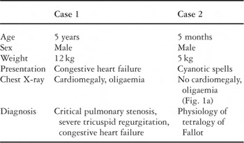
Table 1.

The chest radiograph of our second patient (a) is shown prior to dilation of the pulmonary valve. Subsequent to the dilation (b), there is right-sided pulmonary oedema. This cleared completely (c) after ventilatory support and diuretic therapy.
 You have
Access
- 4
- Cited by