Causes of fetal cardiomegaly include not only fetal cardiac disease, but also extra-cardiac shunt disease.Reference Pedra, Smallhorn and Ryan1 Among these, abnormal fetal circulations such as placental chorioangioma are greatly different from other diseases in terms of the requisite clinical management, because delivery may reduce the cardiac burden. We report a case in which fetal cardiomegaly due to multiple non-giant placental chorioangiomas was unable to be diagnosed prenatally, but was strongly suspected from the dilated umbilical vein.
Case
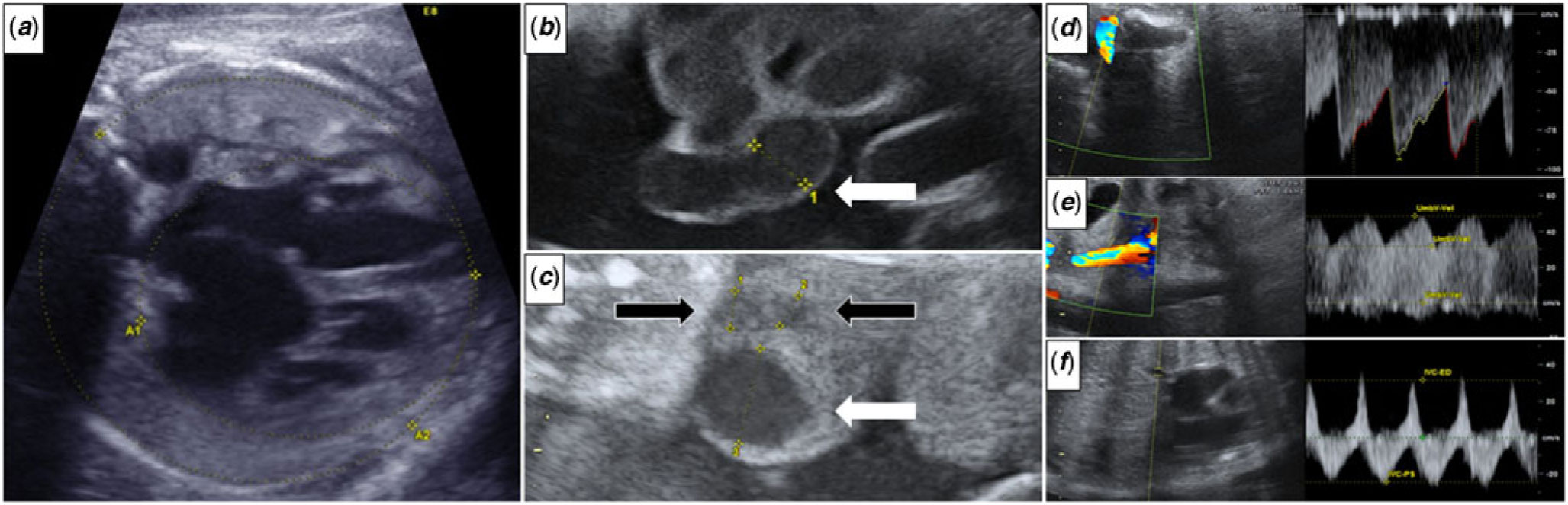
A 39-year-old pregnant woman with no past or family history was referred to our hospital due to fetal cardiomegaly at 31 weeks of gestation. On presentation, fetal echocardiography showed a cardiothoracic area ratio of 0.55 (Fig 1a), but neither structural abnormality nor prominent valve regurgitation were detected, and contractility of both ventricles was maintained. Neither fetal echography nor MRI revealed no extra-cardiac shunt disease. We recognised a thickly expanded umbilical cord and dilated umbilical vein and artery (Fig 1b and c), which raised a suspicion of shunt diseases in the placenta such as placental chorioangioma. However, we were unable to reach a definitive diagnosis. The flow pattern of umbilical artery was normal (Fig 1d). However, the flow pattern of umbilical vein was pulsatile (Fig 1e), and that of inferior vena cava had retrograde component (Fig 1f), so it has been suggested cardiac function declines. We continued careful monitoring for the occurrence of hydrops or fetal heart failure under hospitalisation. Up to the time of delivery, findings from fetal echography did not show any marked changes. No fetal arrhythmia was seen, and anti-SS-A and anti-SS-B antibodies were negative in maternal blood testing. Non-invasive prenatal testing showed no abnormalities.
Fetal echography findings. (a) Cardiothoracic area ratio is 0.55 in fetal echocardiography. (b and c) The umbilical cord is thick, with an umbilical vein diameter of 12 mm (white arrow) and an umbilical artery diameter of 5 mm (black arrow). (d to f) The flow pattern. (d) umbilical artery, (e) umbilical vein, (f) inferior vena cava. Note the pulsatile pattern in the umbilical vein and the retrograde flow in the inferior vena cava, which suggests the decreased cardiac function of the fetus.

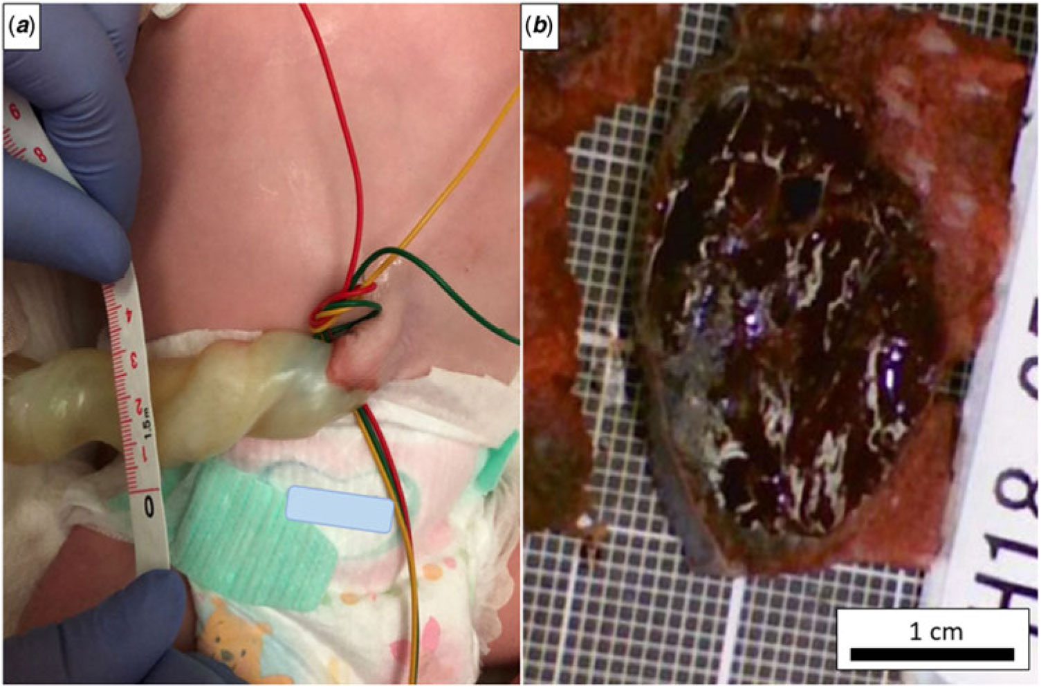
A male neonate was delivered by selective caesarean section at 37 + 1 weeks of gestation with a birth weight of 3114 g without asphyxia. No external malformations were identified, but the umbilical cord was thick with a diameter of about 25 mm (Fig 2a). The cardiothoracic ratio on chest X-ray was 0.63. Echocardiography showed no structural abnormality, left ventricular ejection fraction was 50%, and left ventricular diameter at end-diastole was 20 mm. The value of N-terminal pro-B-type natriuretic peptide was 2156 pg/ml. Haemoglobin level and platelet count were normal. We started continuous intravenous infusion of milrinone after birth, considering the possibility of increased afterload due to withdrawal from the fetal circulation. A few days later, when we tried to reduce milrinone, his cardiac function apparently deteriorated, so it gradually declined over 2 weeks. Although the value of N-terminal pro-B-type natriuretic peptide decreased to 143 pg/ml in 2 weeks, it rose to 236 pg/ml after stopping milrinone, so he was started to take pimobendan and discharged. Cardiothoracic ratio improved to 0.50.
Findings after birth. (a) Diameter of the umbilical cord at birth is about 25 mm. (b) The largest placental chorioangioma is about 3 cm along the major axis.
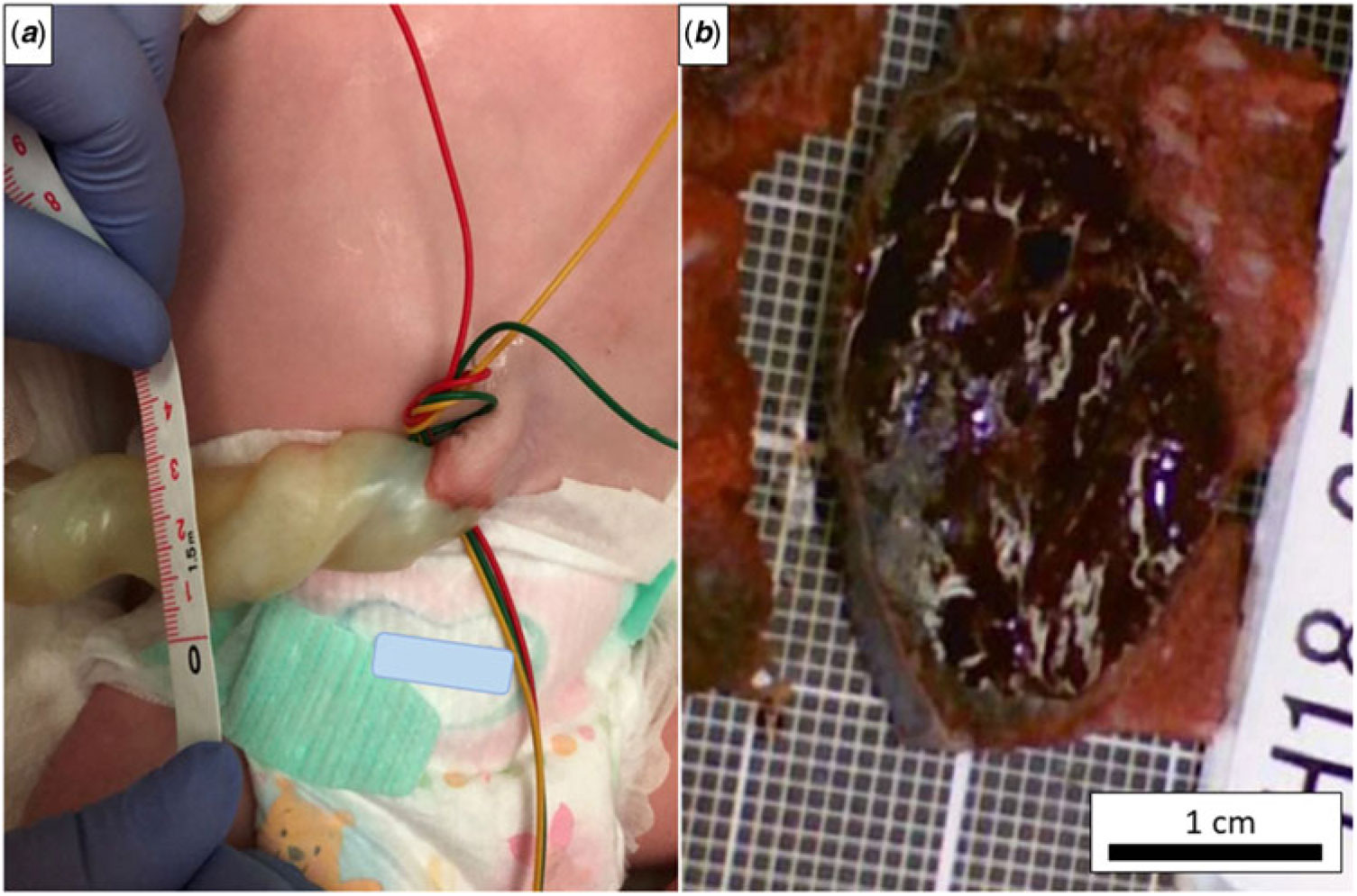
Histopathological examination showed intimal thickening of vessels in the umbilical cord. Five chorioangiomas were identified in the placenta, with a maximum diameter of 3 cm (Fig 2b). We determined that these were the cause of fetal cardiomegaly.
Discussion
Placental chorioangioma is the most common placental tumour, found in 1% of all pregnancies. This disease is reported to cause fetal complications such as growth restriction, anemia, thrombocytopenia, and hydrops, especially in cases with chorioangioma >4 cm in diameter, known as giant chorioangioma. However, most chorioangiomas are small and can be found in postpartum placental pathology examination without any complications ever presenting.Reference Benirschke, Kaufmann, Baergen, Benirschke, Kaufmann and Baergen2, Reference Wu and Hu3
What was noteworthy in our case was that multiple non-giant chorioangiomas may cause fetal complications despite the difficulty of prenatal diagnosis. While colour Doppler echography is considered useful for diagnosing chorioangioma, prenatal diagnosis is sometimes difficult for small lesions.Reference Sepulveda, Alcalde, Schnapp and Bravo4 When fetal cardiomegaly and a dilated umbilical vein are identified, this disease should be suspected even if a definitive diagnosis cannot be reached from echography.
Acknowledgements
The authors would like to thank Dr Yoshinori Moriyama, an obstetrics and gynecologist in our hospital, and Dr Naoki Ohashi, a paediatric cardiologist in Chukyo Children Heart Center, for discussions that led to diagnosis.
Financial Support
This research received no specific grant from any funding agency, commercial, or not-for-profit sectors.
Conflicts of Interest
None.
Ethical Standards
The authors assert that all work reported complies with the ethical standards of the Helsinki convention, and institutional and national research committee.