




No CrossRef data available.
Article contents
Characterization of SSU and LSU rRNA genes of three Trypanosoma (Herpetosoma) grosi isolates maintained in Mongolian jirds
Published online by Cambridge University Press: 07 October 2004
Abstract
Trypanosoma (Herpetosoma) grosi, which naturally parasitizes Apodemus spp., can experimentally infect Mongolian jirds (Meriones unguiculatus). Three isolates from A. agrarius, A. peninsulae, and A. speciosus (named SESUJI, HANTO, and AKHA isolates, respectively) of different geographical origin (AKHA from Japan, and the others from Vladivostok), exhibited different durations of parasitaemia in laboratory jirds (2 weeks for HANTO, and 3 weeks for the others). To assess the genetic background of these T. grosi isolates, their small (SSU) and large subunit (LSU) ribosomal RNA genes (rDNA) were sequenced along with those of 2 other Herpetosoma species from squirrels. The SSU rDNA sequences of these 3 species along with available sequences of 3 other Herpetosoma trypanosomes (T. lewisi, T. musculi and T. microti) seemed to reflect well the phylogenetic relationship of their hosts. Three isolates of T. grosi exhibited base changes at 2–6 positions of 2019-base 18S rDNA, at 5–29 positions of 1817/1818-base 28Sα rDNA, or 1–5 positions of 1557–1559-base 28Sβ rDNA, and none was separated from the other 2 isolates by rDNA nucleotide sequences. Since base changes of Herpetosoma trypanosomes at the level of inter- and intra-species might occur frequently in specified rDNA regions, the molecular analysis on these regions of rodent trypanosomes could help species/strain differentiation and systematic revision of Herpetosoma trypanosome species, which must be more abundant than presently known.
Information
- Type
- Research Article
- Information
- Copyright
- © 2005 Cambridge University Press
INTRODUCTION
Rodent trypanosomes of the subgenus Herpetosoma show rigid specificity of their vertebrate hosts (Hoare, 1972), even though the vectors such as fleas are shared among multiple hosts (Molyneux, 1969a,b; Maraghi & Molyneux, 1989). Due to their narrow host range, only 2 species of Herpetosoma trypanosomes have been studied in detail using common laboratory rodents; i.e., Trypansoma (H.) lewisi and Trypanosoma (H.) musculi, which naturally infect rodents of the genera Rattus and Mus, respectively (reviewed by Dusanic, 1975; Viens, 1985; Albright & Albright, 1991; Monroy & Dusanic, 2000). Experimental observations on other Herpetosoma trypanosomes might be possible only when natural rodent hosts had been prepared with special individual efforts (Molyneux, 1969a,b, 1970). As reported previously (Sato et al. 2003, 2004), Trypanosoma (H.) grosi naturally infecting Apodemus spp. causes persistent infection in laboratory Mongolian jirds (Meriones unguiculatus) as well. In our previous studies (Sato et al. 2003, 2004), we noticed different infection courses of T. grosi isolated originally from Apodemus agrarius (SESUJI isolate) and Apodemus peninsulae (HANTO isolate) in Vladivostok, Russia; the parasitaemia due to these two isolates in jirds terminated at 2 or 3 weeks post-infection (p.i.), respectively.
Morphologically, trypanosomes of the subgenus Herpetosoma resemble closely each other, which is generally called ‘lewisi-like’ after the type species of T. lewisi (Davis, 1952; Hilton & Mahrt, 1972; Hoare, 1972). The rigid ‘host specificity’ of Herpetosoma trypanosomes might provide a clue for differentiation of trypanosome species found in the blood; i.e., it is impossible to infect hosts from different genera or higher taxonomic categories with a singlle Herpetosoma species. However, exceptions to this rigid host specificity are sometimes found in experimental transmission (Culbertson, 1941; Mühlpfordt, 1969; Sato et al. 2003). In this regard, Noyes et al. (2002) detected natural infection with Herpetosoma trypanosomes of a novel genotype distinct from T. grosi from 2 individuals of Apodemus sylvaticus caught in Cheshire, England. In addition, as mentioned above and shown by Chiejina et al. (1993), we know the presence of trypanosome populations of a single species that differ phenotypically e.g. different courses of parasitaemia in the same laboratory host. At present, there are no assays to assess the significance of Herpetosoma trypanosome isolates originating from different host species of the same genus, or showing different biological characters, or to differentiate the observed species with certainty. The present study was designed to assess the genetic background of T. grosi isolates with different origins and/or different biological characters by investigating the small (SSU) and large subunit (LSU) ribosomal RNA genes (rDNA). In addition, 2 squirrel-parasitic species of Herpetosoma trypanosomes including Trypanosoma (H.) otospermophili were similarly characterized. Our results would provide the framework for future studies on abundant Herpetosoma species and/or strains collected from different rodent genera or species in the same or different geographical regions.
MATERIALS AND METHODS
Parasites
Two isolates of T. grosi (SESUJI and HANTO isolates) originated from a striped field mouse, A. agrarius, and a Korean field mouse, A. peninsulae, which were trapped in Vladivostok, Russia, as described previously (Sato et al. 2003). Another isolate of T. grosi (named AKHA isolate after the Japanese name of the host ‘akha-nezumi’) originated from a large Japanese field mouse, A. speciosus speciosus, trapped on 9 June 2003 in Takko, Aomori, Japan. Each isolate was expanded by in vitro cultures on monolayers of jird's renal cells in RPMI 1640 medium (Nissui Pharmaceutical Co., Tokyo, Japan) supplemented with 0·3% L-glutamine, 100 U/ml penicillin, 100 μg/ml streptomycin, 0·25 μg/ml amphotericin B, and 10% foetal calf serum (Sato et al. 2003). Trypanosoma lewisi was isolated from a wild brown rat, Rattus norvegicus, caught in Shihezi, Xinjiang, China on 13 September 2001 as follows. A renal tissue piece was cut aseptically from this rat, immersed in supplemented RPMI 1640 medium, kept at 4 °C for 9 days, and brought to the laboratory. The material was kept for 5 days under sterile conditions and 5% CO2 at 37 °C. Unknown numbers of trypanosomes swimming in the culture medium were inoculated intraperitoneally (i.p.) into a 6-week-old female SD rat injected subcutaneously (s.c.) with 20 mg methylprednisolone acetate (Depo-Medrol® sterile aqueous suspension; Pharmacia & Upjohn Co., Tokyo) simultaneously. Prednisolone treatment of experimental hosts is known to increase the level of parasitaemia (Dusanic, 1975; Viens, 1985; Albright & Albright, 1991; Sato et al. 2003). Nine weeks later, 4×10⁶ trypanosomes taken from the peripheral blood of this rat were injected into another 15-week-old female SD rat treated similarly with prednisolone. Trypanosoma (H.) otospermophili and another Herpetosoma trypanosome species were collected from an imported Richardson's ground squirrel, Spermophilus richardsonii, and Siberian flying squirrel, Pteromys volans, sacrificed on 14 June 2003 for a survey of zoonotic agents in imported pet-rodents. Details of these 2 species will be reported separately. Parasite materials for DNA extraction were preserved at −80 °C until use.
Infection and monitoring of parasitaemia
Mongolian jirds were bred in the Institute for Animal Experiments, Hirosaki University School of Medicine. All animal experiments were performed according to the Guidelines on Animal Experimentation as set out by Hirosaki University.
Trypanosoma lewisi preparations for infection were collected from the peripheral blood of prednisolone-treated rats at days 15 to 77 p.i., and resuspended in supplemented RPMI 1640 medium containing heparin but free of foetal calf serum. Groups of jirds were inoculated i.p. with T. lewisi, and injected s.c. with 10 mg methylprednisolone acetate at infection, or injected i.p. with 0·4 ml of SD rat serum every 1·5 days for 2 weeks of infection. Trypanosoma grosi preparations for infection were described previously (Sato et al. 2003, 2004). After injection of these parasite suspensions in jirds, the course of parasitaemia was monitored by counting the number of trypanosomes in the peripheral blood on a haemocytometer. The blood sample was collected from the orbital venous plexus and diluted in 0·83% NH4Cl-Tris buffer with heparin.
Polymerase chain reaction (PCR) and nucleotide sequencing
Parasite DNA was prepared using GenomicPrep™ Cells and Tissue DNA Isolation Kit (Amersham Pharmacia Biotech, Piscataway, NJ, USA) according to the instructions provided by the manufacturer.
Amplification was performed in 50 μl of a solution containing 10 mM Tris–HCl (pH 8·3), 50 mM KCl, 1·5 mM MgCl2, each dNTP at 0·2 mM, each primer at 0·5 μM (see Fig. 4, Table 1), parasite DNA, and 1·25 U of AmpliTaq Gold® polymerase (PE Applied Biosystems, Inc., Foster City, CA, USA). Each cycle of PCR consisted of denaturation for 45 sec at 94 °C, hybridization for 60 sec at 55–66 °C (Table 1), and extension for 90 sec at 72 °C. This cycle was repeated 30–35 times. Amplified PCR products were analysed by electrophoresis using 1·3% agarose gel and ultraviolet visualization after ethidium bromide staining. Amplicons of interest were semi-purified with QIAquick® PCR purification kit (Qiagen Sciences, Inc., Germantown, MD, USA), and sequenced directly using aforementioned primers (Table 1) on ABI PRISM™ 377 (PE Applied Biosystems, Inc.). DNA sequences were aligned using the CLUSTAL W multiple alignment program (Thompson et al. 1994) availabe on the internet [hypernig.nig.ac.jp/homology/clustalw-e.shtml]. To clarify the relationship of nucleotide sequences of a certain rDNA region, unrooted neighbour-joining (N-J) trees with bootstrap analysis using 1000 steps were prepared on the same web site. The nucleotide sequences reported here are available in the DDBJ/EMBL/GenBank databases under Accession numbers AB175622–AB175626.

Statistical analysis
Differences between two groups were examined for significance using the Student's t-test. A P value less than 0·05 denoted statistical significance.
RESULTS
Course of parasitaemia by T. grosi (AKHA isolate) and T. lewisi in jirds
Parasitaemia due to AKHA isolate of T. grosi was observed for more than 2 weeks but not beyond 3 weeks p.i. (Fig. 1). This indicates that the course of parasitaemia by AKHA isolate resembles that of SESUJI isolate rather than that of HANTO isolate (see Sato et al. 2003). Persistent infection of AKHA isolate in the vasa recta of the kidney was observed as in other isolates of T. grosi, and the infection induced a high resistance to the challenge infection even with a different isolate (SESUJI) of T. grosi.
Parasitaemia by T. lewisi in jirds was observed up to day 9 p.i. regardless of treatment of infected jirds with prednisolone (Fig. 2) or rat serum (Fig. 3). In a preliminary experiment in which 2 each of naive 6-week-old male jirds were inoculated i.p. with 1×10⁷, 1×10⁶, 1×10⁵, 1×10⁴, and 1×10³ T. lewisi, jirds inoculated with 1×10⁷ or 1×10⁶ trypanosomes showed peaks of parasitaemia at day 6 p.i., whereas in jirds inoculated with 1×10⁵ or 1×10⁴ trypanosomes at day 9 p.i., and in jirds with 1×10³ trypanosomes, no parasitaemia was detected. Accordingly, the course of parasitaemia by T. lewisi in jirds was somewhat different in different host-parasite systems, but shorter than that of T. grosi. Without prednisolone treatment, T. lewisi infection in jirds elicited complete resistance to homologous reinfection (Fig. 2), but partial resistance to heterologous challenge infection by T. grosi (Fig. 3).

Fig. 1. Time-course of parasitaemia by AKHA isolate of Trypanosoma grosi in jirds. Naive (closed circles; n=3) and prednisolone-treated (open circles; n=3), 8-week-old female jirds were inoculated i.p. with 2×10⁵ T. grosi (AKHA isolate), and challenged i.p. with 1×10⁶ T. grosi (SESUJI isolate) at day 32 p.i. Blood was examined at days 5, 7, 10, 13, 16, 20, 24, 32, 37 and 41 p.i. No symbol means no detectable trypanosomes in the blood. In this experiment, parasitaemia after the challenge infection was noted in 1 of 3 prednisolone-treated jirds. The same course of primary infection was seen in 6-week-old male jirds inoculated with 4×10⁴ T. grosi (AKHA isolate), albeit the level of parasitaemia was lower. Data are mean+S.D.

Fig. 2. Time-course of parasitaemia by Trypanosoma lewisi in jirds with/without prednisolone treatment. (A) Naive (closed circles; n=3) and prednisolone-treated (open circles; n=3), 10-week-old female jirds were inoculated i.p. with 5×10⁷ T. lewisi from an infected rat. At day 10 p.i., all these jirds were treated with prednisolone and challenged with 2×10⁷ T. lewisi. At day 25 p.i., jirds were re-challenged with 1×10⁷ T. lewisi without prednisolone treatment. (B) Naive 6-week-old male jirds (n=3) were inoculated i.p. with 2×10⁷ T. lewisi, and challenged with 1×10⁷ T. lewisi at day 15 p.i. These two experiments were simultaneously conducted on a time-schedule as illustrated using the same inoculum. Blood was examined at days 3, 6, 9, 13, 16, 20, 25, 28, 31, 34 and 37 p.i. No symbols means no detectable trypanosomes in the blood. Data are mean+S.D.

Fig. 3. Cross-immunity of Trypanosoma lewisi infection against Trypanosoma grosi infection in jirds. Naive 7-week-old male jirds were divided into 3 groups; Group A (closed circles, n=5) was treated at an interval of 1·5 days with rat serum. Groups B (open circles, n=5) and C (open triangles, n=3) were untreated. Groups A and B were inoculated i.p. with 1×10⁷ T. lewisi, and all groups were challenged i.p. with 2×10⁵ T. grosi (SESUJI isolate) at day 12 p.i. Blood was examined at days 3, 6, 9, 12, 16, 21 and 26 p.i. Data are mean+S.D. *P<0·05 compared with Group C.
rDNA sequences of Herpetosoma trypanosomes
Based on the known sequences of SSU and LSU rDNAs of several trypanosomatid species (see footnote for Table 1), primers were selected or designed to amplify and sequence rDNA segments dispersed from the 3′-end region of the intergenic spacer region (IGS) through 18S and 5·8S to the 3′-end region of LSU of 3 T. grosi isolates and 2 Herpetosoma species of squirrels (Fig. 4, Table 2).

Fig. 4. Schematic organization of rDNA of Herpetosoma trypanosomes with segments amplified in this study. The approximate regions of amplified segments are shown by bars, and an attached number indicates the combination of primers shown in Table 1. Any length shown here is not proportional to the substantial length of each rDNA region.

To check the reliability of our sequencing and to visualize the highly variable region of 18S rDNA for differentiation of Herpetosoma trypanosomes, the SSU rDNA sequence of T. grosi with some additional information was plotted as a secondary structure (Fig. 5), based on the provided configuration of Trypanosoma cruzi SSU rDNA secondary structure on the internet (www.rna.icmb.utexas.edu/) (Cannone et al. 2002). Most of the base changes (substitution, deletion or insertion) to T. cruzi sequence by Herpetosoma trypanosomes occurred in the anterior and middle 1/3 parts of the whole SSU sequence, particularly in the region where strands were not coupled. When the change occurred in the coupled region i.e. loops, it often increased the affinity between coupled bases. Similarly, inter- and intra-species changes of Herpetosoma trypanosomes occurred in the anterior 2/3 region; 24 positions in the anterior, 19 positions in the middle, and 2 positions in the posterior 1/3 of SSU rDNA. Subsequent to SSU rDNA, 5·8S and 5 parts of 28S rDNA (α, γ, β, δ and ζ) with internal transcribed spacer (ITS) regions (ITS 1–7) of 3 isolates of T. grosi and 2 Herpetosoma species from squirrels were sequenced and compared (Table 2). More than 70% of all changes within each unit in different species were situated at positions 531–1065 (corresponding to 30% of the whole length) of 28Sα, at positions 817–974 (similarly, 10%) of 28Sβ, and at positions 8–18 (similarly, 6%) of 28Sδ of T. grosi (SESUJI), whereas sequences of 5·8S and 28Sγ of all species were identical. Fig. 6 illustrates the unrooted N-J tree of hitherto known 18S rDNA sequences of Herpetosoma trypanosomes with Trypanosoma rangeli, T. cruzi and Leishmania major, indicating a close association of T. grosi with T. lewisi and T. musculi, but not with T. microti or squirrel Herpetosoma trypanosomes, T. otospermophili and Trypanosoma sp. from a Siberian flying squirrel. The latter 2 species were closely associated with each other. Similarly prepared, 4 N-J trees based on sequences of ITS1 – ITS2, 28Sα, 28Sβ, or all segments of 28S including 4 ITS regions showed an identical schema, although the relationship among the 3 isolates of T. grosi was variably positioned.

Fig. 5. Position of base changes in 18S rDNA among 3 Trypanosoma grosi isolates or among 6 species of Herpetosoma trypanosomes, plotted on the secondary structure of T. grosi (SESUJI isolate). Herpetosoma spp. compared here include T. lewisi (DDBJ/EMBL/Gen Bank Accession no. AJ009156), T. musculi (Accession no. AJ223568), T. microti (Accession no. AJ009158), T. grosi, T. otospermophili, and Trypanosoma sp. from Pteromys volans. Symbols in large letter are conserved bases among Trypanosoma cruzi (Accession no. AF245382) and all Herpetosoma spp. examined here. Symbols in small letter are bases changeable among these Trypanosoma spp.: blue-coloured letter, changed from T. cruzi; red-coloured letter, changeable among different Herpetosoma spp.; violet-coloured letter, changeable bases among T. grosi xenodemes; letter with asterisk, not restricted at the level indicated by colour but also higher level(s); and [utri ] with letter, base insertion by the level indicated by colour. Bases accompanied with yellow marks are the positions of primers used by Noyes et al. (2002).

Fig. 6. Unrooted N-J tree of 18S rDNA sequences of 6 Herpetosoma species and T. cruzi mentioned in the legend for Fig. 5, with T. rangeli (DDBJ/EMBL/GenBank Accession no. AJ009160) and L. major (Accession no. AC005806). Note that Herpetosoma trypanosomes from Murinae, Arvicolinae, and Sciuridae are divided into 2 branches, and located far distantly from other trypanosomatids including T. rangeli. Bootstrap scores expressed as percentages of 1000 replicates are given.
DISCUSSION
Although approximately 40 Herpetosoma trypanosomes that parasitize rodents and insectivores have been named (Hoare, 1972), the number must be too small when considering the huge number and the wide variety of rodents in the world (ca. 2015 species, 443 genera, and 29 families) (see Dr J. Decher's website, University of Vermont [www.uvm.edu/~jdecher/Lecture19.html]; Wilson & Reeder, 1993). As have been actively accomplished in other trypanosomatid species (Maslov et al. 1996; Haag, O'Huigin & Overath, 1998; Stevens & Gibson, 1999; Stevens et al. 1999; Stevens et al. 1999), molecular biological approaches should allow critical differentiation of the species/strains and clarify the relationship between members of the subgenus Herpetosoma. Using such an approach, Stevens et al. (1999) removed T. rangeli that had been classified for a long time in the subgenus Herpetosoma despite of its exceptionally wide host range (Hoare, 1972), from this subgenus. To date, complete 18S rDNA sequences have been reported for only 3 species of Herpetosoma trypanosomes, T. lewisi, T. musculi and T. microti (Haag et al. 1998; Stevens et al. 1999), along with partial 18S rDNA sequences from T. microti, T. evotomys, T. grosi and an unidentified trypanosome species from A. sylvaticus (Noyes et al. 2002). Therefore, differential diagnosis of the Herpetosoma trypanosomes remains essentially in the same state as feared more than 30–50 years ago by Davis (1952) and Hoare (1972). Indeed, nobody could differentiate the Herpetosoma trypanosomes with certainty based on morphological and biological criteria alone.
Hilton & Mahrt (1972) conducted intensive cross-transmission experiments of ‘T. otospermophili’ using 5 Spermophilus squirrels in North America, and observed the course of parasitaemia and the morphology of collected parasites. They found different intensities and durations of parasitaemia after injection of the same inoculum in different host species and significantly different mensural values of trypanosomes collected from different host species used in their experiments. In their experimental cases, we could ascribe these differences to host-induced variations. However, finding these differences in a natural infection makes species differentiation difficult. The presence of Herpetosoma trypanosome isolates showing different durations of parasitaemia, however, could be seen in T. grosi (SESUJI and AKHA vs. HANTO isolates) as shown here and in our previous study (Sato et al. 2003). The course of parasitaemia by all T. grosi isolates in jirds is distinct from the shorter course of parasitaemia by T. lewisi in the same host (Mühlpfordt, 1969; the present study). Furthermore, Chiejina et al. (1993) reported different intensities and durations of parasitaemia by Partinico II and Portuguese isolates of T. musculi that originated from Mus musculus in Sicily, Italy and Mus spretus in Lisbon, Portugal, respectively. Species differentiation of these Herpetosoma trypanosomes originating from different host species of the same genus but showing different biological characters is faced by special difficulties.
Therefore, the main aims of the present study were the following. (1) To clarify the reliability of genotypes on different T. grosi isolates showing different biological characters, that have been seen in experimental infection, or the molecular biological relationships of T. grosi isolates of different origins. (2) To identify the highly variable rDNA region of Herpetosoma trypanosomes where inter- or intra-species differentiations are possible. (3) To demostrate the feasibility of molecular biological approaches, particularly using rDNA sequences, for systemic arrangement of Herpetosoma species or their taxonomic differentiation. Analyses of 2 additional Herpetosoma trypanosomes that originated from ground and flying squirrels (Sciuridae: the subfamilies Sciurinae and Petauristinae, respectively) provided an opportunity to compare the sequences of trypanosomes from phyogenetically distant hosts from Rattus (Muridae: the subfamily Murinae) for T. lewisi, Mus (similarly, Murinae) for T. musculi, Apodemus (similarly, Murinae) for T. grosi, and Microtus (similarly, the subfamily Arvicolinae) for T. microti.
The basic framework of rDNA of Herpetosoma trypanosomes was inferred based on previous studies using other trypanosomatid species (Hasan, Turner & Cordingley, 1984; White, Rudenko & Borst, 1986; Spencer et al. 1987). Prior to this study, complete 18S rDNA sequences are available for only three species of Herpetosoma trypanosomes, while no sequences are available for other parts of Herpetosoma rDNA. The possible positions of the base change in 18S rDNA are plotted on a diagram of the secondary structure (Fig. 5), using these 3 known sequences and newly added 5 sequences of T. grosi and 2 squirrel Herpetosoma species. Most of the changes occurred in uncoupled regions of the strand, particularly the anterior and middle 1/3 of the whole 18S rDNA sequences. Noyes et al. (2002) successfully amplified and sequenced part of this region (a segment between positions 907 and 1475 of 18S rDNA of T. grosi) of several Herpetosoma spp. such as T. lewisi, T. musculi, T. grosi, T. microti and T. evotomys for their characterization. They found a higher homology of the sequences between T. microti and T. evotomys, both of which are parasitic to rodents of the subfamily Arvicolinae (Microtus spp. and Clethrionomys spp., respectively), compared with 3 other species parasitic to rodents of the subfamily Murinae. At the same time, they found occasionally trypanosomes distinct from T. grosi from A. sylvaticus based on sequencing of the aforementioned amplicon, and indicated that the trypanosome closely resembled T. evotomys and T. microti, though it might be different. They emphasized that host range is not a sufficient taxonomic character to erect a new species or to differentiate the species of Herpetosoma trypanosomes.
Based on a molecular biological approach used by Noyes et al. (2002), we recently confirmed that trypanosome isolates (SESUJI and HANTO isolates) from A. agrarius and A. peninsulae in Vladivostok, Russia were T. grosi (Sato et al. 2004). These two isolates showed distinct courses of parasitaemia in experimentally-infected jirds (Sato et al. 2003). With the addition of another isolate of T. grosi from A. speciosus in Japan (AKHA isolate), we compared rDNA sequences of these three isolates in this study. The results showed that these 3 exhibit base changes at 6 positions throughout the whole length of 18S rDNA (2219 bases) as well as several changes in other regions of rDNA including 28S rDNA. This suggests that isolates of the same Herpetosoma species collected in the same geographical region could express minor variation, not inevitably identical, since SESUJI and HANTO isolates collected at the same location express the same amount of differences in rDNA sequences as was found between each of them and a geographically-separated isolate (AKHA isolate).
The highly variable rDNA regions of Herpetosoma trypanosomes where inter- or intra-species differentiation might be possible are not arbitrarily dispersed as supposed. The 18S rDNA region amplified and sequenced by Noyes et al. (2002) is a good candidate for species or strain differentiation. In addition, the sequence of a region prior to this 18S rDNA segment is valuable for checking any base changes at inter- or intra-species levels, since this anterior 1/3 part of 18S rDNA contains all 6 positions where base changes were found among different T. grosi isolates, and almost an equivalent number of base changes among different Herpetosoma species as in the middle 1/3 part part of 18S rDNA targeted by Noyes et al. (2002). The remaining 1/3 of the 18S rDNA contains few base change positions, even when Herpetosoma trypanosomes and T. cruzi are compared. As stated previously, sequences of several specified regions of 28S rDNA segments and multiple ITS regions are similarly highly variable. N-J trees constructed by sequences of 18S rDNA or other regions showed identical configurations. Herpetosoma trypanosome species from hosts of Murinae (T. lewisi, T. musculi and T. grosi), those from Arvicolinae (T. microti), and those from Sciuridae (T. otospermophili and Trypanosoma sp. from Pteromys volans) are clearly separated. With regard to coevolution, accumulated molecular phylogenetic information on Herpetosoma trypanosomes may provide an insight on geographical dispersion or divergence of rodents and ecological interactions of different rodent species. Simultaneously, molecular biological approaches of Herpetosoma trypanosomes such as 18S rDNA sequencing may help the description of a new species beyond the fear of some researchers such as Davis (1952), Hilton & Marhr (1972), or Hoare (1972), since we could assess directly or retrospectively the newly-proposed species regardless of tentative phenotypical characters that might occur often in individual hosts.
This work was supported in part by Grants-in-Aid (Nos. 13460137, 13575039, 13670240, and 15390134) from the Japan Society for the Promotion of Science. In addition, we acknowledge the Japan Health Science Foundation for the scholarship to H. Sato, which allowed invaluable discussion with Professor John R. Kusel, IBLS, University of Glasgow, on our series of work on Herpetosoma trypanosomes.
References
REFERENCES
ALBRIGHT, J. W. &
ALBRIGHT, J. F. (
1991).
Rodent trypanosomes: their conflict with the immune system of the host.
Parasitology Today
7,
137–140.CrossRefGoogle Scholar
CANNONE, J. J.,
SUBRAMANIAN, S.,
SCHNARE, M. N.,
COLLETT, J. R.,
D'SOUZA, L. M.,
DU, Y.,
FENG, B.,
LIN, N.,
MADABUSHI, L. V.,
MULLER, K. M.,
PANDE, N.,
SHANG, Z.,
YU, N. &
GUTELL, R. R. (
2002).
The comparative RNA web (CRW) site: an online database of comparative sequence and structure information for ribosomal, intron, and other RNAs.
BioMed Central Bioinformatics
3,
2. [available on: www.biomedcentral.com/1471-2105/3/2].Google Scholar
CHIEJINA, S. N.,
STREET, J.,
WAKELIN, D. &
BEHNKE, J. M. (
1993).
Response of inbred mice to infection with a new isolate of Trypanosoma musculi.
Parasitology
107,
233–236.CrossRefGoogle Scholar
CULBERTSON, J. T. (
1941).
Trypanosomiasis in the Florida cotton rat, Sigmodon hispidus littoralis.
Journal of Parasitology
27,
45–52.CrossRefGoogle Scholar
DAVIS, B. S. (
1952).
Studies on the trypanosomes of some California mammals.
University of California Publication in Zoology
57,
145–245.Google Scholar
DUSANIC, D. G. (
1975).
Immunosuppression and ablastin.
Experimental Parasitology
38,
322–337.CrossRefGoogle Scholar
HAAG, J.,
O'HUIGIN, C. &
OVERATH, P. (
1998).
The molecular phylogeny of trypanosomes: evidence for an early divergence of the Salivaria.
Molecular and Biochemical Parasitology
91,
37–49.CrossRefGoogle Scholar
HASAN, G.,
TURNER, M. J. &
CORDINGLEY, J. S. (
1984).
Ribosomal RNA genes of Trypanosoma brucei: mapping the regions specifying the six small ribosomal RNAs.
Gene
27,
75–86.CrossRefGoogle Scholar
HILTON, D. F. J. &
MAHRT, J. L. (
1972).
Taxonomy of trypanosomes (Protozoa: Trypanosomatidae) of Spermophilus spp. (Rodentia: Sciuridae).
Parasitology
65,
403–425.Google Scholar
HOARE, C. A. (
1972).
The Trypanosomes of Mammals.
Blackwell Scientific Publications,
Oxford.
MARAGHI, S. &
MOLYNEUX, D. H. (
1989).
Studies on cross-immunity in Herpetosoma trypanosomes of Microtus, Clethrionomys and Apodemus.
Parasitology Research
75,
175–177.CrossRefGoogle Scholar
MASLOV, D. A.,
LUKES, J.,
JIRKU, M. &
SIMPSON, L. (
1996).
Phylogeny of trypanosomes as inferred from the small and large subunit rRNAs: implications for the evolution of parasitism in the trypanosomatid protozoa.
Molecular and Biochemical Parasitology
75,
197–205.CrossRefGoogle Scholar
MOLYNEUX, D. H. (
1969 a).
The morphology and biology of Trypanosoma (Herpetosoma) evotomys of the bank-vole, Clethrionomys glareolus.
Parasitology
59,
843–857.Google Scholar
MOLYNEUX, D. H. (
1969 b).
The morphology and life history of Trypanosoma (Herpetosoma) microti of the field vole, Microtus agrestis.
Annals of Tropical Medicine and Parasitology
63,
229–244.Google Scholar
MOLYNEUX, D. H. (
1970).
Developmental patterns in trypanosomes of the subgenus Herpetosoma.
Annals de Societe Belge de Medecine Tropicale
50,
229–238.Google Scholar
MONROY, F. P. &
DUSANIC, D. G. (
2000).
The kidney form of Trypanosoma musculi: a distinct stage in the life cycle?
Parasitology Today
16,
107–110.Google Scholar
MÜHLPFORDT, H. (
1969).
Die Entwicklung von Trypanosoma lewisi in Meriones unguiculatus bei zusätzlicher Bartonella-Infection.
Zeitschrift für Tropenmedizin und Parasitologie
20,
136–144.Google Scholar
NOYES, H. A.,
AMBROSE, P.,
BARKER, F.,
BEGON, M.,
BENNET, M.,
BOWN, K. J. &
KEMP, S. J. (
2002).
Host specificity of Trypanosoma (Herpetosoma) species: evidence that bank voles (Clethrionomys glareolus) carry only one T. (H.) evotomys 18S rRNA genotype but wood mice (Apodemus sylvaticus) carry at least two polyphyletic parasites.
Parasitology
124,
185–190.Google Scholar
SATO, H.,
ISHITA, K.,
MATSUO, K.,
INABA, T.,
KAMIYA, H. &
ITO, M. (
2003).
Persistent infection of Mongolian jirds with a non-pathogenic trypanosome, Trypanosoma (Herpetosoma) grosi.
Parasitology
127,
357–363.CrossRefGoogle Scholar
SATO, H.,
ISHITA, K.,
OSANAI, A.,
YAGISAWA, M.,
KAMIYA, H. &
ITO, M. (
2004).
T cell-dependent elimination of dividing Trypanosoma grosi from the bloodstream of Mongolian jirds.
Parasitology
128,
295–304.CrossRefGoogle Scholar
SPENCER, D. F.,
COLLINGS, J. C.,
SCHNARE, M. N. &
GRAY, M. W. (
1987).
Multiple spacer sequences in the nuclear large subunit ribosomal RNA gene of Crithidia fasciculata.
EMBO Journal
6,
1063–1071.Google Scholar
STEVENS, J. R. &
GIBSON, W. (
1999).
The molecular evolution of trypanosomes.
Parasitology Today
15,
432–437.CrossRefGoogle Scholar
STEVENS, J. R.,
NOYES, H. A.,
DOVER, G. A. &
GIBSON, W. C. (
1999).
The ancient and divergent origins of the human pathogenic trypanosomes, Trypanosoma brucei and T. cruzi.
Parasitology
118,
107–116.CrossRefGoogle Scholar
STEVENS, J. R.,
TEIXEIRA, M. M. G.,
BINGLE, L. E. H. &
GIBSON, W. C. (
1999).
The taxonomic position and evolutionary relationships of Trypanosoma rangeli.
International Journal for Parasitology
29,
749–757.CrossRefGoogle Scholar
THOMPSON, J. D.,
HIGGINS, D. G. &
GIBSON, T. J. (
1994).
CLUSTRAL W: Improving the sensitivity of progressive multiple sequence alignment through sequence weighting, positions-specific gap penalties and weight matrix choice.
Nucleic Acids Research
22,
4673–4680.CrossRefGoogle Scholar
VAN DE PEER, Y.,
DE RIJK, P.,
WUYTS, J.,
WINKELMANS, T. &
DE WACHTER, R. (
2000).
The European small subunit ribosomal RNA database.
Nucleic Acids Research
28,
175–176.CrossRefGoogle Scholar
VIENS, P. (
1985).
Immunology of nonpathogenic trypanosomes of rodents. In
Immunology and Pathogenesis of Trypanosomiasis
(ed. Tizard, I.), pp.
201–223.
CRC Press,
Boca Raton, Florida.
WHITE, T. C.,
RUDENKO, G. &
BORST, P. (
1986).
Three small RNAs within the 10 kb trypanosome rRNA transcription unit are analogous to Domain VII of other eukaryotic 28S rRNAs.
Nucleic Acids Research
14,
9471–9489.CrossRefGoogle Scholar
WILSON, D. E. &
REEDER, D. M. (
1993).
Mammal Species of the World: a Taxonomic and Geographic Reference, 2nd Edn.
Smithsonian Institution,
Washington.
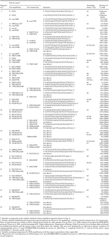
Table 1. Primers used to amplify and sequence a special segment of rDNA of Herpetosoma trypanosomes

Fig. 1. Time-course of parasitaemia by AKHA isolate of Trypanosoma grosi in jirds. Naive (closed circles; n=3) and prednisolone-treated (open circles; n=3), 8-week-old female jirds were inoculated i.p. with 2×10⁵ T. grosi (AKHA isolate), and challenged i.p. with 1×10⁶ T. grosi (SESUJI isolate) at day 32 p.i. Blood was examined at days 5, 7, 10, 13, 16, 20, 24, 32, 37 and 41 p.i. No symbol means no detectable trypanosomes in the blood. In this experiment, parasitaemia after the challenge infection was noted in 1 of 3 prednisolone-treated jirds. The same course of primary infection was seen in 6-week-old male jirds inoculated with 4×10⁴ T. grosi (AKHA isolate), albeit the level of parasitaemia was lower. Data are mean+S.D.

Fig. 2. Time-course of parasitaemia by Trypanosoma lewisi in jirds with/without prednisolone treatment. (A) Naive (closed circles; n=3) and prednisolone-treated (open circles; n=3), 10-week-old female jirds were inoculated i.p. with 5×10⁷ T. lewisi from an infected rat. At day 10 p.i., all these jirds were treated with prednisolone and challenged with 2×10⁷ T. lewisi. At day 25 p.i., jirds were re-challenged with 1×10⁷ T. lewisi without prednisolone treatment. (B) Naive 6-week-old male jirds (n=3) were inoculated i.p. with 2×10⁷ T. lewisi, and challenged with 1×10⁷ T. lewisi at day 15 p.i. These two experiments were simultaneously conducted on a time-schedule as illustrated using the same inoculum. Blood was examined at days 3, 6, 9, 13, 16, 20, 25, 28, 31, 34 and 37 p.i. No symbols means no detectable trypanosomes in the blood. Data are mean+S.D.

Fig. 3. Cross-immunity of Trypanosoma lewisi infection against Trypanosoma grosi infection in jirds. Naive 7-week-old male jirds were divided into 3 groups; Group A (closed circles, n=5) was treated at an interval of 1·5 days with rat serum. Groups B (open circles, n=5) and C (open triangles, n=3) were untreated. Groups A and B were inoculated i.p. with 1×10⁷ T. lewisi, and all groups were challenged i.p. with 2×10⁵ T. grosi (SESUJI isolate) at day 12 p.i. Blood was examined at days 3, 6, 9, 12, 16, 21 and 26 p.i. Data are mean+S.D. *P<0·05 compared with Group C.

Fig. 4. Schematic organization of rDNA of Herpetosoma trypanosomes with segments amplified in this study. The approximate regions of amplified segments are shown by bars, and an attached number indicates the combination of primers shown in Table 1. Any length shown here is not proportional to the substantial length of each rDNA region.
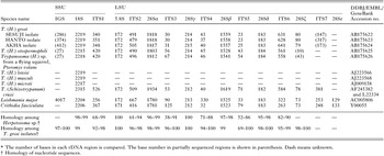
Table 2. Comparison of SSU and LSU rDNA organization of trypanosomatids including 6 Herpetosoma trypanosomes*
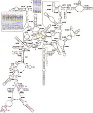
Fig. 5. Position of base changes in 18S rDNA among 3 Trypanosoma grosi isolates or among 6 species of Herpetosoma trypanosomes, plotted on the secondary structure of T. grosi (SESUJI isolate). Herpetosoma spp. compared here include T. lewisi (DDBJ/EMBL/Gen Bank Accession no. AJ009156), T. musculi (Accession no. AJ223568), T. microti (Accession no. AJ009158), T. grosi, T. otospermophili, and Trypanosoma sp. from Pteromys volans. Symbols in large letter are conserved bases among Trypanosoma cruzi (Accession no. AF245382) and all Herpetosoma spp. examined here. Symbols in small letter are bases changeable among these Trypanosoma spp.: blue-coloured letter, changed from T. cruzi; red-coloured letter, changeable among different Herpetosoma spp.; violet-coloured letter, changeable bases among T. grosi xenodemes; letter with asterisk, not restricted at the level indicated by colour but also higher level(s); and [utri ] with letter, base insertion by the level indicated by colour. Bases accompanied with yellow marks are the positions of primers used by Noyes et al. (2002).

Fig. 6. Unrooted N-J tree of 18S rDNA sequences of 6 Herpetosoma species and T. cruzi mentioned in the legend for Fig. 5, with T. rangeli (DDBJ/EMBL/GenBank Accession no. AJ009160) and L. major (Accession no. AC005806). Note that Herpetosoma trypanosomes from Murinae, Arvicolinae, and Sciuridae are divided into 2 branches, and located far distantly from other trypanosomatids including T. rangeli. Bootstrap scores expressed as percentages of 1000 replicates are given.
 You have
Access
- 23
- Cited by