Sinus of Valsalva aneurysms are uncommon congenital heart defects (CHD) accounting for < 1% of all CHD, they more commonly involve right coronary sinus (70%), non-coronary sinus (29%), and rarely involve the left coronary sinus (1%). Reference Sinha, Sujatha and Mahapatro1 The malformation arises due to an improper or incomplete fusion of the distal bulbar septum. Reference Kakos, Kilman, Williams and Hosier2
Its rupture usually presents from the second to the fourth decade of life with a wide spectrum of presentation, from asymptomatic continuous murmur to heart failure, cardiogenic shock, and death. Reference Sinha, Sujatha and Mahapatro1
Surgery has been the mainstay treatment for these CHDs, however, the morphological features of ruptured sinus of Valsalva aneurysms make them susceptible to percutaneous closure and new techniques are emerging. Reference Sinha, Sujatha and Mahapatro1
We highlight this case since there are few reports on this approach with an AmplatzerTM Duct Occluder II Additional Sizes device in children.
Case report
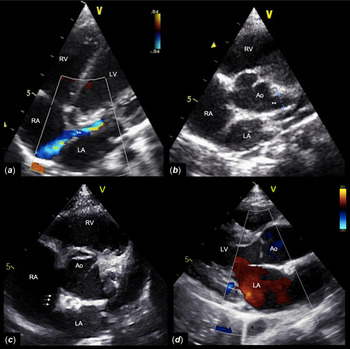
An 18-month-old full-term male with no significant past medical history was referred to our department due to an incidentally detected continuous murmur audible over the precordium. The electrocardiogram was normal. Echocardiography and colour Doppler study on admission showed an intracardiac shunt with a “wind-sock” appearance, arising from the left sinus of Valsalva and creating an inward movement towards the right atrium during systole and diastole (Fig 1a). The aortic valve was trileaflet and there was no regurgitation. The left main coronary artery had normal dimensions (Fig 2b).
Echocardiogram with colour Doppler study before ( a and b ) and 12 months after ( c and d ) the transcatheter closure of the ruptured sinus of Valsalva aneurysm. A- 5-chamber view, shows an intracardiac shunt with a “wind-sock” appearance (**) between the left sinus of Valsalva and the right atrium (RA). B - Short-axis view, shows an aneurysmatic left sinus of Valsalva (**). C - Short-axis view shows a well-placed device (arrows). D - Long-axis view shows no aortic regurgitation. Ao = Aorta; LA = left atrium; RA = right atrium; RV = right ventricle.

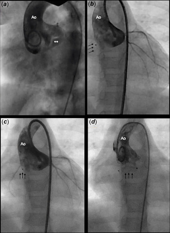
( a ) Aortogram, left anterior oblique projection, shows opacification of the right atrium through an aneurysmatic fistula (**) of the left sinus of Valsalva. ( b and c ) Aortogram, anteroposterior projection, done immediately before the definitive release of the AMPLATZERTM Duct Occluder II Additional Sizes show the device (arrows) inside the fistula with good opacification of the left coronary arteries ( b ) and the right coronary artery ( c ), without opacification of the right cavities, revealing no residual leakage. ( d ) Post- procedure control aortogram, anteroposterior projection, showing the device (arrows) in a good position with no residual shunt.

The aortogram confirmed the presence of an aneurysmatic fistula of the left sinus of Valsalva (Fig 2a). Since the mean pulmonary artery pressure on cardiac catheterisation was 22 mmHg, on the limit for normality, we decided to close the defect to prevent pulmonary overloading secondary to left-to-right cardiac shunt.
Through a 4-F end-hole catheter, a 0.035-inch exchange guide wire was passed through the fistula into the right atrium, then a continuous arteriovenous loop was created. Over the wire, a Mullins sheath was advanced across the fistula from the aortic side. A 5mm×4mm AmplatzerTM Duct Occluder II Additional Sizes device was advanced and deployed in the fistula. An angiogram using the end-hole catheter was done prior to the release of the device and the final control angiogram showed that the device was in situ with no residual leakage, no significant aortic regurgitation, or any encroachment on coronary arteries (Fig 2b and c). The patient was discharged 24 hours after the procedure with a normal electrocardiogram and completed 6 months of treatment with acetylsalicylic acid. Follow-up 12 months later revealed no residual shunt or aortic regurgitation and a progressively less pronounced dilatation of the left sinus of Valsalva (Fig 1c and d).
Discussion
Since the first report of transcatheter closure of a ruptured sinus of Valsalva aneurysm in 1994, these techniques using different devices are growing in popularity. However, no dedicated closure devices exist yet and open surgical repair remains the mainstay of treatment, mainly when ruptured aneurysms are associated with other intracardiac abnormalities. Reference Cullen, Somerville and Redington3
Although the mortality from surgery is low (<2%), some problems limit the application of surgical repair, for instance, haemodynamic instability. In fact, in patients with poor general condition and morbidities, cardiac surgery is high risk, and transcatheter closure can be life-saving. Also, it spared patients from the morbidity related to sternotomy and use of cardiopulmonary bypass. Reference Sinha, Sujatha and Mahapatro1
A wide variety of devices have been used to close ruptured sinus of Valsalva aneurysms depending on the anatomy of the lesion and the experience of each centre. The most commonly used in children has been the AmplatzerTM Duct Occluder I, with reports in the literature between the ages of 5 and 17 years. Reference Kerkar, Lanjewar and Mishra4–Reference Santoro, Pacileo and Bigazzi7 It is often favoured because its shape is suitable for closing these saccular aneurysms with high occlusion rates and stability through the retention disc and the short waist. Also, it has the distinct advantage of being user-friendly and of easy retrievability. Reference Kerkar, Lanjewar and Mishra4
AmplatzerTM Muscular Ventricular Septal Defect Occluder theoretically has the same advantages as the AmplatzerTM Duct Occluder I with even more stability through the right and left-sided umbrellas, however, there are few paediatric reports concerning the closure of ruptured sinus of Valsalva aneurysms with this device. Reference Schaeffler, Sarikouch and Peuster8 Other devices less used in children were the Coccon Duct Occluder, Reference Sinha, Sujatha and Mahapatro1 the Amplatzer Septal Occluder, Reference Arora, Rangasetty and Thakur9 and coils, which should be implemented only in cases of minor connections.
Due to the small size, coils were an option for the treatment of the ruptured aneurysm in our patient, however, they have a higher risk of embolisation, therefore, and considering the aforementioned advantages, including our familiarity with this device, we chose to use a Duct Occluder. As the AmplatzerTM Duct Occluder II Additional Sizes has a narrow waist diameter, making it less bulky, we believe that this device is more suitable for closing smaller defects like the one we report. In addition to ours, there is only one case in the literature referring to closure of a paediatric ruptured sinus of Valsalva aneurysm with an AmplatzerTM Duct Occluder II Additional Sizes in a four-year-old child. Reference Capogrosso, Santoro and Giordano10
So far, there is no commonly accepted standard for selecting the size or type of Duct Occluder. The authors chose devices 1–5 mm larger than the maximal diameter of the aortic opening of the aneurysm. Reference Kerkar, Lanjewar and Mishra4,Reference Zhao, Yan and Zhu5 In our patient, the opening diameter measured by aortic root angiography was 3 mm, so we used a 5mm×4mm AmplatzerTM Duct Occluder II Additional Sizes.
Although some concerns need to be clarified, we conclude that, in short term, transcatheter closure with the AmplatzerTM Duct Occluder II Additional Sizes was a safe and effective treatment for isolated ruptured sinus of Valsalva aneurysms and could be a cost-effective alternative to surgery in small children.
Acknowledgements
None.
Financial support
This research received no specific grant from any funding agency, commercial, or not-for-profit sectors.
Conflicts of interest
None.
Ethical standards
No specific ethical approval from Institutional Reviews Boards are necessary for this type of publication. The authors assure the patients data provided in this case report are anonymized.