Introduction
Toxic shock syndrome is a rare but life-threatening bacterial infection, named by Todd et al. in 1978.Reference Todd, Fishaut, Kapral and Welch1 It is an acute disease caused by superantigens released by bacteria (mainly Staphylococcus aureus and streptococcus spp.), and is characterised by shock and multiple organ failure. Bacterial superantigens can directly bind T lymphocytes and the major histocompatibility complex class II (MHC-II) molecules of antigen-presenting cells, causing inflammatory cytokine storms and a subsequent series of pathological processes.
Although toxic shock syndrome is more common in women who use tampons during menstrual periods, non-menstrual-associated toxic shock syndrome can also occur secondary to an operation, burns (in children), flu and other diseases.Reference Low2 The annual incidence of toxic shock syndrome is approximately 0.5 out of 100 000, and the reported incidence of non-menstrual toxic shock syndrome mortality varies from 5 per cent to 22 per cent.Reference Burnham and Kollef3 Most otologists are unfamiliar with toxic shock syndrome, whilst it is one of the few fatal complications that an ENT doctor may encounter in his or her career.
Although the relationship between toxic shock syndrome and nasal packing has been widely described,Reference Abram, Bellian, Giles and Gross4,Reference Nahass and Gocke5 only one case of toxic shock syndrome related to otological surgical procedures has been reported in the literature so far.Reference Kotlarz and Crane6 Here, we report a case of toxic shock syndrome after surgery for a discharging ear with chronic otitis media. This case emphasises the importance of early diagnosis of toxic shock syndrome, even when the patient has no signs of local infection, and the cautious surgical approach required when dealing with discharging ears.
Case report
A 43-year-old man was admitted for surgical treatment of chronic otitis media. He had a history of steady hypertension and a 10-day history of conjunctivitis. The eye infection improved with the use of levofloxacin ointment. He had no diabetes or immune deficiency, and pre-operative laboratory test results were unremarkable. Samples of ear discharge were collected for culture and sensitivity analyses. The results were not available pre-operatively.
Canal wall down mastoidectomy and tympanoplasty were performed in the discharging ear. A titanium ossicular prosthesis and tragus cartilage were used in the procedure. Gelfoam® and tela iodoform were packed into the auditory meatus and mastoid cavity. Moxifloxacin was administered pre- and post-operatively based on the local empirical antibiotic policy.
Approximately 12 hours post-operatively, the patient developed a high fever (39.5°C), vomiting and watery diarrhoea. Examination revealed bilateral conjunctival congestion deteriorating worse than before surgery, and hyperactive bowel sounds without abdominal tenderness. He had no headache or meningeal irritation signs. Given that the patient had eaten a takeaway meal, bacteraemia and gastrointestinal infection were considered, without excluding influenza or intracranial complications. He was treated with rehydration, moxifloxacin (400 mg) for suspected bacteraemia and symptomatic cooling.
Subsequent investigations showed a decreased white blood cell count of 1.92 × 10⁹/l with 83.4 per cent neutrophils, a platelet count of 121 × 109/l within normal limits, and a normal procalcitonin level of 0.053 ng/ml. The blood urea nitrogen level (225 μmol/l) and the creatinine level (7.36 mmol/l) were elevated. A slight increase was noted in the bilirubin level, whilst liver enzymes were normal. The results of stool analysis, an influenza test and blood electrolytes were unremarkable.
Five hours later, the patient developed hypotension (blood pressure of 90/53 mmHg). He looked lethargic and complained about generalised myalgia. He had only haemorrhagic effusions in the operated ear, with no redness or tenderness. A diagnosis of septic shock was considered. Aggressive fluid resuscitation and noradrenaline were delivered, to maintain blood pressure. The ear discharge culture grew methicillin-resistant Staphylococcus aureus (MRSA), and the antibacterial regimen was changed to vancomycin based on a drug sensitivity test. His condition improved over the next 2 hours, with blood pressure rising and body temperature dropping.
The patient was then transferred to the intensive care unit. He received vigorous fluid resuscitation (10 l/day), albumin, plasma therapy and intravenous immunoglobulin. The antibiotic regimen was changed to meropenem upon intensive care unit admission, and then switched to linezolid and vancomycin.
At this time, a definitive infection site had not yet been identified, so the mastoid cavity was opened for drainage. All packing and avascular graft material were removed from the cavity, and no signs of infection were discovered. However, his condition deteriorated soon afterwards, and he needed high doses of inotropes to maintain blood pressure. His blood coagulation function deteriorated, and the platelet count decreased progressively, alongside a sharp increase in the levels of blood urea nitrogen, creatinine, lactic acid and liver enzymes. The patient was intubated. Haemofiltration was commenced to alleviate the cardiac load, and remove toxins and inflammatory mediators.
Two days post-operatively, the patient developed a macular rash over his chest and abdomen. At this point, a clinical diagnosis of toxic shock syndrome could be made. The patient died of multiple organ dysfunction and ventricular fibrillation 47 hours after surgery. Multiple faecal and blood cultures all demonstrated negative results. The toxic shock syndrome process is summarised in Figure 1.
Development and progression of toxic shock syndrome. DIC = disseminated intravascular coagulation; h = hours

Discussion
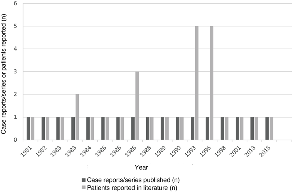
Otological surgical procedures related to toxic shock syndrome have rarely been reported. We reviewed the literature on toxic shock syndrome related to otolaryngology. As shown in Figure 2, most of the literature was published 30 years ago, and was associated with nasal surgery. This phenomenon may be linked to periodic changes in bacterial virulence.Reference Meehan, Murchan, Gavin, Drew and Cunney7 It may also reflect the incidence or current studies regarding toxic shock syndrome.
Reports of toxic shock syndrome related to otolaryngology by year.

The onset of toxic shock syndrome requires four conditions: bacteria, toxins, skin or mucosal barrier destruction, and lack of antibodies in the host. In previous reports, toxic shock syndrome was mostly found after nasal surgery in which Merocel® sponges or gauze were usually used as packing materials.Reference Abram, Bellian, Giles and Gross4 Research by Todd et al . revealed that the high carbon dioxide, anaerobic and protein-rich local environments constructed by packing materials facilitate the production of bacterial toxins.Reference Todd, Todd, Franco-Buff, Smith and Lawellin8 In the present case, the ear discharge culture grew MRSA, the Gelfoam packed in the mastoid cavity might have promoted the proliferation of bacteria and the production of toxins, and the surgical wound may have facilitated toxins entering the body. When reviewing this case, the pre-operative conjunctivitis might have been caused by MRSA and suggested the colonisation of MRSA in this patient. However, no bacteriological examination of eye discharge was performed to confirm this speculation.
• Toxic shock syndrome is a rare but life-threatening complication after surgery
• Toxic shock syndrome may not demonstrate local signs of infection
• Risk of this syndrome must be considered when performing otological surgery, especially when otorrhoea is persistent
• Prompt identification and proper treatment are vital for the survival of patients with toxic shock syndrome
The principle of a treatment approach is to maintain haemodynamic stability and, if present, to eliminate the bacteria producing superantigens. These are objectives achieved through the use of aggressive fluid resuscitation, vasoactive agents, antibiotics, and surgical abscess drainage or debridement. Early diagnosis and proper treatment are crucial to the disease prognosis. McAllister et al. reviewed the data of burned children with toxic shock syndrome, and implied that if the shock occurs before treatment is initiated, the mortality rate is as high as 11–50 per cent.Reference McAllister, Mercer, Morgan and Sanders9 If a patient survives intensive life-sustaining treatment, a ‘cytokine storm’ will result in activation-induced cell death in T cells as the body restores its balance.Reference Lussow and Macdonald10 Clindamycin and linezolid are recommended for these patients, as these can inhibit toxic protein synthesis.Reference Stevens, Ma, Salmi, McIndoo, Wallace and Bryant11
There were several distinctive characteristics in our case: acute onset, rapid deterioration, obvious gastrointestinal symptoms and the lack of local signs of infection. These characteristics could delay the diagnosis of toxic shock syndrome for relatively unfamiliar clinicians.
Several suggestions can be summarised when we consider this case. First, any suspicious signs of toxic shock syndrome should be treated as toxic shock syndrome. Patients who develop unexpected gastrointestinal symptoms shortly after surgery should raise suspicion of a potentially serious underlying cause. Although the Centers for Disease Control proposed the diagnostic criteria of toxic shock syndrome (Table 1), fully meeting these criteria seems to be impractical in a clinical setting. For the present case, the macular rash appeared 2 days post-operatively when the condition was irreversible. Second, for a discharging ear with chronic otitis media caused by MRSA, there is evidence that mastoidectomy may incur benefits for graft survival and reduction of complications.Reference Mutoh, Adachi, Tsuji, Okunaka and Sakagami12 However, in light of our experience with this case, the risk of toxic shock syndrome must be balanced when performing surgery on an ear with lasting purulent otorrhoea. The basal high level of inflammatory mediators may predispose to a ‘cytokine storm’. Third, Gelfoam and tela iodoform were used as packing materials in the procedure to provide support and haemostasis, and aid wound healing. In addition, tela iodoform has antibacterial effects. Given that packing of the mastoid cavity may be associated with toxic shock syndrome, we would recommend reducing unnecessary packing after otological surgical procedures. Remove packing materials as early as possible after surgery to reduce bacterial reproduction. In addition, topical antibiotics used according to local protocols and sensitivities may prevent surgical site infection.
Diagnostic criteria of toxic shock syndrome

CNS = central nervous system
In summary, our case reminds clinicians that toxic shock syndrome is a potentially lethal complication in patients after otological surgical procedures, especially when a patient develops unrelated symptoms, such as high fever (38.9°C or higher), vomiting, diarrhoea and rash, even though there are no local signs of infection. In addition, for discharging ears with chronic otitis media, surgery should be performed after controlling otorrhoea as much as possible. If surgery is necessary during the infection period, pre-operative bacterial culture testing and toxin identification are recommended. For patients colonised with toxin-producing bacteria, we would recommend the use of appropriate systemic and topical antibiotics, as per local protocols and sensitivities. However, these suggestions should be interpreted cautiously, and further clinical trials are still needed to confirm these findings.
Competing interests
None declared