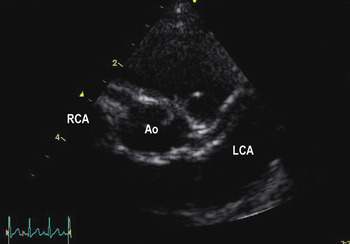
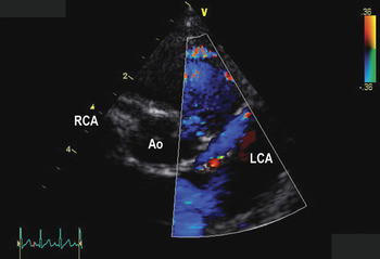
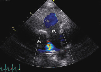
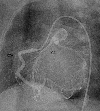
A 6-month-old male infant was referred to us after 5-months of treatment for mitral regurgitation. Echocardiography confirmed severe mitral valvar regurgitation, due to prolapse of the valvar leaflets, mild dilation of the left ventricle, the end-diastolic dimension of 33.3 millimetres being 3.6 standard deviations greater than the normal values for the population, but with preserved systolic ventricular function, the left ventricular ejection fraction being 63%. On initial examination of the short axis views (Fig. 1), the coronary arteries appeared to be normally located (abbreviations: Ao = aorta; LCA = left coronary artery; RCA = right coronary artery), albeit with mild dilation of the orifice of the right coronary artery to 2.9 millimetres, this being +3.3 standard deviations from the norm, with the diameter of the left coronary artery, at 2.5 mm, being within 1.2 standard deviations of the norm. It was only with colour Doppler flow mapping (Fig. 2) that retrograde flow in the left coronary artery revealed the true diagnosis.Reference Yang, Nanda and Wang1 When the transducer was then tilted cranially, maintaining the short axis (Fig. 3), we noted that the left coronary artery changed direction acutely alongside the aorta, and colour Doppler interrogation revealed flow into the proximal pulmonary trunk. Selective right coronary angiography (Fig. 4) then showed the flow from the right coronary artery into the left through multiple collateral vessels, confirming the diagnosis of anomalous origin of the left coronary artery from the pulmonary trunk. This experience illustrates the importance of colour Doppler flow mapping to visualize the direction of flow of blood in what might otherwise appear to be a normal coronary artery.
Figure 1

Figure 2

Figure 3

Figure 4