Non-technical Summary
This study describes an unknown fauna of extinct echinoderms from the Canadian Rockies. They were part of a complex community of echinoderms that, when fossilized, resulted in thick layers of sediment. We report new details of their anatomy paleogeography, and paleoecology. Comparisons are made with echinoderms of the same age from Montana, which are very different, indicating provinciality.
Introduction
In western Canada, Mississippian rocks occur in the Canadian Rockies in Alberta and British Columbia (see Richards et al., Reference Richards, Henderson, Hinds, Glossop and Shetson1994). The initial Mississippian deposits are the Exshaw and the Banff formations. The Banff Formation was characterized as an upward-shallowing, prograding, carbonate ramp by Chatellier (Reference Chatellier1988). Overlying the Banff Formation is the massive Pekisko or Livingstone Formation of the Rundle Group. Chatellier (Reference Chatellier1988) characterized the Exshaw-Banff intervals as representing an argillaceous recessive interval wedged between two massive carbonate sequences, the Devonian Wabamum-Palliser and the Mississippian Rundle Group. The Banff Formation has an argillaceous and rhythmic lower member, a crinoidal grainstone supported middle member, and a bioclastic argillaceous and dolomitic upper member at the top. Thicknesses range from 300–600 m.
Blastoids are widely occurring in the Banff Formation, but poorly known. We have brought together all known material to document their taxonomy, paleoecological, and paleogeographic relationships.
Records of blastoids in Canada
The oldest record of blastoids in the Banff Formation is by Shimer (Reference Shimer1926) in the Lake Minnewanka Section near Banff, Alberta. He reported a Metablastus sp. indet. Warren (Reference Warren1927) described and illustrated two very poorly preserved blastoids of two species of Pentremites Say, Reference Say1820, subgenus Strongyloblastus? Fay, Reference Fay1962, from the Rundle Limestone on Cascade Mountain and Stoney Squaw Mountain adjacent to the Banff town site.
The view of Mount Rundle south of Banff is iconic. There, the stratigraphic triad of the Palliser, Banff, and Rundle formations can be seen. Cascade Mountain and the neighboring Stoney Squaw Mountain, where Warren’s (Reference Warren1927) specimens were found, are north of Banff. Shimer’s location, Lake Minnewanka, lies northeast of Banff. The steepness of the terrain shows the difficulty of collecting at these sites.
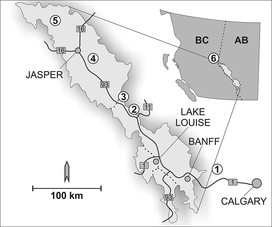
The next record of blastoids was from Mount Coleman east of the Icefields Highway, 156 km NNW of Banff. Fritz and Cline (Reference Fritz and Cline1937) described their specimens of Mesoblastus haynesi (Clark, Reference Clark1917), comparing their specimens to a blastoid described as Schizoblastus haynesi by Clark (Reference Clark1917) from southwestern Montana. From the matrix, they assigned the specimens to the Banff Formation. We have reidentified these specimens as Cryptoblastus canadensis new species (see Fig. 1 for localities).
Locality map, Provinces of Alberta and British Colombia, Canada. Enlarged inset outlines Banff National Park (51°10’40"N, 115°34’25"W) and Jasper National Park (52°52’40"N, 118°04’59"W): 1, sites near Banff (Lake Minnewanka, Cascade Mountain, and Squaw Mountain); 2, Mount Coleman; 3, Nigel Peak at Sunwapta Pass; 4, Sampson Peak at Lake Maligne; 5, Mount Simla; 6, Wapiti Lake Provincial Park.

Figure 1. Long description
A geographical map with a regional inset and a main detailed view.
Regional Inset (Top-Right): Shows the border between British Columbia (B C) and Alberta (A B). A dashed line represents the provincial border. Site 6 is located on this border, with a callout box expanding the park regions to the main map.
Main Map (Center and Left):
- A scale bar at the bottom left indicates 100 kilometers. A north arrow points upward.
- Major roads are marked with square highway shields for routes 1, 11, 16, and 93.
- Site 1 is located furthest Southeast, near Calgary and Banff, connected by Highway 1.
- Moving Northwest along Highway 93, Site 2 (Mount Coleman) and Site 3 (Nigel Peak) are positioned near the center of the park chain.
- Further Northwest, Site 4 (Sampson Peak) is located near the Jasper townsite at the junction of Highways 93 and 16.
- Site 5 (Mount Simla) is located in the far Northwest corner of the Jasper region.
- Site 6 (Wapiti Lake Provincial Park) is shown in the regional inset, situated North of the main park cluster on the B C and A B border.
In the late 1940s, Lowell Laudon, then a professor at the University of Wisconsin, began a series of studies of the Mississippian rocks of Alberta and British Columbia with a team of graduate students. Fieldwork was conducted in 1952 on Nigel Peak and Nigel 2 to the south, located at Sunwapta Pass at the Columbia Icefields, at the juncture of Banff and Jasper National Parks. They discovered a deposit of smothered crinoids on Nigel 2. Thirty genera and 52 species of crinoids were described (Laudon et al., Reference Laudon, Deidreick, Grey, Hamilton, Lewis, McBee, Spreng and Stoneburner1952). The discovery was in the Banff Formation at the northern end of Nigel 2 (see also Spreng, Reference Spreng1953, fig. 2). Blastoids listed by Laudon et al. (Reference Laudon, Deidreick, Grey, Hamilton, Lewis, McBee, Spreng and Stoneburner1952) were Metablastus sp. indet., Cryptoblastus sp. indet., and three species of Pentremites but they were not illustrated. This site is only 26 km from Mt. Coleman to the south.
In 1947, Laudon took a field party into Wapiti Lake in British Columbia, 348 km northwest of Sunwapta Pass. Field conditions were difficult because of the weather. Their publication (Laudon et al., Reference Laudon, Spreng and Stoneburner1952) described the Devonian and Mississippian stratigraphy of the Wapiti Lake area, which is now a park (Wapiti Lake Park). Illustrations in the 1949 article showed the complex thrust faulting of this area. The study area lies east of Wapiti Lake. The Banff Formation was cited as thinning southward from 212 m (697 ft) in the north to 150 m (495 ft) in the south. A new unit, the Desa Dawn Formation, was introduced for the massive limestone above the Banff Formation. This term was also used in the 1952 article (Laudon et al., Reference Laudon, Spreng and Stoneburner1952) on Sunwapta Pass. This was not recognized by Canadian geologists. Nomenclature developed at Banff to the south was used for this interval at Sunwapta Pass.
Two species of Pentremites were listed from the Banff Formation at Wapiti Lake. Pentremites was also listed from members 2 and 4 of the Desa Dawn Formation at Wapiti Lake, and said to be similar to the smaller species found in the underlying Banff Formation.
Reports of Pentremites from Sunwapta Pass, Wapiti Lake, and other places cited below could actually be one of three different blastoid species: Pentremites jasperenis new species (herein), Strongyloblastus petalus, or Strongyloblastus recurvatus new species (herein).
In the 1950s, geologists of Shell Oil undertook a regional study of Mississippian outcrops in Alberta and British Columbia. The blastoids collected by the Shell geologists were assembled from the Shell Canada collections by the late Gill Raasch of Canada. He made this material available to Macurda. It comprises 23 individual occurrences that are a part of this study. Unfortunately, the locality nomenclature was of internal Shell Canada origin. Efforts to better localize these have only been partially successful, despite inquiries at Shell Canada and the University of Alberta where the Shell Canada collections are housed.
Following the large field parties assembled by Laudon in the 1940s, individual students conducted further studies of the Mississippian at Sunwapta Pass, the Lake Maligne area southeast of Jasper, and elsewhere. These are the most detailed studies of the Mississippian of these areas. Hall (Reference Hall1952) listed and illustrated five species of Pentremites from two levels of the Rundle Limestone at Crowsnest Pass. Paul (Reference Paul1953) illustrated a ‘Pentremites sp. A’ and Orbitremites norwoodi (Owen and Shumard, 1852); the latter we assign to Globoblastus bamberi new species (herein). Paul’s (Reference Paul1953) study area was Lake Maligne. Spreng (Reference Spreng1953) found Mesoblastus haynesi (nee Cryptoblastus canadensis n. sp., herein), and Pentremites cf. Pentremites godoni (Defrance, Reference Defrance1819)—most likely Strongyloblastus petalus—at Sunwapta Pass and Mt. Coleman. Howe (Reference Howe1965) was the last of Laudon’s students to study the Mississippian of Canada. He examined the paleontology of Sunwapta Pass; two species of Pentremites were listed from the Banff Formation.
Additional occurrences of blastoids were noted by other geologists. Raasch (Reference Raasch, Goodman and Warren1958) listed blastoids from his Meramecian Member 4 of the Mount Head Formation at Beechwood Pass. Patton (Reference Patton, Goodman and Warren1958) identified Pentremites from the Meramecian succession in the South Yahani River area 500 km west of Yellowknife in the Northwest Territories. Harker (Reference Harker1963) recorded Pentremites from the Meramecian Flett Formation in the southwestern McKenzie Mountains in the Yukon. Thus, blastoids are widely distributed in western Canada, but details of their identity are lacking. Mississippian blastoids are also known from Alaska.
In 1966, Macurda and Sprinkle made a cursory examination of the Banff Formation at Sunwapta Pass on the boundary between Banff and Jasper National Parks. In 1970, under a permit granted by Parks Canada, Macurda and three graduate students from the University of Michigan studied and collected from the Banff Formation at Sunwapta Pass and exposures at or near Lake Maligne south of Jasper, Jasper National Park. We were aided by maps from Laudon. One publication resulted from this work, a redescription of the genus Strongyloblastus Fay, Reference Fay1962 (see Macurda and Breimer, Reference Macurda and and Breimer1977).
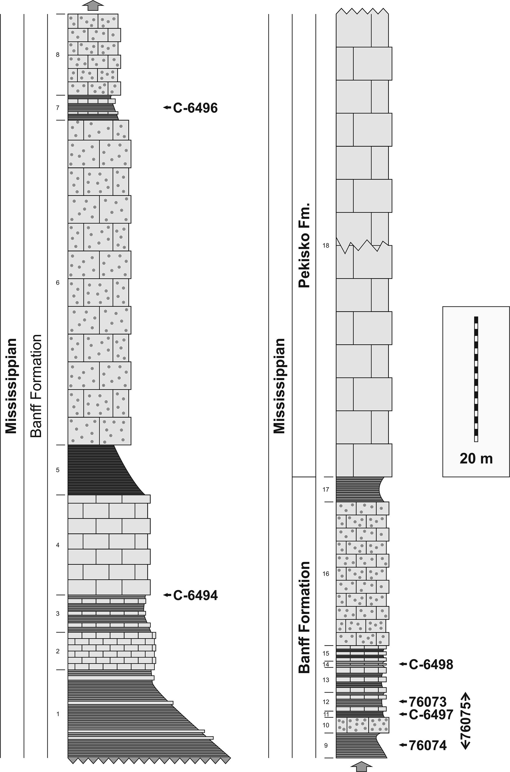
Sampson Peak, ~70 km north-northeast of Sunwapta Pass, on the eastern side of Lake Maligne, south of Four Mile Creek, is our single most important locality for the number of specimens, the diversity, and relating the blastoids to specific stratigraphic horizons. It was suggested to Macurda by Laudon (personal communication, 1966). The spur extending northward from the peak is the site of the 250 m section given in Figure 2. It is an isolated fault block. It is interpreted as the upper Banff Formation with the massive limestone at the top to be the Pekisko Formation. Stratigraphically, there is a lower section of gray shale with occasional limestone ledges. The strike is 80° southeast; the dip is 40° southwest. Except for a collection of Strongyloblastus petalus (GSC locality C-6494), blastoids were scarce in this unit. Above the massive crinoidal limestone (49 m), there is an interval in which Lophoblastus neglectus (Meek and Worthen, Reference Meek and Worthen1869) and Cryptoblastus canadensis n. sp. were found together. This co-association was repeated in the layers above. The upper part of this interval was the most prolific in terms of the number of blastoids found. GSC localities 76073 and 76074 repeated the Lophoblastus-Cryptoblastus association. Occasional Strongyloblastus petalus specimens were found with these. GSC locality 76074 is the only horizon from which a definite stratigraphic location can be established for Tholoblastus raaschi new genus new species. A definite stratigraphic level for Globoblastus bamberi n. sp. is tied down at GSC locality C-6498. Pentremites jasperensis new species occurred with Lophoblastus Rowley, Reference Rowley1901 and Cryptoblastus Etheridge and Carpenter, Reference Etheridge and Carpenter1886 in GSC locality 76074. Pentremites occurred by itself at GSC locality C-6497.
Columnar section of the Mississippian Banff Formation on the crest of the ridge running northwest from Sampson Peak above Four Mile Creek, eastern side of Maligne Lake, Jasper National Park, Alberta, Canada: Universal Coordinates 11UMJ628379, Athabaska Falls 83C12 Sheet. Collections from 1966 are indicated by a five-digit number; collections from 1970 have C as a prefix followed by four-digit number. Intervals with dashed lines represent calcareous shales, squares represent limestones, and squares with circles represent crinoidal limestones. Small numbers in the third column represent bed numbers.

Figure 2. Long description
A stratigraphic diagram consisting of two vertical columns. A scale bar to the right indicates 20 meters.
Left Column (Banff Formation):
* Base: Bed 1 consists of thick calcareous shales (dashed lines).
* Beds 2 and 3: Alternating layers of limestone (squares) and shales.
* Bed 4: A thick unit of limestone. An arrow points to collection C-6494 at the top of this bed.
* Bed 5: A layer of calcareous shale.
* Bed 6: A very thick unit of crinoidal limestone (squares with circles).
* Bed 7: A thin shale layer with an arrow pointing to collection C-6496.
* Bed 8: Crinoidal limestone at the top of the left column.
Right Column (Banff and Pekisko Formations):
* Base: Bed 9 starts with shale, labeled with collection 76074.
* Bed 10: Crinoidal limestone.
* Bed 11: Shale with collection C-6497.
* Bed 12: Shale with collection 76073. A vertical bracket to the right of beds 9 through 12 is labeled 76075.
* Beds 13, 14, and 15: Alternating thin layers of limestone and shale. Collection C-6498 is located at bed 14.
* Bed 16: A thick unit of crinoidal limestone.
* Bed 17: A shale layer marking the top of the Banff Formation.
* Bed 18: A very thick, continuous unit of limestone representing the Pekisko Formation, extending to the top of the diagram.
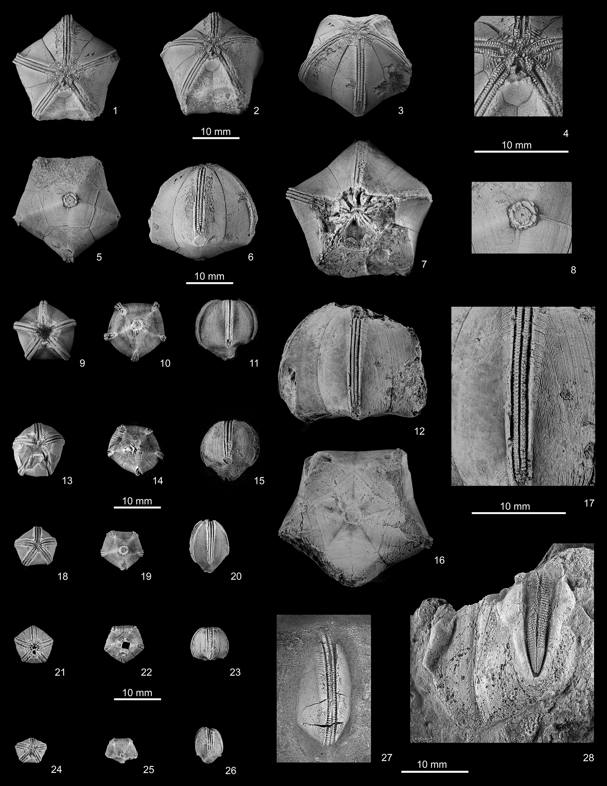
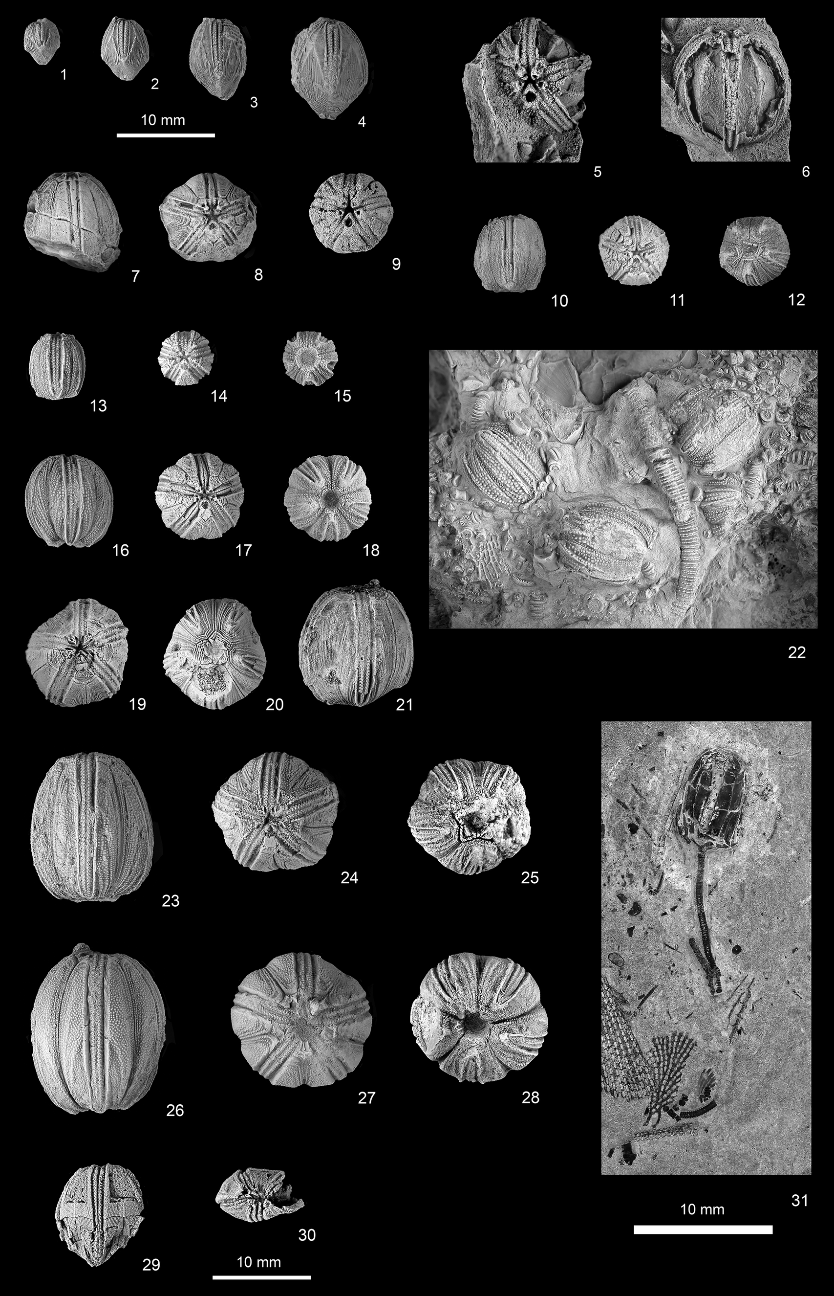
The preserved blastoids are basically isolated thecae. Stems and brachioles are not preserved except in two exceptional specimens (Fig. 3.22, 3.31).
Blastoids from Banff Formation, Lower Mississippian, Canadian Rockies, western Canada. (1–27) Tholoblastus raaschi n. gen. n. sp.: (1–6, 8) holotype, UA UA 14398, a relatively large and mostly well-preserved theca: (1) oral view showing ornamented regular deltoids, and cover plates over most of mouth and oral ambulacra; (2) oblique CD side of summit showing hypodeltoid larger than regular deltoids (but unornamented), and slight caliche at bottom of this interray; (3) oblique E-ray view showing fairly complete narrow ambulacrum and slightly damaged interray areas; (4) enlargement of (2) summit showing ornamented deltoids and larger, nearly smooth hypodeltoid; (5) basal view with large cicatrix and well-preserved basals, including small azygous basal at lower right; (6) side view of A-ray ambulacrum and radial with well-preserved growth lines and slightly convex basals; (8) enlargement of cicatrix and fine basal ornament; (7, 12, 16, 17) paratype, GSC 23199, largest specimen known but with badly damaged summit and CD interray: (7) oral view showing summit with damage extending down CD and BC sides; (12) E-ray side view showing fairly well-preserved ambulacrum, growth lines in DE interray, and slightly crushed basals; (16) basal view showing faint growth lines, slight banding on lower radials and large cicatrix in center; (17) enlargement of well-preserved E ambulacrum and right-side radial growth lines; (9–11) paratype, GSC 142985, a medium-sized, fairly well-preserved theca: (9) oral view showing projecting ambulacra, ornamented deltoids, but unfortunately depressed summit; (10) basal view showing basals and medium-sized central cicatrix; (11) side view showing globular shape, protruding basals, well-preserved thin ambulacrum, and pushed-in lower right edge; (13–15) paratype, GSC 142984, slightly smaller than (9–11): (13) slightly damaged oral view; (14) basal view showing slightly protruding ambulacra and damaged cicatrix; (15) side view showing radial ornament and projecting basals; (18–20) paratype, GSC 142983, a smaller elongate theca: (18) oral view showing well-preserved summit; (19) basal view showing basals and protruding cicatrix; (20) side view showing elongate shape, thin ambulacrum, and protruding basals; (21–23) paratype, GSC 142981, a smaller squat theca: (21) oral view showing summit with eight spiracles and anispiracle; (22) basal view showing basals except for missing azygous basal in lower right; (23) side view showing globular shape, flat basals (unusual for a small theca, possibly crushed in), and radial ornament; (24–26) paratype, GSC 142982, smallest elongate specimen: (24) slightly crushed oral view; (25) basal view with tiny cicatrix; (26) side view with protruding basals and thin ambulacrum; (27) paratype, GSC 143033, a relatively large apparent D-radial and nearly complete thin ambulacrum on a slab showing radial ornament. (28) Strongyloblastus petalus? Fay, Reference Macurda1962, GSC 143016, a very large partial blastoid with a fairly wide ambulacrum and parts of two radials with bulged edges. Scale bar at (2) for (1–3); at (4) for (4, 8); at (6) for (5–7, 12, 16); at (14) for (9–11, 13–15); at (17) for (17); at (22) for (18–26); at (28) for (27, 28).

Figure 3. Long description
A multi-panel photographic plate containing 28 numbered views of blastoid fossils.
* Panels 1 through 8 show the holotype U A 14398. 1 is an oral view with a five-pointed star shape. 2 is an oblique summit view with a 10 millimeter scale bar. 3 is an oblique E-ray view. 4 is a close-up of the summit. 5 is a basal view showing the circular cicatrix. 6 is a side view of the A-ray with a 10 millimeter scale bar. 8 is an enlargement of the basal cicatrix.
* Panels 7, 12, 16, and 17 show paratype G S C 23199. 7 is a damaged oral view. 12 is an E-ray side view. 16 is a basal view. 17 is a high-magnification view of the E ambulacrum with a 10 millimeter scale bar.
* Panels 9 through 11 show paratype G S C 142985. 9 is oral, 10 is basal, and 11 is a side view showing a globular profile.
* Panels 13 through 15 show paratype G S C 142984. 13 is oral, 14 is basal with a 10 millimeter scale bar, and 15 is a side view.
* Panels 18 through 20 show paratype G S C 142983. 18 is oral, 19 is basal, and 20 is an elongate side view.
* Panels 21 through 23 show paratype G S C 142981. 21 is oral, 22 is basal with a 10 millimeter scale bar, and 23 is a side view.
* Panels 24 through 26 show paratype G S C 142982. 24 is oral, 25 is basal, and 26 is a side view.
* Panel 27 shows paratype G S C 143033, a radial and ambulacrum on a rock slab.
* Panel 28 shows Strongyloblastus petalus, G S C 143016, a large partial specimen with a 10 millimeter scale bar.
The fossils generally exhibit pentametric symmetry, with narrow, grooved ambulacra radiating from a central point, and fine growth lines visible on the calcitic plates.
Paleoecology of the Banff blastoids
The stratigraphic section measured at Sampson Peak is interpreted to be the transition from the argillaceous deposits of the lower and middle Banff Formation into the massive limestones of the overlying Rundle Group. The lowest formation of the Rundle Group is the Pekisko Formation, which is at the top of the section in Figure 2. In the studied section, there are massive ledges of crinoidal limestone in the upper Banff Formation interspersed with argillaceous units. Most of the specimens were recovered from the latter facies.
One aspect of the Banff Formation and the Rundle Group commented on by observers is the apparent cyclic nature of the strata. Spreng (Reference Spreng1953) studied this at Sunwapta Pass, carefully measuring each bed. However, he could not find the same repetitive small-scale cycles over a distance of 20+ km; rather, he defined megasequences. We take this as a key to the environments of the carbonates versus the argillaceous sediments. Is the bedding in the Banff Formation and Rundle Group a product of cyclic or stochastic depositional processes? Bruce Wilkinson documented the development of carbonate facies in many stratigraphic intervals as being the result of stochastic rather than cyclic processes (Wilkinson et al., Reference Wilkinson, Diedrich and Drummond1996, Reference Wilkinson, Drummond and Rothman1999; Wilkinson and Drummond, Reference Wilkinson and Drummond2004).
Chatellier (Reference Chatellier1988) showed an assemblage of microfacies from the Banff Formation and overlying encrinal limestones of the Rundle Group. His figures 9, 13, and 17 showed the transition from the eastern to western domain of the Banff Formation. The Banff Formation is upward shallowing and westward stepping, as is the Rundle Group during the Lower Mississippian. The ‘crinoidal meadows’ of Chatellier (Reference Chatellier1988) produced the encrinal limestones, which are a product of mosaic development over time. The Banff blastoids were found in the western domain on the outer ramp.
In 1970, Macurda and three accompanying graduate students found an in situ deposit of crinoids with preserved stems and arms in the laterally equivalent Lodgepole Formation of Montana. Excavation of this colony showed that the peripheral members were crinoids with shorter stems but longer stemmed crinoids were in the center. The lowest members were interpreted as Tier 1 filter feeders on the periphery, whereas those in the center wwere Tier 3 filter feeders (Ausich, Reference Ausich1990). They were buried in situ. This is an example of the ‘crinoidal meadows’ of Chatellier (Reference Chatellier1988). Views of modern stemmed crinoids from submersibles show groups of crinoids with spaces between (personal communication, C. Messing, 2021).
We envision the deposition of the encrinal limestones and calcareous shales of the Banff Formation to be a result of stochastic processes. Carbonate deposition started with the localized development of crinoid gardens that were ultimately destroyed by high energy events and deposited as traction deposits. Transport was facilitated by the low specific gravity of carbonate particles. These deposits had relative continuity at the outcrop scale but not regionally. The interspersed calcareous shales in the upper Banff Formation represent the intrusion of argillaceous sediments carried by storms or currents from an external source. The upper Banff Formation represents the transitional stage from a siliciclastic-dominated to a carbonate-dominated environment.
Meyer and Waters (Reference Meyer and Waters2022) have recently suggested that there was a fundamental difference between the plankton available for filter feeders, e.g., crinoids and blastoids, between the Upper Devonian and Lower Mississippian. The concept is that the mass extinctions of the Upper Devonian eliminated the larger planktonic elements of the Upper Devonian and plankton of a smaller size were utilized in the Mississippian. The latter is the acme of stemmed pelmatozoans in the Paleozoic. Encrinites as the Burlington Limestone of Iowa, Missouri, and Illinois represent a shelf deposit that is tens of meters thick that spread over thousands of square kilometers; these rocks are comprised of almost nothing but the remains of crinoids and blastoids. The dominant particles are comprised of stem ossicles; calyces and thecae are rare. Sedimentary structures suggest tractional transport.
The blastoids of the Upper Banff Formation present at Sampson Peak are a mixture of several genera of relatively small thecae. Some associations are repetitive. Blastoids, because of their size, are interpreted as Tier 1 filter feeders, being up to 20 cm above the substratum. We did not find any preserved crinoid calyces at Sampson Peak. Laudon (personal communication to Macurda, Reference Macurda1966) noted that the main occurrence of blastoids was lower than the crinoid colony that he described from Sunwapta Pass (Laudon et al., Reference Laudon, Spreng and Stoneburner1952).
Conceivably, we envision a cluster of several rooted blastoid genera with brachiolar fans outspread in the currents. Growth lines in blastoid thecae suggest life spans of 5–10 years. Individual beds a few meters thick represent tens of years of accumulation based on a life span of this length. The blastoids flourished in the environment of the upper Banff Formation. Many blastoid gardens developed on the Banff ramp. Locations fluctuated at any given time interval; stochastically, these add up to a significant volume of rock. The limestones of the Rundle Group represent a clearer water environment with a more complex assemblage of crinoids with blastoids in the minority.
Comparative faunas
In 1990, Sprinkle and Gutschick published an account of the Early Mississippian blastoids from western Montana. They found > 1,400 specimens representing nine genera and 16 species from the Lodgepole and Allan Mountain limestones, distributed between three different zones, Specimens were found in 31 localities. This was one of the largest blastoid collections from western North America. The two lower zones are in the Paine Member of the Lodgepole Formation; the upper zone is in the Woodhurst Member of the Lodgepole. The age of both members was given as Tournaisian by Sprinkle and Gutschick (Reference Sprinkle and Gutschick1990).
It is instructive to compare the similarities and differences between the Banff and Lodgepole blastoid faunas, which lie ~1,000 km apart. The Lodgepole Formation has both fissiculate and spiraculate blastids. The former are lacking in the Banff Formation. Among the spiraculates, only the genera Cryptoblastus and Strongyloblastus were found in common. These differ at the species level. Some of the crinoids found in the Lodgepole Formation also occur in the Banff Formation (Laudon and Severson, Reference Laudon and Severson1953, p. 513).
A third major occurrence of Lower Mississippian blastoids is in the Chouteau Limestone of Missouri, ~2,000 km from those of Montana. It has a small fauna of spiraculates (Peck, Reference Peck1938) and a fissiculate (Macurda, Reference Macurda1964). Tanaoblastus Fay, Reference Fay1961a co-occurs in Montana and Missouri; they are separate at the species level.
If our assessments of the taxonomic differences at the species level are correct, a high degree of provinciality is implied. The only other major occurrences of Tournaisian blastoids is in Belgium and Ireland.
Stratigraphic age of the Banff Formation
Mamet (Reference Mamet, MacQueen, Bamber and Mamet1972), based on the microfauna (Foraminifera), and Bamber (Reference Bamber1972), based on the megafauna, were in agreement that the biostratigaphic age of the Banff Formation is Tournaisian. Chatellier (Reference Chatellier1992) illustrated the comparative foraminiferal zonation of Mamet and Skipp (Reference Mamet and Skipp1970) with the conodont biozones based on Sandberg et al. (Reference Sandberg, Ziegler, Leuterwitz and Brill1978) and Lane et al. (Reference Lane, Sandberg and Ziegler1980). The Banff Formation is in pre-zone 7 and zone 7 of Mamet and Skipp (Reference Mamet and Skipp1970) and the lower and upper Isotricha crenulata conodont biozones of Sandberg et al. (Reference Sandberg, Ziegler, Leuterwitz and Brill1978) and Lane et al. (Reference Lane, Sandberg and Ziegler1980). The age given for the Pekisko and Shunda formations was foraminiferal zones 8 and 9, and typicus and anchoraliatus conodont zones. Both are Tournaisian in age.
Richards et al. (Reference Richards, Henderson, Hinds, Glossop and Shetson1994) presented a composite correlation chart extending from southwestern Manitoba to the southwestern district of McKenzie. In the areas of the Rocky Mountains of southwestern Alberta, the age of the Banff Formation was given as Lower Tournaisian (Hasterian) and the Pekisko Formation as Upper Tournaisian (Ivorian).
Materials and methods
Measurement abbreviations
Blastoids, compared to many other echinoderms, usually have beautifully preserved growth lines on their thecal plates. New growth calcification usually occurred along the plate margins, called holoperipheral growth (Sprinkle, Reference Sprinkle1973), resulting in the growth increments of a plate being clearly visible.
A basal view of Tholoblastus raaschi n. gen. n. sp. (Fig. 3.5) illustrates the growth lines very clearly. Macurda (Reference Macurda1965a, Reference Macurda1966) developed the concept of using growth axes of a plate to record ontogenetic growth. This was clearly shown in a series of photographs in Part S of the Treatise (Macurda, Reference Macurda and Moore1968) and has been widely utilized in monographic studies of the fissiculate blastoids (Breimer and Macurda, Reference Breimer and Macurda1972; Macurda, Reference Macurda1983) and by other authors (e.g., Sprinkle and Gutschick, Reference Sprinkle and Gutschick1990).
Using the azygous basal in Figure 3.5 as an example, a growth axis as the basal-radial (BR) is measured from the origin of the plate at the stem cicatrix to the basal-radial suture. The basal-radial suture is the growth front of the BR growth axis (BRF). The area defined by BR and BRF is the BR sector. The same concept is applied to the radials and deltoids. The RB (radial-basal), RR (interradial), and RD (radial-deltoid) define the growth axes of the plate; RBF, RRF, and RDF define the growth fronts (Fig. 3.4–3.6, 3.17). Together, these form the RB, RR, and RD sectors. In the deltoid, only one growth axis is usually defined, the DR (deltoid-radial) growth axis and RDF (radial-deltoid growth front) (Fig. 3.4). When values of the growth axes and growth fronts are plotted for a series of specimens, the ontogenetic development is clearly visible.
For explanation of other abbreviations used in the text, see Macurda (Reference Macurda and Moore1968).
Repositories and institutional abbreviations
Types, figured, and other specimens examined during this study are deposited in the following institutions. Buffalo Museum of Science, Buffalo, New York, USA (BMS); Geological Survey of Canada, Ottawa (GSC); Museum of Comparative Zoology, Cambridge, Massachusetts, USA (MCZ); University of Alberta, Edmonton, Alberta, Canada (UA; UA UA indicates a holotype).
Collections made by members of the Geological Survey of Canada, the late L.R. Laudon and his students from the University of Wisconsin, Shell Oil Company geologists, and Macurda and his former students when at the University of Michigan have been brought together herein.
Systematic paleontology
Subphylum Blastozoa Sprinkle, Reference Sprinkle1973
Class Blastoidea Say, Reference Say1825 (emended by Donovan and Paul, Reference Donovan and Paul1985)
Subclass Eublastoidea Bather, Reference Bather1899
Superorder Stomatoblastida Waters and Macurda, Reference Waters and Macurda2026
Diagnosis
Blastoids possessing hydrospire pores that margin the ambulacra; new pores are added at the aboral end of the ambulacra during ontogeny. Pores bordered by side plates, outer side plates, and radials and deltoids.
Occurrence
Silurian-Permian; North America, South America, Europe, Africa, Asia, and Australia.
Order Cribroblastida new order
Diagnosis
Blastoids with hydrospire pores, ten spiracles or eight spiracles plus an anispiracle. Ten hydrospire groups.
Occurrence
Devonian-Permian; North America, Europe, and Asia.
Family Cribroblastidae new family
Diagnosis
Globose blastoids with hydrospire pores, eight spiracles, plus an anispiracle. Theca globose to oblate spheroid; ambulacra usually extending to base of theca. Deltoids short. Partial or complete exposure of lancet plate. Ten hydrospire groups, one to four hydrospires per group.
Occurrence
Mississippian-Pennsylvanian; North America, Europe, and Asia.
Genus Tholoblastus new genus
Type species
Tholoblastus raaschi n. gen. n. sp., by original designation.
Diagnosis
As for the type species by monotypy.
Occurrence
Mississippian Tournaisian; Canada.
Etymology
The name of the genus is derived from tholos, the Greek word for a dome.
Remarks
Tholoblastus n. gen. differs from other genera in the Cribroblastidae n. fam. by having four hydrospires per group, a domal (vs. a globose or oblate) spheroidal theca, two anal deltoids, an epi- and a hypodeltoid, and pores along both the radial and deltoid margins.
Other genera assigned to the Cribroblastidae n. fam. herein are: Arcuoblastus Macurda, Reference Macurda1977; Decemoblastus Macurda, Reference Macurda1977; Euryoblastus Ausich and Meyer, Reference Ausich and Meyer1988; Cribroblastus Hambach, Reference Hambach1903; Heteroblastus Etheridge and Carpenter, Reference Etheridge and Carpenter1886; and Iranoblastus Flügel, Reference Flügel1966.
Tholoblastus raaschi new genus new species
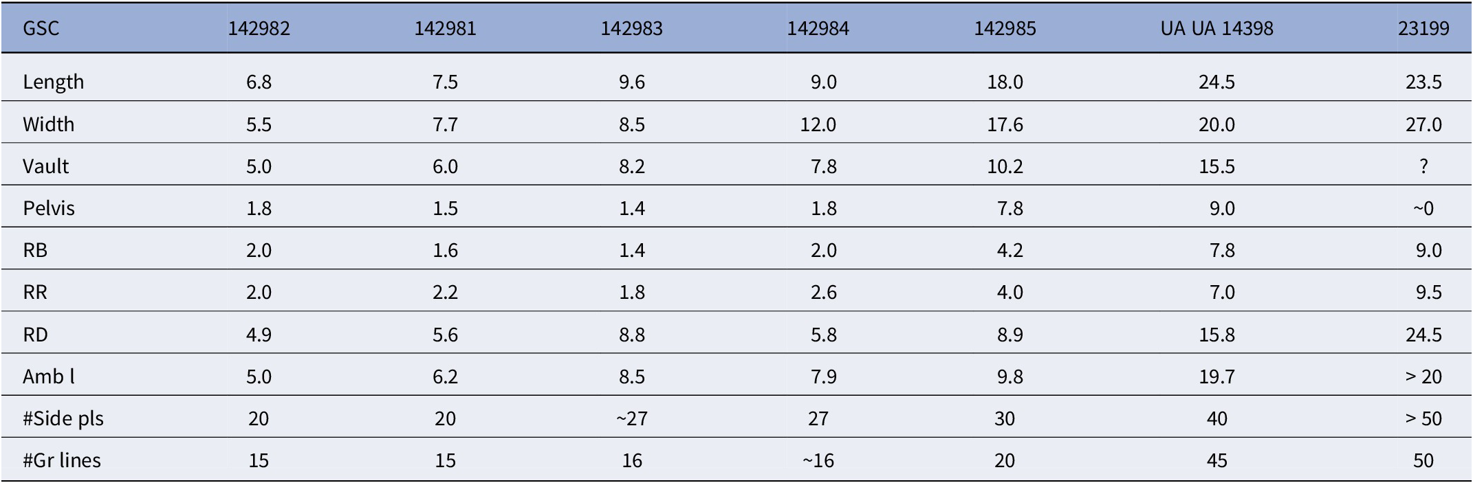
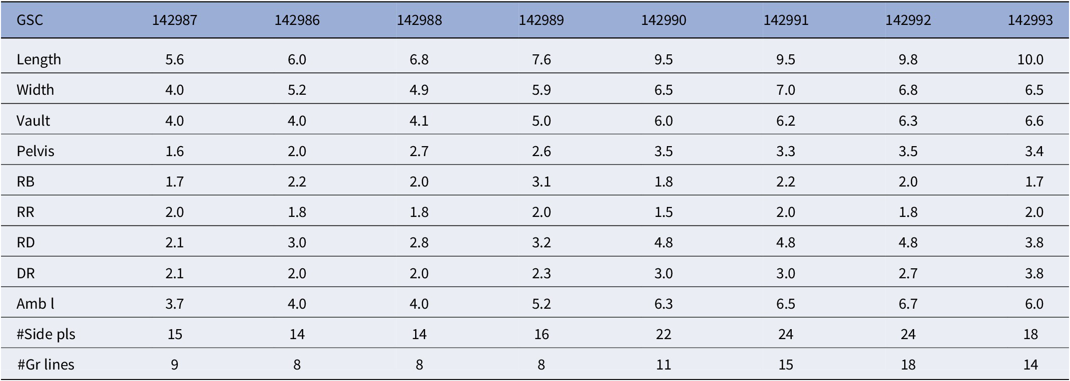
Measurements (in mm except for #) of Tholoblastus raaschi n. gen. n. sp., holotype (UA UA 14398) and paratypes (GSC 23199, 142981–142985). Amb l, ambulacral length; #Gr lines, number of growth lines; RB, radial-basal axis; RD, radial-deltoid axis; RR, radial-radial axis; #Side pls, number of side plates

Table 1. Long description
The table contains 8 columns and 11 rows. The header row lists specimen identifiers: G S C 142982, G S C 142981, G S C 142983, G S C 142984, G S C 142985, U A U A 14398 (holotype), and G S C 23199.
Measurements are in millimeters except for counts marked with #. The rows are as follows:
* Length: ranges from 6.8 to 24.5.
* Width: ranges from 5.5 to 27.0.
* Vault: ranges from 5.0 to 15.5; G S C 23199 is unknown.
* Pelvis: ranges from 1.4 to 9.0; G S C 23199 is approximately 0.
* R B (radial-basal axis): ranges from 1.4 to 9.0.
* R R (radial-radial axis): ranges from 1.8 to 9.5.
* R D (radial-deltoid axis): ranges from 4.9 to 24.5.
* Amb l (ambulacral length): ranges from 5.0 to greater than 20.
* #Side pls (number of side plates): ranges from 20 to greater than 50.
* #Gr lines (number of growth lines): ranges from 15 to 50.
Type specimens
Holotype, UA UA 14398, from Shell Canada collection, University of Alberta, Mississippian Banff Formation, Spirifer missouriensis biozone? SS1019F58; talus; length 24.5 mm; width 20.0 mm. Paratype GSC 23199, Geological Survey of Canada, Upper Banff Formation, Upper Wapiti Group, Big Horn Range, collector W.B. Brady, 1953. The Big Horn Range is in Clearwater County, Alberta. The Big Horn District occurs northeast of Banff and includes Lake Minnewanka. Paratype GSC 142981, Banff Formation, southern end of Maligne Lake along first gully along Maligne Trail, collector P. H. (Peter Harker?), 1951, M81976. Paratypes, GSC 142982–142984, GSC locality C-6496, from 4 m recessive unit just above massive limestone (see Fig. 2), collected by Macurda party, 1970. Paratype GSC 142985, northern side of Sampson Peak above Four Mile Creek, Maligne Lake, collector L.R. Laudon.
Diagnosis
Theca hemispherical in lateral view with broadly convex base. Greatest width aborally, three-quarters of thecal length. Pentagonal in oral view with ambulacra forming the points of the pentagon. Slightly concave interambulacra. Ambulacra narrow, extending far down on theca. Basals broadly convex. Lancet slightly exposed upper two-thirds of ambulacrum. Differential growth along interradial suture, more pronounced adorally. Radials slightly overlapping deltoids. Deltoids confined to summit. Anal deltoids an epi- and a hypodeltoid. Eight spiracles plus an anispiracle. Ten hydrospire groups. Four hydrospires per group. One hydrospire pore per side plate/outer side plate along radial and deltoid margins.
Occurrence
Tholoblastus raaschi n. gen. n. sp. is known only to occur in the Lower Mississippian of Alberta. Our best stratigraphic locality information is in the measured section on Sampson Peak (Fig. 2). These are specimens (paratypes) GSC 142982–142984.
Description
Theca with prominent hemispherical vault and a broadly convex pelvis. Ambulacra arcuate, extending well below midline. V:P 3:1. Theca pentalobate in oral view, with ambulacra forming the points of the pentagon. Interambulacral areas slightly concave.
Three basals (Fig. 3.5) with azygous basal in AB interambulacral area. Basals slightly convex in lateral view, with prominent stem facet formed of secondary calcite on each basal (Fig. 3.8). Cicatrix 2.0 mm in diameter, with 57–60 crenellae in outer part of the facet. Central lumen at junction of the three basals. Secondary deposit of calcite 3.5 mm wide, lobate. Azygous basal quadrate in plan view, BR sectors gently convex parallel and normal to BR axes. Very narrow BB sectors along lateral margins of plate. With ~40 slightly arcuate growth lines parallel to ABF. Width of growth lines ~0.125 mm. AB 6.0 mm; ABF 5.0 mm. Zygous basals pentagonal, with both central and lateral BR sectors gently convex parallel to ZB axes and normal to them. ZB 4.6 mm; ZBF 4.2 mm. Again, small BB sectors on lateral margins of plates. With ~40 growth lines ornamenting BR sectors.
Radials quadrate in plan view, with convex or angular lower edges, convex lateral edges, narrowing adorally (Fig. 3.6). Deep, narrow radial sinus extending four-fifths of length of plate. Radial triangular in lateral view, with a very convex upper edge, slightly recurved aboral edge, concave lower edge (Fig. 3.12). RB sectors almost straight parallel to RB axes; slight negative curvature; RB sectors convex normal to RB axes. Two sectors merging smoothly. RR sector slightly convex parallel to RR axis, strongly convex normal to RR axis. RD sector narrow, strongly convex parallel to RD axis; convex normal to it. RR sectors ornamented by fine growth lines. With ~40 per RR growth axis. Growth lines in the RR sectors fanning out adorally, due to differential rate of addition of calcite in the adoral portion of the RR sector; this allowed the vault to expand differentially (Fig. 3.17). RD sector elevated above RR sector. Aborally, the ornament is knobby; middle and upper parts with evident growth lines. Each growth line in the RD sector is twice as wide as those in the RR sector; they are more ornamented than those in the RR and RB sectors (Fig. 3.17). The growth rate per increment on the RD axis was twice that of the RR and RB axes. RB 4.0 mm; RBF 7.8 mm; RR 7.0 mm; RRF 15.0 mm (linear length); RD 15.8 mm; RDF 1.5 mm.
Deltoids rhombic, with broad V-shaped, slightly concave radiodeltoid suture. Greatest width aborally. Length 2.8 mm; width 1.3 mm. Deltoids confined to uppermost part of theca. Adoralmost part V-shaped toward peristome. Knobby, then with discontinuous transverse ornament on the adoral and medial parts of the deltoid. Faint growth lines on the aboral portion (Fig. 2.4 Slightly concave lateral edges bordering the ambulacra. Small ridges along those sides of plate that border the ambulacra.
Two anal deltoids, an epi- and a hypodeltoid. Epideltoid horseshoe shaped; V-shaped toward peristome. Prongs extending aborally under hypodeltoid leading to anal hydrospires. Facets on upper surface of epideltoid suggest an anal chimney. Length 1.3 mm; width 1.2 mm. Hypodeltoid pentagonal, with adoral edge embayed by anispiracle. Hyporadial suture convex compared to concave suture on regular deltoids. Upper surface of hypodeltoid ornamented by aborally directed convex growth lines (Fig. 3.4). Length 5.6 mm; width 3.9 mm.
Ambulacra long, convex, slightly recurved aborally (Fig. 3.17), convex in cross section. Lancet exposed in upper two-thirds of ambulacrum. Side grooves of central ambulacral tract preserved. Ambulacral length 19.7 mm (chord); width 2.3 mm. With ~40–45 side plates per ambulacral side. Each side plate/outer side plate combination 1.3 mm wide. One hydrospire pore per side plate/outer side plate. Brachiolar facets visible on D ambulacrum of holotype (UA UA 14398). Radial forming thickened rim at aboral end of ambulacrum. Oral area of holotype obscured by small cover plates over peristome and adoral parts of ambulacral tract (Fig. 3.4).
A growth series of seven specimens was studied to reveal the ontogenetic development (Table 1). GSC 142981 shows that the oral area has eight spiracles plus an anispiracle. GSC 142995 shows the presence of four hydrospires per group on each side of an ambulacrum.
Etymology
The species is named in honor of Gilbert Raasch, who did much to bring together many of the blastoids from the Geological Survey of Canada and the Shell Oil collections used in this study.
Significance of growth lines in Tholoblastus n. gen
The excellent preservation of growth lines in the holotype (Fig. 3.1) allows a semiquantitative estimate of the volume of calcite secreted by the mesoderm in a growth increment. The total length of the adoral edges of the basals is 27 mm. The total length of the edges of the radials is 210 mm. The sum of both is 237 mm. (Deltoids are not considered here because they are small and slow growing.) Each growth line is 0.15 mm wide. Using a plate thickness of 1.0 mm, 237 x 0.15 mm x 1. 0 mm yields a total of ~35 cubic mm per growth increment for the latest increment. This is a conservative estimate because ambulacra and brachioles are not included. Earlier growth increments would be proportionately smaller.
The growth rate on the RD axis is actually two times greater per increment. The RD axis is 15.8 mm. Dividing this by the number of growth lines (40) yields a value of nearly 0.4 mm per growth increment.
The number of growth lines in the radial is almost the same as the number of side plates per ambulacral side. For every growth increment, a new set of side plates, and thus a new brachiole, was added to the 10 sides of the ambulacra. As the radial grew, the origin of the radial at the end of the ambulacrum was pushed outward. This created space at the end of the ambulacrum for the addition of new side plates. There are 2.5 side plates/mm, each of which is 0.4 mm wide. This correlates to the growth increment on the RD axis of 0.4 mm/increment (discussed above). Once the side plate/outer side plate combination was formed, brachiole growth must have been fairly rapid. In most blastoids with preserved brachioles, the length of all is about equal.
Study of the growth lines in the Devonian Belocrinus Munier-Chalmas, Reference Munier-Chalmas1881, suggests the possibility of annual groups (see Macurda, Reference Macurda and Moore1968, fig. 20). Reimann (Reference Reiman1961) illustrated a specimen of the Devonian blastoid Hyperoblastus alveata (Reimann, Reference Reimann1945). His plate 2 shows color markings in the basals and radials. There are six bands in the RR sectors, each with ~12 growth lines from band to band. The six bands suggest that the blastoid is six years old. If each growth line represents a monthly addition, the holotype of Tholoblastus raaschi n. gen. n. sp. is at least four years old.
Life mode
The smaller individuals of Tholoblastus raaschi n. gen. n. sp. illustrated in Figure 3 have a conventional globose theca, suggesting that they were supported above the sea floor by their stems. The shape of the largest individuals with their flat bases might not be hydraulically stable in a current.
Remarks
The description given above is based primarily on the holotype (UA UA 14398) except where noted.
Measurements of the growth series are given in Table 1. Comments on variations in the specimens follow. The largest specimen (paratype GSC 23199, which is damaged) is shown in Figure 3.7, 3.12, 3.16, and 3.17. GSC 142985 is a smaller paratype with good locality information (Fig. 2.9–2.11). The base is more protuberant. Growth differentially inflates the vault of the theca, thus accentuating the development of the ambulacra. There are 20 growth lines in the RR sector. The hypodeltoid has a suggestion of an aboral protuberance.
Three other smaller specimens are also included (GSC 142982–142984). The smallest, GSC 142982 (Fig. 3.24–3.26), has ~15 growth lines in the RR sector. GSC 142983 (Fig. 3.18–3.20) is larger, well preserved, with 16 growth lines in the RR sector; outer side plates are visible. GSC 142984 (Fig. 3.13–3.15) has a more inflated theca; the ambulacra are more arcuate. Developmental anomalies are present in some of the upper parts of the radials in several interareas.
A definitive feature of Tholoblastus raaschi n. gen. n. sp. is the change in thecal shape from juvenile to adult specimens. Differential growth along the interradial suture inflates the vault as does rapid growth on the RD axis. This reshapes the theca from globose to a dome-shaped outline.
Family Schizoblastidae Etheridge and Carpenter, Reference Etheridge and Carpenter1886
Diagnosis
Theca bud-like or globose in lateral view; ambulacra usually extending to base of theca. Lancets exposed. Ovoid, pentagonal, or decalobate in oral view. Spiracles 10 plus one; separate from anal opening in anal interarea. Two or four anal deltoids. Deltoids short to long, usually one hydrospire pore per side plate. Two to four hydrospires per group.
Occurrence
Mississippian-Permian; North America, Asia, and Europe.
Genus Lophoblastu s Rowley, Reference Rowley1901
Type species
Codonites inopinatus Rowley and Hare, Reference Rowley and Hare1891, by original designation.
Diagnosis
Theca bipyramidal in lateral view with pelvis being shorter. Typical theca relatively small. Theca pentagonal in oral view, with ambulacra forming the points of the pentagon. Lateral view characterized by prominent stem attachment area. Basals conical, small, with prominent growth lines. Radials elongate, with strong ridge between RR and RD sectors. Prominent radial growth lines. Deltoids prominent in lateral view, occupying one-third of vault. Two anal deltoids, an epi- and ahypodeltoid, the latter hooded. Ambulacra prominent, relatively narrow. Ten spiracles plus an anispiracle. Ten hydrospire groups; three hydrospires per group.
Occurrence
Mississippian, Lower to Upper Tournaisian; North America.
Lophoblastus neglectus (Meek and Worthen, Reference Meek and Worthen1869)
Blastoids from Banff Formation, Lower Mississippian, Canadian Rockies, western Canada. (1–4) Lophoblastus neglectus (Meek and Worthen, Reference Meek and Worthen1869), GSC 142986, 142987, 142992, and 142993, small to medium-sized side views, respectively, showing growth ornament and small anal hoods. (1–12) Globoblastus bamberi n. sp.: (5, 6) holotype, UA UA SH 32494-19, oral and side views showing four spiracles and anispiracle on summit and badly damaged side view of large globular theca with thin ambulacra in small slab; (7, 8) paratype, UA SH 3337-19B, damaged side view and better oral view; (9) paratype, GSC 143020, nearly round oral view better showing four spiracles and anispiracle; (10–12) paratype, GSC 143021, side, oral, and basal views, respectively, of better preserved small specimen showing nearly flat basals. (13–28) Cryptoblastus canadensis n. sp.: (13–15) GSC 143009, small well-preserved specimen showing elongate thecal shape and slightly convex basal cavity partly filled with matrix; (16–18) holotype, GSC 143013, side, oral, and basal views, respectively, of more rounded theca showing spiracles on summit and concave basal cavity; (19–21) GSC 143017, summit, basal, and side views, respectively, showing summit and nearly flat basals; (22) GSC 143014, crowded slab of medium-sized to large thecae, three of which (lower center, upper left, and partly buried right) with short stem segments attached to their basals; (23–25) GSC 143018, fairly large theca showing side, oral, and basal views, respectively, with well-preserved summit and partial concave base; (26–28) GSC 143019, one of largest theca showing, respectively, elongate side view with projecting hypodeltoid (top left), summit view with short deltoids and depressed interray sutures, and basal view with depressed basals. (29, 30) Fragmentary blastoid, Stomastoblastida indet., UA SH 40712-19, showing side view with distinctive long deltoids, thin ambulacra, and partly crushed and damaged summit view. (31) Cryptoblastus sp. indet. (possibly), crushed black blastoid in slab, GSC 143015, with fairly long but thin attached stem plus two detached segments near bryozoans. Scale bars at (2, 14, 22, 24) and (29) are equal and apply to (1–30); at (31) for (31).

Figure 4. Long description
The image contains 31 numbered panels of blastoid fossils.
Panels 1 through 4 show side views of Lophoblastus neglectus, increasing in size from left to right, with a 10 mm scale bar positioned below panel 2.
Panels 5 through 12 display Globoblastus bamberi. Panels 5 and 6 show oral and side views of the holotype. Panels 7 and 8 show a damaged side and oral view of a paratype. Panel 9 is a round oral view. Panels 10, 11, and 12 show side, oral, and basal views of a small specimen with flat basals.
Panels 13 through 28 show Cryptoblastus canadensis. Panels 13 to 15 show an elongate specimen in side, oral, and basal views. Panels 16 to 18 show the holotype in side, oral, and basal views. Panels 19 to 21 show summit, basal, and side views. Panel 22 is a large slab containing multiple thecae with attached stem segments. Panels 23 to 25 show a large theca in side, oral, and basal views. Panels 26 to 28 show the largest specimen in elongate side, summit, and basal views.
Panels 29 and 30 show a fragmentary Stomastoblastida indet. in side and damaged summit views, with a 10 mm scale bar below.
Panel 31 shows a crushed black blastoid, possibly Cryptoblastus sp. indet., with a long thin stem attached and a separate 10 mm scale bar at the bottom right.
The fossils generally exhibit pentameral symmetry, ribbed thecal plates, and delicate ambulacral structures.
Measurements (in mm except for #) of Lophoblastus neglectus (Meek and Worthen, Reference Meek and Worthen1869); growth series of Canadian specimens. Amb l, ambulacral length; DR, deltoid-radial axis; #Gr lines, number of growth lines; RB, radial-basal axis; RD, radial-deltoid axis; RR, radial-radial axis; #Side pls, number of side plates

Table 2. Long description
The table contains 11 rows of data for eight G S C specimens identified as 142987, 142986, 142988, 142989, 142990, 142991, 142992, and 142993.
Measurements in millimeters include:
* Length: ranges from 5.6 to 10.0.
* Width: ranges from 4.0 to 7.0.
* Vault: ranges from 4.0 to 6.6.
* Pelvis: ranges from 1.6 to 3.5.
* R B (radial-basal axis): ranges from 1.7 to 3.1.
* R R (radial-radial axis): ranges from 1.5 to 2.0.
* R D (radial-deltoid axis): ranges from 2.1 to 4.8.
* D R (deltoid-radial axis): ranges from 2.0 to 3.8.
* Amb l (ambulacral length): ranges from 3.7 to 6.7.
Count data includes:
* Number of Side plates: ranges from 14 to 24.
* Number of Growth lines: ranges from 8 to 18.
Reference Meek and Worthen1869 Granatocrinus neglectus Meek and Worthen, p. 90.
Reference Meek and Worthen1873 Granatocrinus neglectus; Meek and Worthen, p. 471, pl. 9, fig. 3a, b.
Reference Rowley and Hare1891 Codonites inopinatus Rowley and Hare, p. 100, pl. 2, figs. 12, 13.
Reference Rowley1901 Lophoblastus inopinatus; Rowley, p. 344.
Reference Fay1961a Lophoblastus inopinatus; Fay, p. 75, pl. 45, figs. 1–9.
Reference Macurda1962 Lophoblastus inopinatus; Macurda, p. 370, text-fig. 3.
Reference Macurda and Moore1968 Lophoblastus neglectus; Macurda, fig. 274.1, subsequent designation of type species.
Type specimens
Lectotype, MCZ 378 (in part), Mississippian, Lower Burlington Limestone, Burlington, Iowa. The species was described from five specimens by Meek and Worthen (Reference Meek and Worthen1869). These were from Worthen’s collection, no. 396. Four are still extant. The largest of the four is designated the lectotype; the other three as paralectotypes.
Diagnosis
A species of Lophoblastus characterized by theca ovoid in lateral view, pentagonal in oral view, with prominent stem attachment and prominent growth lines on radials and basals.
Occurrence
Mississippian, Lower to Upper Tournaisian; USA and Canada. The 20 best preserved specimens of Lophoblastus came from the lower part of a 3.6 m recessive unit above a 13 m ledge in the Banff Formation on Sampson Peak above Maligne Lake, GSC locality 76074 (Fig. 2). These include those discussed in the text. Other specimens with less specific locality information are deposited in the GSC.
Description
Theca bipyramidal in lateral view, with pelvis shorter than vault. Typical theca relatively small, length 10.0 mm; width 6.5 mm; vault 6.6 mm; pelvis 3.4 mm. Theca pentagonal in oral view, with ambulacra forming the points of the pentagon. Interambulacral areas convex.
Basals three, conical in lateral profile. Prominent stem cicatrix, 1.0 mm in diameter, rimmed by strong, foliate secondary deposits of calcite. Width ~1.9 mm in diameter. Azygous basal quadrate. BR sectors convex parallel and perpendicular to BR axes. Narrow BB sectors present on lateral edges. BR axes 2.0 mm in length. Plate ornamented by ~18 strong growth lines. Zygous basals pentagonal. BR sectors convex parallel and perpendicular to BR axes. Ornament as for azygous basal.
Radial quadrate in plan view with convex lower and lateral edges. Narrow ambulacral sinus on upper two-thirds of plate. Radial triangular in lateral view, with convex adoral facing edge, straight aboral edge, and concave lower edge. RB axes straight, RB sectors convex perpendicular to RB axes. RB sectors merging over a convex surface. RR axes straight; RR sectors convex perpendicular to RR axes. Prominent growth lines in RR and RB sectors. Additional growth lines inserted in upper parts of RR sectors ontogenetically, resulting in fanning of growth lines adorally. This differentially expanded the vault. With 14 growth lines in RR sector. RD sectors triangular in plan view, with slightly convex RD axes; slightly concave perpendicular to the RD axes. Growth lines subdued compared to those of RR and RB sectors. Prominent ridge separating RD sector from RR sector. Upturned rim borderng ambulacrum. RB 1.7 mm; RR 2.0 mm; RD 3.8 mm.
Deltoid quadrate, arrowhead-shaped in plan view. Deltoid extending downward one-third length of theca. Prominent ridge with tubercles dividing each deltoid medially into two DR sectors. Ridge meeting converging ridges of radials below them. Each DR sector slightly concave in cross section as are RD sectors; merging smoothly. Narrow rim turning up against ambulacrum. DR axis 3.0 mm. Two anal deltoids, an epi- and a hypodeltoid, the latter with prominent projection along adoral edge above anal opening, producing a very characteristic hooded appearance to anal opening.
Ambulacra lanceolate, convex both lengthwise and in cross section. Adoral portion projecting above surrounding radial and deltoids. Lancet narrowly exposed along main ambulacral tract. Side grooves along main ambulacral tract and adoral edge of side ambulacral tract. Prominent ellipsoidal, downward-sloping brachiolar facets along outer margins of ambulacrum. With ~17 or 18 side plates per ambulacral side, and one pore per side plate along radial; closed along deltoid margins. Ambulacral length 6.0 mm; width 2.0 mm.
Ten spiracles around peristome on summit, plus a separate anal opening between the epideltoid and hypodeltoid. Anal opening ovoid.
Ten hydrospires groups; three hydrospires per group in Upper Tournaisian forms; presumably same in Canadian specimens.
Remarks
Lophoblastus is a small blastoid, the largest preserved specimens being 10–11 mm in length. The above description with the relevant dimension was based on specimen GSC 142993. Measurements of eight specimens (GSC 142986–142993) are given in Table 2. Features of these specimens were also incorporated into the above discussion. The ornament is very consistent from specimen to specimen. A ninth specimen (GSC 143038) clearly shows the separation between the 10 spiracles and the adjacent anal opening.
Fay’s ‘Blastoid Studies’ (Reference Fay1961a) described Lophoblastus as having eight spiracles and an anispiracle. This is different from Rowley’s (Reference Rowley1901) definition that this genus has 10 spiracles and a separate anus. Macurda (Reference Macurda1962) discussed Lophoblastus and showed Rowley’s (Reference Rowley1901) original description to be correct; the generic definition was then amended.
The tuberculate ornament between the RD and RR sectors is interpreted to have a hydrodynamic function. The blastoid would be bent to a nearly horizontal position in the currents in a Type 1 feeding posture (Breimer and Macurda, Reference Breimer and Macurda1972). The tubercles would set up turbulent eddies that would impinge on the brachiolar array, thus enhancing food-gathering opportunities, The median tuberculate ridge on the deltoids would direct the eddies across the spiracles and anal opening, to remove waste and deoxygenated water.
There is a nearly one to one correspondence of the number of growth lines in the RR sector and the number of brachioles. Each time a growth increment occurred in the radial, this provided space for the addition of a new set of side and outer side plates in the 10 ambulacra. Thus, 10 new brachioles were added to the filtration fan with each growth increment.
Morphologically, our specimens from Canada do not seem to show any differences in external form and ornamentation from those that occur in the Lower Tournaisian Burlington Limestone in Missouri (see Macurda, Reference Macurda and Moore1968, fig. 224.1). We therefore think that the Canadian and Missouri specimens belong to the same species.
Order Pentremitida Matsumoto, Reference Matsumoto1929
Diagnosis
Theca with five spiracle openings and hydrospire pores on the ambulacra.
Occurrence
Devonian-Pennsylvanian; Europe, North America, South America, and Africa.
Superfamily Pentremitoidea new superfamily
Diagnosis
Theca convex, pyriform, ellipsoidal, or godoniform. Ambulacra lanceolate to petaloid; lancet broadly exposed. Hydrospire pores on radials only or on radials and deltoids. Deltoid crest only or crest and body as well. Anal deltoids two or four. Ten hydrospire groups, two to nine hydrospires per group. Four spiracles plus an anispiracle. Some spiracles split by a deltoid septum.
Occurrence
Devonian-Pennsylvanian; North America, South America, Asia, and Australia.
Family Pentremitidae d’Orbigny, Reference Orbigny1851
Diagnosis
Theca pyriform to godoniform, pentagonal to decagonal in oral view. Ambulacra extending well down on theca. Lateral lancet growth during ontogeny. Ambulacra lanceolate to petaloid. Four spiracles plus an anispiracle (variable in oldest species). Aboral deltoid a crest, then forming a prominent deltoid body. One hydrospire pore per side plate on radial; can be absent on deltoid. Ten hydrospire groups.
Occurrence
Mississippian to Pennsylvanian; USA, Canada, Colombia, and Australia.
Genus Pentremites Say, Reference Say1820
Type species
Encrina godonii DeFrance, Reference Defrance1819, by original designation.
Diagnosis
Theca ellipsoidal, bipyramidal to godoniform. V:P varying from 1:2 to 12:1. Pelvis conical to broadly convex to nearly flat; most broader pelvises flaring outward. Vault parabolic, narrow to wide, with slightly flattened summit. Oral outline pentagonal to decagonal with slightly concave interambulacral areas. Ambulacral sinus usually long and wide, filled by lanceolate to petaloid ambulacra. Ambulacra even with or uncommonly slightly depressed below the level of radial and deltoid plates surrounding the ambulacral sinus. Ambulacra slightly convex to concave in cross section. Median portion of lancet widely exposed. RB sector usually shorter than RD sector, which is long and narrow. Deltoids four plus an epi- and a hypodeltoid. Deltoid crest continuing a short distance aboral to the spiracle, then flaring into a deltoid body that forms one-quarter to one-third of the vault. Radials overlapping deltoids; deltoid prongs extending aborally beneath the radial. Four spiracles plus an anispiracle. Hydrospire pores along radial and deltoid margins. Ten hydrospire groups, with three to nine hydrospires per group.
Occurrence
Mississippian Tournaisian to Pennsylvanian Baskirian; USA, Canada, Colombia, and Australia.
Key references
Galloway and Kaska (Reference Galloway and Kaska1957); Fay (Reference Fay1961b); Macurda (Reference Macurda1975); Waters et al. (Reference Waters, Horowitz and Macurda1985).
Pentremites jasperensis new species
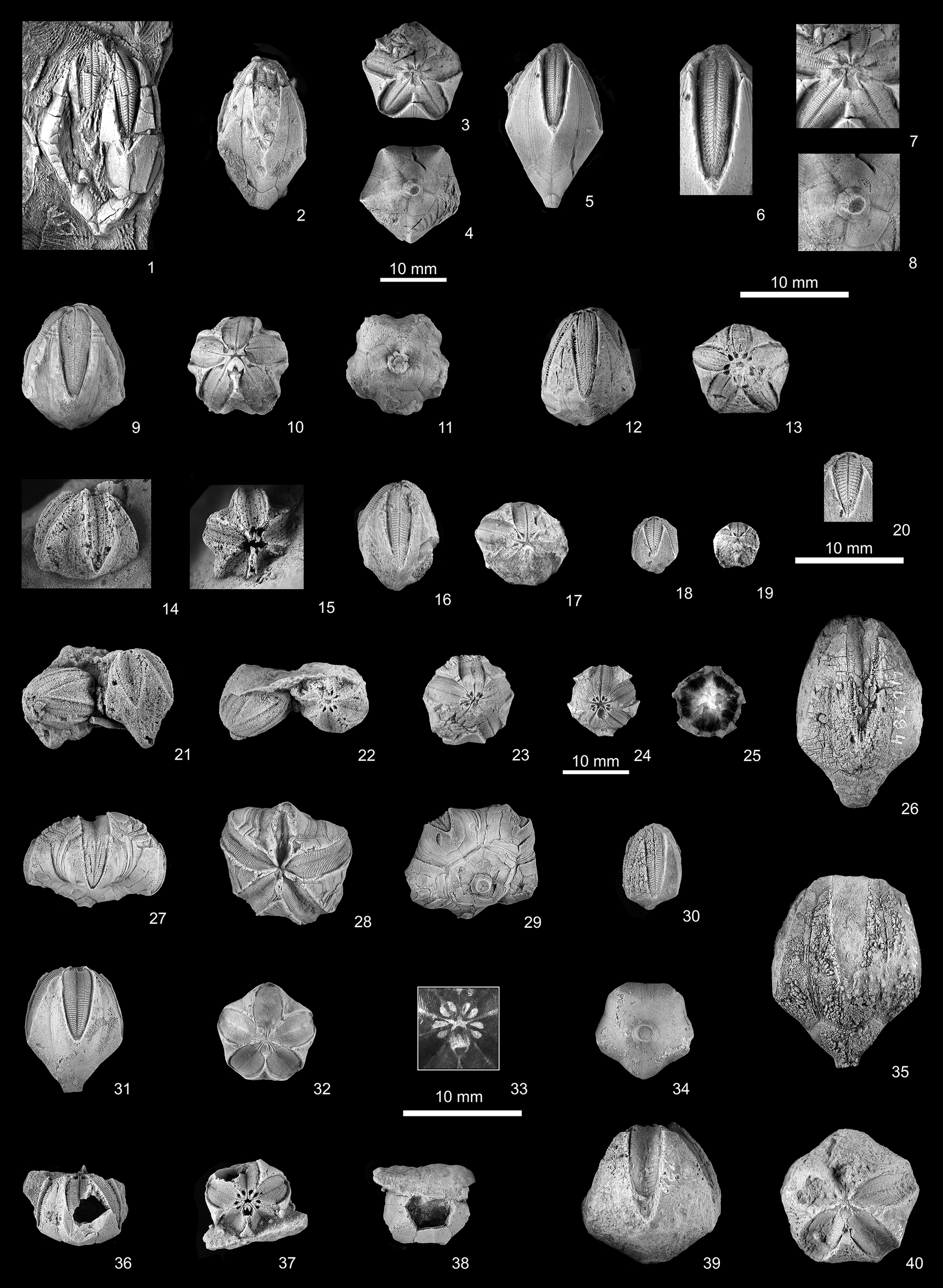
Blastoids from Banff Formation, Lower Mississippian, Canadian Rockies, western Canada. (1–8) Pentremites jasperensis n. sp.: (1) paratype, GSC 142997, a very large but crushed theca on the edge of a slab showing wide lancets and long pelvis; (2) paratype, GSC 142996, slightly damaged specimen showing broken basals and fairly long deltoids; (3–5) holotype, GSC 142994, oral, basal, and D-ray side views, respectively, showing five spiracles, short deltoids, and small cicatrix at base; (6–8) enlargements of (3–5): ambulacrum (D ray), summit with deltoid tips surrounding five spiracles, and basals surrounding cicatrix, respectively. (9–13, 16–20, 23–25) Strongyloblastus petalus Fay, Reference Macurda1962: (9–11) GSC 143022, well-preserved theca in side, summit, and basal views, respectively, showing wide ambulacra and protruding basals, 10 spiracles, and larger hypodeltoid and epideltoid surrounding anus, and indented radials, large basals, and central cicatrix; (12, 13) GSC 143023, slightly deformed godoniform side and oral views with divided spiracles on pentagonal summit; (16, 17) GSC 143025, elongate specimen in side and oral views, respectively, showing split spiracles; (18–20) GSC 143026, small specimen showing side, summit, and enlarged best ambulacrum, respectively; (23) GSC 143029, medium oral view showing slightly convex ambulacra and few paired spiracles; (24, 25) GSC 143030, sectioned theca showing paired spiracles around summit and slightly convex ambulacra in oral view, and five internal hydrospires per group in polished section. (14, 15, 21, 22, 26–29, 31–40) Strongyloblastus recurvatus n. sp.: (14, 15) GSC 143024, squat specimen in side and oral views, respectively, in small slab, with concave ambulacra and medium-length deltoids; (21, 22) two attached pyriform blastoids, in side and oral views, respectively; GSC 143027 (on right) most likely Strongyloblastus recurvatus n. sp. with divided spiracles and concave ambulacra; GSC 143028 (on left) most likely Pentremites sp. indet. with five spiracles and slightly convex ambulacra; (26) GSC 143031, large pyriform specimen in side view, showing damaged thecal plates and moderately wide but concave ambulacra; (27–29) UA P1780, somewhat squat and perhaps abnormal specimen in side, summit, and basal views, respectively, showing relatively long deltoids, larger radials, and fairly large basals with cicatrix, mostly wide concave ambulacra, except for short and very narrow upper ambulacrum in (28); (31–34) holotype, UA UA P1779, well-preserved medium-sized specimen with side, oral, summit enlargement, and basal views, respectively, showing pyriform side view with wide sightly concave ambulacra, paired spiracles when summit unsmoked, and base with fairly large cicatrix; (35) GSC 143032, very large pyriform specimen in side view, showing sutures for small deltoids, large radials, and medium basals, and mostly covered ambulacra; (36–38) GSC 143035, relatively small, somewhat damaged specimen in side, summit, and basal views, respectively, showing squat thecal shape and slightly concave ambulacra, summit with divided spiracles, and missing basals beside attached matrix; (39, 40) GSC 143036, fairly large godoniform specimen in side and oral views, respectively, showing thecal shape and wide, moderately concave ambulacra. (30) Pentremites sp. indet., GSC 143034, small but elongate specimen, similar to Pentremites elongatus Shumard, 1855, with moderately wide but slightly convex ambulacra, and a short godoniform base. Scale bars at (4) and (24) are equal and apply to (1–5, 9–19, 21–32, 34–40); at (6) for (6–8); at (20) for (20); and (33) for (33).

Figure 5. Long description
The collection is organized into seven rows of varying specimen sizes and orientations.
Row 1 (1-8): Features Pentremites jasperensis. 1 is a large crushed theca on a slab. 2 shows a side view with long deltoids. 3, 4, and 5 are oral, basal, and side views of the holotype. 6, 7, and 8 are close-up enlargements of the ambulacrum, summit, and base. A 10 mm scale bar is positioned below 3 and 4.
Row 2 (9-13): Features Strongyloblastus petalus. 9, 10, and 11 show side, summit, and basal views of specimen G S C 143022. 12 and 13 show side and oral views of a godoniform specimen. A 10 mm scale bar is located above 13.
Row 3 (14-20): 14 and 15 show Strongyloblastus recurvatus in side and oral views on a slab. 16 and 17 show an elongate specimen. 18 and 19 show a small specimen in side and summit views. 20 is an enlargement of the ambulacrum for 18, with a 10 mm scale bar below it.
Row 4 (21-26): 21 and 22 show two attached blastoids. 23 is an oral view of a medium specimen. 24 and 25 show a sectioned theca in oral view and polished section. 26 is a large pyriform specimen in side view. A 10 mm scale bar is below 24.
Row 5 (27-30): 27, 28, and 29 show side, summit, and basal views of a squat specimen. 30 is a small elongate Pentremites specimen.
Row 6 (31-35): 31, 32, and 34 show side, oral, and basal views of the holotype UA P 1779. 33 is a summit enlargement with a 10 mm scale bar below. 35 is a very large pyriform specimen in side view.
Row 7 (36-40): 36, 37, and 38 show side, summit, and basal views of a damaged squat specimen. 39 and 40 show side and oral views of a large godoniform specimen.
Type specimens
Holotype, GSC 142994, Mississippian Banff Formation, spur north of Sampson Peak, Lake Maligne, Jasper National Park, Alberta, Canada; length 23.5 mm; width 15.0 mm; V: 12.5 mm; P: 11.0 mm. Paratypes, GSC 142996 and 142997.
Diagnosis
A species of Pentremites with a bipyramidal theca; vault parabolic, pelvis conical; nearly equidimensional. Strongly pentagonal in oral view. Lanceolate ambulacra, set slightly below surrounding plates, convex in cross section. Sharp deltoid crest; very small deltoid body present only in largest individuals. RD sector confined to ambulacral sinus. Hydrospire pores lacking along deltoid crest. Ten hydrospire groups; five hydrospires in each group.
Occurrence
Mississippian Tournaisian, Banff Formation, Alberta, Canada. Our best site for collection of Pentremites jasperensis n. sp. was on the spur north of Sampson Peak at three different levels (Fig. 2): GSC localities 76074, 76073, and C 6497. In GSC localities 76073 and 76074, they co-occured with Crypyoblastus canadensis n. sp. and Lophoblastus neglectus. Specimens of Pentremites jasperensis n. sp. have also been found in other Banff exposures around Lake Maligne, but locality data is poor. Two excellent specimens of Pentremites jasperensis n. sp. are also known from Mt. Coleman, Alberta (GSC 18563).
Description
Theca bipyramidal in lateral view (Fig. 5.5), with parabolic vault and conical pelvis. Pelvis longer than vault in small specimens (e.g., length 8.5 mm; width 5.2 mm; V 3.5 mm, P 5.0 mm). Nearly equidimensional in larger specimens with larger vault (e.g., holotype, GSC 142994). Largest specimens exceeding 30 mm in length. Theca strongly pentagonal in oral view (Fig. 4.3), with ambulacra forming the points of the pentagon. Ambulacra set slightly lower than surrounding radials.
Basals three; azygous basal in normal position. Basals conical in lateral profile; pelvic angle ~45°. Pentagonal in basal view with concavities along the basal sutures. Stem attachment area covering origins of the basal plates, conical, 2.0 mm long (Fig. 5.8). Cicatrix at aboral end, 1.5 mm in diameter. Crenellae at outer edge of cicatrix, ~42 in number. Secondary deposits extending higher along interbasal sutures. Secondary calcite of stem attachment forming early in ontogeny. Relatively large in small specimens; increasing in size ontogenetically. Stem plates discoidal.
Azygous basal quadrate in plan view. BR sectors straight parallel to BR axes, convex normal to them. Sectors merging smoothly over convex surface. Surface ornamented by closely spaced growth lines. AB 6.8 mm; ABF 3.2 mm. Zygous basals pentagonal in plan view. ZB sectors straight parallel to BR axes; convex normal to them. Sectors merging over convex surfaces. Lateral sectors more convex than medial sectors. ZB 8.0 mm; ZBF 3.0 mm.
Radials five, elongate, fork-shaped with slightly convex interradial sutures in plan view. Long lanceolate sinus embaying plate adorally. Radial triangular in lateral view. Adoral edge convex, aboral facing edge straight, lower edge concave. RB sectors straight or slightly convex parallel to RB axes, gently convex normal to them. Two sectors merging over convex surface. RR sectors straight parallel to RR axes, convex normal to them. RB and RR sectors ornamented by closely spaced growth lines. Those in RB sectors fine; those in RR sectors can be more prominent. With ~33 growth lines in RR sector of holotype. Some fanning of growth lines adorally in specimens > 30 mm in length. In another individual, number of growth lines equaling number of side plates, suggesting synchronization of the development of a new set of side plates on each ambulacral side with each growth increment. RB 6.2 mm; RBF 1.6 mm; RR 4.5 mm; RRF 14.8 mm. RD sectors confined to ambulacral sinus. RD suture very narrow, on strongly sloping sides of ambulacral sinus (Fig. 4.6). Faint growth lines can be visible in RD sectors, otherwise smooth. RD 10.0 mm; RDF 0.9 mm.
Deltoids four, plus an epi- and a hypodeltoid. Adoral part of deltoid arrowhead-shaped bordering peristome. Aboral side of proximal part indented by spiracle. Trough on edge facing spiracle. Deltoid crest extending aborally, sharp, gently sloping downward. DR sectors on surfaces of adjacent ambulacral sinuses. Junction of aboral tip of deltoid crest and upper tips of radials forming a pyramid-shaped area. Deltoid crest of variable height; can completely divide the spiracle. Ambulacra extending over medial part of deltoid crest; aboral part exposed. Internally, deltoid with two prongs that extend under the radial next to the ambulacrum, as in Pentremites elongatus Shumard, Reference Shumard1855 (see Macurda, Reference Macurda1975, pl. 2, fig. 1). Small deltoid body forming adoral to the crest in largest specimens (~3.0 mm in length). Deltoid 4.0 mm long.
Anal deltoids an epi- and a hypodeltoid. Adoral part of epideltoid as for regular deltoid. Epi-hyporadial sutures visible internally in anispiracle. Hypodeltoid forming aboral edge of anispiracle. Aboral part of hypodeltoid with crest, as does regular deltoid; adoral part covered by ambulacra.
Ambulacra five, lanceolate (Fig. 5.6). Lancet broadly exposed adorally and medially, tapering toward aboral end of ambulacrum. Maximum exposed width 2.2 mm. Adoral tip of lancet extending almost to peristome. Elliptical brachiolar facets developed on side and outer side plates, > 50 side plates per ambulacral side. Well-preserved main and side ambulacral tracts. Side grooves bordering main and side ambulacral tracts; side grooves on both adoral and aboral sides of side ambulacral tract. Hydrospire pores at margin of ambulacrum on radial, between lateral edges of side and outer side plates. Pores absent along deltoid crest. Ambulacral length 14.0 mm; maximum width 4.5 mm.
Spiracles four plus anispiracle. Spiracles can be divided by deltoid crest, giving appearance of eight spiracles. Spiracles relatively large. Adoral border is an acute sharp angle; aboral border arcuate. Peristome ovoid.
Hydrospires 10 groups, five per group under each side of ambulacrum. Length of brachioles at least equals length of theca.
Etymology
After Jasper National Park, one of the places that the species was found.
Remarks
There are many reports of ‘Pentremites’ from the Banff Formation. Almost all of those we have been able to study are identifiable as belonging to the genus Strongyloblastus. The description and measurements are based on the holotype (GSC 142994). Comparative information was derived from paratypes GSC 142996 and 142997.
There is a dramatic change in the growth ratios of the RB and RD axes as the blastoid grew. In order of increasing size: RB 4.2 mm, RD 2.8 mm; RB 4.8 mm, RD 8.2 mm; RB 6.2 mm, RD 10.0 mm. It is growth on the RD axis that accelerates during ontogeny, thus providing space for additional side plates and brachioles.
The hydrospires were studied by thin sectioning 10 specimens (GSC 142999–143008). The number of hydrospires per group (five) was consistent among the specimens. The specimens are from GSC localities C-6497 and 76073 on Sampson Peak (Fig. 2).
This is the oldest known species of Pentremites sensu stricto. The pyramidal shape mimics the shape of pyramidal phaenoschismatids from which they were derived by closure of the ambulacral sinuses by the ambulacra. Waters et al. (Reference Waters, Horowitz and Macurda1985, figs. 3, 4) provided two figures that summarized the ontogeny, phylogeny, and systematics of the genus Pentremites. Their figure 3 shows the stratigraphic range of pyriform Pentremites, which are regarded as more primitive, and figure 4 shows the godoniform species, which are regarded as a more advanced form.
Pentremites kirki Hambach, Reference Hambach1903 from the Tournaisian Burlington Limestone in Iowa is an early pyramidal form. It was discussed and illustrated by Macurda (Reference Macurda1975, pl. 1, fig. 4, pl. 2, fig. 4, pl. 3, figs. 3, 9, 10). Pentremites kirki is a narrow, elongate blastoid with a sharply parabolic vault that extends to an equatorial position. It has a much more rounded profile than Pentremites jasperensis n. sp. Pentremites kirki has three hydrospires per group, whereas Pentremites jasperensis n. sp. has five. Pentremites kirki has hydrospire pores along the radial and deltoid margins, whereas they are present only on the radials in Pentremites jasperensis n. sp. This is unique to this species of Pentremites.
Family Strongyloblastidae new family
Diagnosis
Theca bud-shaped to godoniform, with wide lanceolate to petaloid ambulacra. Widely exposed lancet occupying half of ambulacral width. Anal deltoids much more pronounced than regular deltoids. Ten spiracles plus a separate anal opening.
Occurrence
Mississippian Tournaisian; USA and Canada.
Remarks
The type species of Strongyloblastus, Strongyloblastus petalus, strongly resembles a Pentremites except for two essential characteristics: 10 spiracles and a separate anal opening (as opposed to four spiracles and an anispiracle) and much more massive anal deltoids.
The development of the configuration of the spiracles in Pentremites involved the internalization of the deltoid crest within the spiracle itself. The earlier configuration was a septum separating two spiracles. One of the earliest species of Pentremites, Pentremites elongatus, has a thin septum that divides a spiracle as opposed to a complete spiracle in 20 of 29 specimens in a growth series (Macurda, Reference Macurda1975). In younger species, the septum is within the interior of the spiracle. In Strongyloblastus, there is a flattened crest that clearly separates adjacent spiracles. Waters and Horowitz (1993) defined this as a more primitive character in Strongyloblastus, a genus in transition to pentremitid morphology.
Sprinkle and Gutschick (Reference Sprinkle and Gutschick1990) suggested placing Pentremites elongatus and Pentremites kirki in Strongyloblastus. Because the deltoid septum is sharp in both and the regular and anal deltoids are very similar, these species are retained in Pentremites.
Genus Strongyloblastus Fay, Reference Fay1962
Type species
Strongyloblastus petalus Fay, Reference Fay1962, by original designation.
Diagnosis
Theca elongate ellipsoidal to godoniform in lateral view. V:P varying from 4:1 to 6.5:1. Pelvis convex, broader in godoniform species. Vault narrow to broadly convex, flattened in oral area. Oral view pentagonal to decagonal; interambulacral areas slightly concave. Ambulacral sinus long and deep, lanceolate. Ambulacra depressed below level of surrounding radials and deltoids. Ambulacra convex to concave in cross section. Lancet broadly exposed. RB axis much shorter than RD axis. Strong rugose ornament in RD section of godoniform species. Deltoids four plus an epi- and a hypodeltoid. Adoral part of deltoid bordering peristome triangular, followed by deltoid septum. Deltoid body aboral to deltoid septum. Epideltoid can be very pronounced. Eight spiracles on either side of regular deltoid septa. Spiracles in anal area can be variable. The spiracle on the D side is sometimes not as strongly exposed as that on C side, which is separate from the anal opening. Hydrospire pores one per side plate along radial; absent from deltoid except aboralmost part. Ten hydrospire groups; three to five hydrospires per group.
Occurrence
Mississippian Tournaisian; USA and Canada.
Key references
Macurda and Breimer (Reference Macurda and and Breimer1977); Sprinkle and Gutschick (Reference Sprinkle and Gutschick1990).
Remarks
The original description of Strongyloblastus by Fay (Reference Fay1962) was based on a mislabeled specimen in the Buffalo Museum of Science that was obtained from a geological supply house. It was said to be from the Devonian of New York. Using specimens collected by Macurda and his party in 1970 from the Banff Formation near Medicine Lake south of Maligne Lake, Macurda and Breimer (Reference Macurda and and Breimer1977) showed that the holotype had to be from the Mississippian of Canada. In 1990, Sprinkle and Gutschick described two additional species from the Mississippian Lodgepole Formation in Montana.
The origin of Strongyloblastus is clearly related to the origin of Pentremites. The Canadian specimens co-occurred with pyriform Pentremites. Both of these genera are part of the lanceolate-petaloid explosion of blastoids in the Famennian with very broad, exposed lancet plates. This was a new evolutionary development during the Famennian. Pentremites ranges into the Pennsylvanian and Strongyloblastus is restricted to the Tournaisian of Alberta and Montana. We think that these blastoids were derived from a fissiculate ancestor that grew broad lancet plates through lateral ontogenetic growth to fill in the deep ambulacral sinuses found in some fissiculates. Mississippian forms developed deltoid bodies, in which there is aboral growth outside the ambulacral sinus.
Strongyloblastus petalus Fay, Reference Fay1962
Figure 5.9–5.13, 5.16–5.20, 5.23–5.25
Reference Fay1962 Strongyloblastus petalus Fay, p. 135, pl. 1, fig. 1, pl. 2, figs. 1–4.
Reference Macurda and and Breimer1977 Strongyloblastus petalus; Macurda and Breimer, pl. 1, figs. 1–7, text-fig. 1.
Holotype
BMS E13297, original locality and age were errant (see above); Mississippian Banff Formation, Alberta, Canada.
Diagnosis
Theca godoniform, rounded pentagonal in oral view. Broad, convex ambulacra; broadly exposed lancet. Strongly ornamented RD sector of radial and DR sector of the deltoid, the latter with transverse ridges. Massive horseshoe-shaped epideltoid. Massive secondary basal deposits. Ten closely spaced spiracles plus a separate anal opening. Five hydrospires per group. Hydrospire pores absent on ambulacra on deltoid margins.
Occurrence
Mississippian Tournaisian, Banff Formation, Alberta, Canada. Strongyloblastus petalus is one of the more common and widely occurring blastoids in the Banff Formation. Its distribution on Sunwapta Pass was discussed earlier. Strongyloblastus was also found on Nigel Peak to the north of the pinnacle containing Laudon’s crinoid fauna (Laudon et al., Reference Laudon, Spreng and Stoneburner1952). Specimens have also occurred on the exposure north of Sampson Peak (GSC locality C-6494; Fig. 2). Strongyloblastus usually does not co-occur with other Banff blastoids. It has been found at other Banff exposures around Maligne Lake and to the north southeast of Medicine Lake (Macurda and Breimer, Reference Macurda and and Breimer1977). Individual specimens were also found during earlier work by geologists of the GSC: (a) Banff Limestone, Athabasca Point, 16 km north of Jasper, GSC locality 18566 (b) Banff Limestone, just west of highway bridge across Athabasca River, GSC locality 49965; (c) Shell Canada collection, catalog no. 18489, ‘Pentremites n. sp.’ MO5, Wapiti Pass, British Columbia, GP 38, N61, 280 ft (85.3 m), now UA SH 36088-19b. It also appears to be at Wapiti Lake in brown crinoidal limestone (Fig. 1.28), Jasper, GSC locality 18566.
Description
Theca godoniform in lateral view with ambulacra extending four-fifths of lateral view. Pelvis broadly convex, with arcuate profile with negative curvature. Vault nearly hemispherical, forming very broad dome. Theca pentalobate in oral view, with ambulacra forming the points of the pentagon. Interambulacral areas slightly convex to concave.
Basals prominent in basal view, forming half of the pelvis. Azygous basal rhombic in plan view; zygous basals pentagonal. BR sectors of azygous basal are convex parallel to and at right angles to the BR axes. Zygous basal sectors as for azygous basal. Strong growth lines paralleling upper margins of basals. Narrow BB sectors present along interbasal sutures. Prominent secondary deposits covering origins of the basals, forming petaloid pattern. Prominent stem cicatrix at basal center of secondary deposits.
Radials quadrate, with convex or angular base, convex sides, and deep lanceolate-petaloid ambulacral sinus extending almost to base of radial. Radial triangular in lateral view, with convex adoral facing edge, slightly convex aboral facing edge, and concave lower edge. RB sectors moderately convex both parallel and normal to RB axes. RR sectors straight parallel to RR axes, and convex normal to them. Growth lines paralleling margins of RB and RR sectors. RD sectors very prominent, with broad rugose growth lines or nodose deposits, forming prominent elevations that border each side of an ambulacrum.
Deltoids four, plus an epi- and a hypodeltoid. Deltoids prominent in lateral view, extending one-quarter of the length of the vault. Adoral edge of deltoid arrowhead-shaped, giving rise to prominent septum, which sets off the closely spaced adjacent spiracles. Aborally, there is a prominent deltoid body with very strong transverse ribbing, which parallels the radiodeltoid suture. Deltoid body convex axially, slightly convex in cross section. Radiodeltoid suture convex toward radial.
Two anal deltoids. Epideltoid a very prominent horseshoe-shaped plate with raised upper surface that is elevated above the peristome and anal opening. Aboral septa extending externally to hypodeltoid, separating anal spiracles from anal opening. Adoral part of hypodeltoid forming elevated concave rim to anal opening. Ornament of hypodeltoid as for regular deltoid.
Ambulacra five, lanceolate-petaloid, set below margins of neighboring radials and deltoids, extending four-fifths of length of theca. Ambulacra convex axially, with low convexity in cross section. Upper surface of lancet broadly exposed, forming medial half of ambulacrum. Laterally, hydrospire pores along ambulacral margin bordering the radial, present only on part of ambulacrum bordering aboral part of deltoid body; absent adorally above this.
Ten teardrop-shaped spiracles separate from anal opening. Ten hydrospire groups; five hydrospires per group.
Remarks
This species was discussed and described by Macurda and Breimer (Reference Macurda and and Breimer1977). Peels were made to study the interior. Statistics were given for a growth series of 10 individuals that ranged from 9.5 to 23.8 mm in length and 7.5 mm to 20.2 mm in width. The above description was drawn from that work and was provided for comparison with the description of Strongyloblastus recurvatus n. sp. (below). Comparison with the other Strongyloblastus species described by Sprinkle and Gutschick (Reference Sprinkle and Gutschick1990) from the Lodgepole Formation of Montana is given following the description of Strongyloblastus recurvatus n. sp.
Study of additional specimens revealed several details that were not previously noted. First was the recognition of the BB sector in the basals. Secondly, one specimen (GSC 143039) preserves some of the proximal discoidal columnals and ambulacral cover plates on the adoral part of the ambulacra. It also has polygonal plates above the anus, the lower part of an anal chimney.
Where preservation of radial growth lines in the RR sector extends almost to the origin of the radial at the aboral end of the ambulacrum, there is a 1:1 correspondence between the number of growth lines and the number of ambulacral side plates. Each growth increment increases the number of brachioles by a factor of 10. The spacing of the growth lines is 8 per mm. Thus, each is ~0.125 mm wide, a number present in other blastoids as well. Some growth lines are stronger than others. Some adoral fanning of growth lines occurs in the upper part of the RR sectors when there is a differential addition of calcite adorally along an individual growth line.
The stem cicatrix is relatively large in the early stages of the blastoid growth, being 1.5 mm in a specimen 10 mm in length. In the ambulacra, side grooves are developed along the main ambulacral groove and the adoral and aboral edges of the ambulacral side grooves.
Strongyloblastus petalus ? Fay, Reference Fay1962
Occurrence
Mississippian Wapiti Lake in brown crinoidal limestone (GSC 143016).
Description
One fragmentary blastoid in the material that we have studied is larger than any others. It consists of two partial radials with the ambulacra. Preserved length of the radials is 27 mm; width of the specimen is ~30 mm. The RB sector is well developed on the left radial. The RB axis is ~10 mm in length. The RB sector is convex both parallel and normal to the RB axis. The RR sector is gently convex both axially and normal to the RR axis, the length of which is 7 mm. The preserved RD sector is ~15 mm in length. It is characterized by a massive buildup of calcite surrounding the ambulacrum that widens abruptly adorally. Growth lines are present in the massive deposits. Lanceolate ambulacra are set below this massive rim. Maximum preserved ambulacral length is 16 mm; maximum width is 5 mm. The lancet is exposed in the medial part of the ambulacrum. Side plates are 3 per mm, bordered on their outer edge by hydrospire pores.
Remarks
We think that this is a hyperadult specimen of Strongyloblastus petalus, larger than any other known individual. Macurda and Breimer’s (Reference Macurda and and Breimer1977, pl. 1, figs. 2, 4) lateral views of two specimens of Strongyloblastus petalus show the massive buildups in the RD sectors with growth lines present. The RR and RB axes of the specimen in figure 4 are 5 mm and 7 mm, respectively. In this individual, these values are 7 m and 10 mm, respectively. The length of the individual in Macurda and Breimer’s (Reference Macurda and and Breimer1977) figure 4 is 21 mm. The ratio of the RB and RR axes is 1:1.4. Applying this to the partial specimen suggests a possible length of > 30 mm and a similar width.
Strongyloblastus recurvatus new species
Holotype
UA UA P1779 (Fig. 5.31–5.34), Shell Canada collection, no. 18490, locality MO(5), Wapiti Pass, British Columbia, GP 38, N 61, 290 feet (88.4 m), Mississippian. Measurements in the species description are from this specimen.
Diagnosis
Theca godoniform or bud-shaped. Ambulacra broad, concave in cross section. Lancet broadly exposed. Growth lines with equal expression in all radial sectors. Ten closely spaced spiracles plus separate anal opening. Ten hydrospire groups; four hydrospires per group.
Occurrence
Mississippian Tournaisian, Banff Formation; Rundle Formation, Alberta, British Columbia, Canada.
Description
Theca godoniform or bud-shaped, with vault and pelvis of equal length in lateral view. Pelvis conical. Vault parabolic in lateral profile; deltoid crests highest points on theca. Theca ovoid, pentagonal in oral view with ambulacra forming the points of the pentagon. Ambulacra concave in cross section. Interambulacral areas slightly concave. Protuberant stem attachment at base of theca, comprised of three parts of secondary calcite covering origins of the basals, 4.0 mm in diameter. Stem cicatrix at base, 2.0 mm in diameter, with 54 crenellae at outer edge of cicatrix. Pelvic profile above secondary deposit conical, with straight edges; pelvic angle 80°.
Basals three, in normal position. Azygous basal quadrate; zygous basals pentagonal in plan view. Azygous basal sectors straight axially; convex in cross section. Zygous basal sectors straight parallel to ZB axes; convex normal to them. Central sectors greater in convexity than lateral sectors. Very fine growth lines ornamenting basals parallel to BR sutures. AB 4.9 mm; ABF 3.8 mm; ZB 5.0 mm; ZBF 3.0 mm.
Radials five, quadrate in plan view with convex lateral edges and convex or angular lower edges. Deep parabolic ambulacral sinus occupyng two-thirds of profile. Radial triangular in lateral view, with concave lower edge, convex upper adoral edge, and straight upper aboral facing edge. RB sectors straight parallel to RB axes, convex normal to them. Adjacent sectors merging over convex surface. RR sectors gently convex parallel to RR axes, convex normal to them. RB and RR sectors ornamented by fine growth lines. RD sector narrow, convex parallel to RD axis, slightly convex normal to it. RD sector ornamented by low, lumpy, irregular ornament. RB 5.0 mm; RBF 2.9 mm; RR 4.0 mm; RRF 10.0 mm; RD 8.3 mm; RDF 1.1 mm.
Deltoids four, together with epideltoid forming border to peristome. Adoral part of deltoid arrowhead-shaped with rim facing peristome. Sharp prominent crest extending aborally, separating two adjacent spiracles. Crest extending aboral to spiracles and flattening to form deltoid body that slopes sharply downward, forming upper lateral profile of vault. Deltoid body concave in cross section, ornamented by growth lines. Radial overlapping deltoid. Deltoid length 7.0 mm. Deltoid crest 2.8 mm. Deltoid body length 4.0 mm, width 1.8 mm.
Anal deltoids two, an epi- and a hypodeltoid. Adoral part of epideltoid as for regular deltoid. Two-bladed prongs extending aborally, separating spiracles from anal opening. Adoral part of hypodeltoid concave, elevated to form backstop to the anal opening. Adorally, a spine extending 1.8 mm before broadening into a rhombic hypodeltoid body. Epideltoid length 1.8 mm. Hypodeltoid length 5.2 mm; width 2.1 mm.
Ambulacra five, broadly lanceolate petaloid in plan view, concave in cross section. Set below surrounding radial and deltoids. Lancet prominently exposed in middle half of ambulacrum. Minute side grooves ornamenting main ambulacral tract. Side ambulacral grooves slightly sinusoidal. Brachiolar facets at outer edges of ambulacrum. Ambulacral length 10.3 mm; width 4.1 mm. Maximum width of exposed lancet 2.2 mm. Side plates 3 per mm. Hydrospire pores one per side plate along margin with radial, absent along margin with deltoid.
Oral openings 10 spiracles plus anal opening. Spiracles teardrop-shaped, widest adorally. Length 1.0 mm; width 0.5 mm. Length of anal opening 1.2 mm, width 1.5 mm. Ten hydrospire groups; four hydrospires per group.
Etymology
The new species name is derived from the Latin recurvous, meaning bent back. This captures the negative concave profile of the ambulacra, differing from other species in the genus.
Remarks
The new species has the generic characteristics of Strongyloblastus but with one salient difference: concave ambulacra as opposed to convex as in Strongyloblastus petalus, and two species from the Lodgepole Limestone in Montana: Strongyloblastus breimeri Sprinkle and Gutschick, Reference Sprinkle and Gutschick1990 and Strongyloblastus laudoni Sprinkle and Gutschick, Reference Sprinkle and Gutschick1990; and also Strongyloblastus sp. indet. of Sprinkle and Gutschick, Reference Sprinkle and Gutschick1990 (see Sprinkle and Gutschick, Reference Sprinkle and Gutschick1990, pl. 6, figs. 1–32).
The development of concave ambulacra in Strongyloblastus recurvatus n. sp. is a fundamental morphological difference from the other species of Strongyloblastus. Similar differences exist in Pentremites in some of the godoniform species (e.g., Pentremites spicatus Ulrich, Reference Ulrich1917 and Pentremites sulcatus Roemer, Reference Roemer1851) that have concave ambulacra (see Galloway and Kaska, Reference Galloway and Kaska1957 and Waters et al., Reference Waters, Horowitz and Macurda1985 for these comparative forms).
A second specimen (UA P1780, Shell Canada collection no. 18493; Fig. 4.27–4.29) is assigned to this species. It is from Wapiti Pass, with the same GP and M coordinates, 420 ft (128.0 m). The specimen has been flattened diagenetically. It is similar in size to the holotype. The deltoid crests are much more pronounced than in the holotype. The A ambulacrum is not fully developed (6.7 mm vs. ~12.0 mm), and the AB and EA deltoids are asymmetric. The deltoid bodies are larger. The peristome has been pushed downward, and oral details are not clear. Ambulacral details and growth lines are well preserved. Those on the basals and RB and RR sectors of the radials are often very broad. Some granulose ornament is preserved on the inner parts of the basals.
A third specimen (GSC 143035; Fig. 5.36–5.38) is completely silicified, and the basals are missing. It is smaller than the previous two (12.5 mm width; 8.0 mm ambulacral length). The oral details are very clear. The locality is given as Penny Mountain, southwestern fork of the Narraway River, LS 20923, F 60, 435–460 ft (132.6–140.2 m).
In 1961, Eric Mountjoy collected 14 blastoids from the Rundle Formation on the southwestern ridge of Mt. Simla, Alberta. This is a wilderness peak at 53°16’48.04"N, 118°39’18.00"W, northeast of Mt. Robson. The specimens are now in the Geological Survey of Canada, field number MJ-355, catalog no. 46784. The specimens are partially silicified and encased in a siliceous siltstone. The godoniform shape is clear in 13 specimens (e.g., Fig. 5.26, 5.35, 5.39). One specimen was weathered enough to clearly establish its identity. The specimen has 10 spiracles, a separate anal opening, and lacks hydrospire pores on the margins of the ambulacra by the deltoids. We assign the 13 specimens to Strongyloblastus recurvatus n. sp. Some comparative growth measurements of GSC 143036 (Fig. 4.39, 4.40) are: length 21.7 mm; width 20.2 mm; RB 6.8 mm; RBF 4.8 mm; RR 5.0 mm; RRF 16.3 mm; RD 15.1 mm; RDF 1.8 mm. Deltoid length 8.0 mm; ambulacral length 17.0 mm; maximum width 4.5 mm. GSC 143035 exposes four hydrospire slits at the end of an ambulacrum.
The fourteenth specimen (Pentremites sp. indet., GSC 143034; Fig. 3.30) has a different thecal form. It has an ellipsoidal theca and convex ambulacra. Length 13.4 mm; width 9.5 mm. It resembles a small Pentremites elongatus but is not well preserved.
The occurrence of the specimens of Strongyloblastus recurvatus n. sp. in the Rundle Formation is important stratigraphically. The age of the Banff Formation containing Strongyloblastus petalus and the two species of Strongyloblastus from Montana (Sprinkle and Gutschick, Reference Sprinkle and Gutschick1990) is Tournaisian. Mountjoy (Reference Mountjoy1962) published a paper on the Mount Robson Southeast Map Area (83 SE/SE). He divided the Rundle Group into four formations. He gave the thickness of the Rundle Group on Mount Simla as ~300 m. It is not known from which of the four formations he collected his specimens in the Rundle Group. Petryk et al. (Reference Petryk, Mamet and MacQueen1970) discussed the age of the Mount Rundle Group in southwestern Alberta. The age given for the Mount Rundle Group ranges from Tournaisian to Lower Visean. This occurrence is possibly a range extension for Strongyloblastus.
As mentioned earlier, Wapiti Pass, the locality given for the holotype of Strongyloblastus recurvatus n. sp. is inexact. Laudon et al. (1949) mapped the nearby Wapiti Lake geology. They recognized three formations in the Mississippian: Banff, Dessa Dawn, and Rundle. They collected ‘Pentremites species’ from each of the three units. The whereabouts of these specimens is unknown; they are probably a combination of specimens of Pentremites sensu stricto and Strongyloblastus.
Superorder Tubuloblastida Waters and Macurda, Reference Waters and Macurda2026
Diagnosis
Blastoids with hydrospire tubules, which provide entrance to the hydrospires, formed along the radiodeltoid sutures, penetrating radial or radial and deltoid plates.
Occurrence
Devonian to Mississippian; North America, Europe, and Asia.
Order Granatocrinida Bather, Reference Bather1899
Diagnosis
Thecae globose to bell-shaped. Eight spiracles plus an anispiracle. Two to four anal deltoids. Ten hydrospire groups, one to four hydrospires per group. Hydrospire tubules in radials, occasionally in deltoids. Part of lancet usually exposed.
Occurrence
Mississippian Tournaisian to Namurian; North America, Europe, and Asia.
Family Granatocrinidae Fay, Reference Fay1961a (sensu stricto)
Diagnosis
Thecae globose; eight spiracles plus an anispiracle. Ten hydrospire groups, one to four hydrospires per group. Hydrospire tubules in radials, occasionally in deltoids. Part of lancet usually exposed.
Occurrence
Mississippian Tournaisian to Viséan; North America and Europe.
Family Nodoblastidae new family
Diagnosis
Thecae bell-shaped, with flat base; eight spiracles plus an anispiracle. Deltoids with median crest, longer than radials. Ten hydrospire groups, one hydrospire per group. Two anal deltoids. Hydrospire tubules in radials only. Lancet partly exposed.
Occurrence
Mississippian Namurian; Asia: Kazakhstan.
Remarks
This family contains the single genus Nodoblastus Fay, Reference Fay1963, which differs from other Granatocrinidae genera in thecal shape, long deltoids, hydrospire tubules in the radials only, and one hydrospire per group.
Genus Cryptoblastus Etheridge and Carpenter, Reference Etheridge and Carpenter1886
Type species
Pentremites melo Owen and Shumard, Reference Owen and Shumard1850, by original designation.
Diagnosis
Theca ellipsoidal-globose in lateral view, ovoid pentagonal in oral view, with ambulacra forming the points of the pentagon. Ambulacra narrow, extending to base of theca. Basals concave. Lancet exposed except at aboral end of ambulacrum. Deltoids small, confined to upper surface of theca. Radials overlapping deltoids. Four anal deltoids, an epi-, two crypto-, and a hypodeltoid. Ten hydrospire groups, two hydrospires per group. Absent on internal surface of deltoids. Hydrospire tubules piercing radial; approximately two tubules per side plate. Tubules absent on deltoids. Usually granular ornamentation on growth lines.
Occurrence
Mississippian Tournaisian and Lower Viséan; USA and Canada.
Key references
Cline (Reference Cline1937); Fay (Reference Fay1961a); Macurda (Reference Macurda1962).
Remarks
In his 1961 description of Cryptoblastus, Fay (Reference Fay1961a) had some uncertainty about whether cryptodeltoids were present in the type species, Cryptoblastus melo. Macurda (Reference Macurda1962, fig. 2) illustrated a definitive specimen showing the internal cryptodeltoids in Cryptoblastus melo. Fay and Wanner (Reference Fay, Wanner and Moore1968) defined the genus Cryptoblastus as having four anal deltoids.
Cryptoblastus canadensis new species
Measurements (in mm except for #) of Cryptoblastus canadensis n. sp.; growth series of specimens drawn from a collection of 50 specimens from GSC locality 76074. Amb l, ambulacral length; Amb w, ambulacral width; Del l, deltoid length; DR, deltoid-radial axis; RB, radial-basal axis; RBF, radial-basal growth front; RD, radial-deltoid axis; RDF, radial-deltoid growth front; RR, radial-radial axis; RRF, radial-radial growth front; #Side pls, number of side plates

Table 3. Long description
The table contains 14 rows of measurements for G S C specimens 143009, 143010, 143011, 143012, and 143013. All measurements are in millimeters except for the number of side plates.
* Length: 6.8, 8.5, 8.0, 9.3, 9.2.
* Width: 5.3, 6.0, 7.5, 7.5, 9.0.
* Basal Area: 2.1, 2.0, 1.5, 2.0, 1.8.
* R B (radial-basal axis): 2.0, 1.5, 1.3, 2.0, 1.5.
* R B F (radial-basal growth front): 1.7, 1.3, 1.5, 1.2, 1.3.
* R R (radial-radial axis): 2.0, 1.8, 1.5, 1.3, 1.5.
* R R F (radial-radial growth front): 5.8, 6.0, 8.0, 7.5, 8.0.
* R D (radial-deltoid axis): 5.5, 6.8, 7.5, 7.5, 8.2.
* R D F (radial-deltoid growth front): 1.0, 1.3, 1.0, 1.1, 1.0.
* D R (deltoid-radial axis): 1.0, 0.8, 1.5, 1.5, 1.2.
* Del l (deltoid length): 1.8, 1.5, 1.8, 1.8, 2.0.
* Amb l (ambulacral length): 5.5, 6.5, 8.5, 8.0, 8.8.
* Amb w (ambulacral width): 1.0, 1.0, 1.0, 1.0, 1.2.
* #Side pls (number of side plates): 23, 26, 20, 29, 34.
Holotype
GSC 143013, Mississippian Banff Formation, Sampson Peak above Lake Maligne, Jasper National Park, Alberta, GSC locality 76074; 9.2 mm in length, 9.0 mm in width.
Diagnosis
A species of Cryptoblastus in which the interradial and radiodeltoid sutures are less depressed than those of the type species, Cryptoblastus melo. In that species, the plates in the RR sectors are thick and robust, whereas they are thinner in Cryptoblastus canadensis n. sp. In the latter, the ambulacra are not depressed below the surrounding plates. The ornament is also finer.
Occurrence
Mississippian Tournaisian; Alberta, Canada. Cryptoblastus canadensis n. sp. has been found at several localities in the Banff Formation. The most prolific occurrence is in the section on the northern side of Sampson’s Peak (Fig. 2) where it occurs at several levels (GSC localities 76073, 76074, and C-6498). Lophoblastus neglectus is a commonly co-occurring blastoid at these same levels.
Cryptoblastus canadensis n. sp. is found at several levels at Sunwapta Pass where Laudon et al. (Reference Laudon, Spreng and Stoneburner1952) discovered the crinoids that were from a smothered crinoid community. Specimens from GSC locality 76078 occur below the crinoid zones. Specimens were also collected by Laudon and his group from the Lower Crinoid Zone (GSC 143041) and the Middle Crinoid Zone (GSC 143030). Brachioles were preserved in the latter example. Specimens were also found on Nigel Peak to the north. There are scattered individuals that have been found at other localities around Maligne and Medicine Lakes.
Fritz and Cline (Reference Fritz and Cline1937) described Mesoblastus haynesi (i.e., Cryptoblastus canadensis n. sp.) from Mt. Coleman, south of Sunwapta Pass (see following discussion).
Sprinkle and Gutschick (Reference Sprinkle and Gutschick1990) described three different Cryptoblastus from the Lodgepole Limestone in Montana as Cryptoblastus A, B, and C. None of these appear co-specific with Cryptoblastus canadensis n. sp.
Description
Theca oblate spheroid in lateral views, becoming more laterally inflated in larger specimens. Longer than wide. Summit rounded; base flatter with slightly concave profile. Ambulacra reaching very base of lateral profile. In oral view, theca rounded pentagonal. Radial plates are the most protuberant part of the profile, indented along interradial sutures. Studied specimens range from 6.8–9.2 mm in length; 5.3–9.0 mm in width.
Profile in basal view as for oral view. Basals in a small, concave depression, 1.8 mm in diameter. Ambulacra curvng inward toward depression. Basals three, occupying innermost part of basal concavity. Azygous basal quadrate; BR sectors concave parallel and normal to BR axes. Zygous basals pentagonal in plan view; BR sectors concave parallel and normal to BR axes although medial sectors are flatter. Small lumen at center of junction of the three basals.
Stem composed of two types of discoidal plates that alternate with each other. One is thicker and wider; the other is narrower and has a sharp keel. Crenellae are located on the upper and lower surfaces of the stem plates near the edges of the plates.
Radials quadrate in plan view, with narrow upper and lower edges, and convex lateral edges, embayed to the base by narrow ambulacral sinus. In lateral view, radials very arcuate, with convex upper edge, concave lower edge, and straight aboral facing edge. RB sectors highly arcuate, bending into basal concavity. Sharp edge in RB sectors usually bordering basal concavity. RB sectors convex parallel to RB axes outside basal concavity, convex normal to them. Rugose growth lines ornamenting RB sectors outside basal concavity. Width of basal concavity expanding ontogenetically. RR sectors straight parallel to RR axes, convex normal to them. Growth lines in RR sectors highly arcuate, recording strong upward and outward growth of theca ontogenetically. With 15 growth lines in each RR sector of holotype. Each growth line stacking adorally onto its predecessor; youngest ones present only in adoral parts of RR sectors. Granular ornament on growth lines. RD sector forming narrow, elevated rim to the ambulacrum. Growth lines in RD sector crossing rim and sloping down toward ambulacrum. Radials overlapping deltoids. RB 1.5 mm; RBF 1.3 mm; RR 1.5 mm; RRF 8.0 mm; RD 8.2 mm; RDF 1.0 mm.
Deltoids four, plus four anal deltoids, confined to the summit. In plan view, deltoid rhombic, arrowhead-shaped, convex in lateral view. Adoralmost portion small, forming broad ‘V’ that borders the peristome as a rim; can have knobby ornament. Aboral edges of adoral portion embayed by two spiracles, separated by narrow septum. Deltoid body expanding aborally. Lateral edges straight bordering ambulacrum; aboral edges concave in a gullwing-shaped suture. Upper surfaces ornamented by growth lines and nodose ornament. Deltoid length 2.0 mm; DR 1.2 mm.
Superdeltoid relatively large, triangular, with aboral edge notched by the anispiracle. Cryptodeltoids hidden internally. Hypodeltoid pentagonal; adoral edge concave due to anispiracle. Adoral edge forming elevated area, a backstop to anispiracle. Hypodeltoid extending farther aborally than a regular deltoid. Nodose ornament on surface of hypodeltoid.
Ambulacra long, narrowly lanceolate, reaching base of theca. Convex axially and in cross section, set slightly below level of surrounding radials and deltoids. Lancet narrowly exposing adoral one-third of ambulacrum. Internal nerve canal in lancet. Elliptical brachiolar facets comprised of side and outer side plates on downward sloping lower parts of the ambulacrum. Main and adoral edge of ambulacral tracts ornamented by side grooves. Thirty-four brachiolar facets per ambulacral side in holotype. Ambulacral length 8.8 mm; width 1.2 mm. Brachioles preserved on a specimen from Sunwapta Pass (GSC 143030).
Ten hydrospire groups, two hydrospires per group. Entrance to hydrospires via hydrospire tubules that pierce the radials marginal to the ambulacra. Numbering the same as the number of side plates. Tubules absent on deltoids.
Eight spiracles small, ovoid, bordered adorally and aborally by deltoid as is its inner edge, and ablaterally by the lancet. Anispiracle ovoid, bordered adorally by superdeltoid, aborally by hypodeltoid, and laterally by lancet and side plates of C and D ambulacra. Anispiracle length 1.2 mm, width 1.3 mm. Peristome pentagonal.
Etymology
The species name honors the country where it occurs.
Cryptoblastus versus Tanaoblastus
In 1917, Clark described Schizoblastus haynesi from the Madison Limestone of southwestern Montana. In 1937, Fritz and Cline identified eight blastoid specimens from the Banff Limestone on Mt. Coleman as belonging to Mesoblastus haynesi.
Fay (Reference Fay1961a) created the blastoid genus Tanaoblastus with the type species Tanaoblastus roemeri (Shumard, Reference Shumard1855) from the Chouteau Limestone of Missouri. It was described as a spiraculate blastoid with eight spiracles and an anispiracle, a super- and a hypodeltoid with two hidden cryptodeltoids, moderately short deltoids, the absence of hydrospire ‘pores’ along the deltoid margin, with a ‘hydrospire plate’ and pores (also called hydrospire tubules) bordering the radials, two per side plate along the radial margins and two hydrospire folds on each side of an ambulacrum, an exposed lancet along the main ambulacral groove, even interradial sutures, a base that is flat to convex outwardly, radials overlapping deltoids, and a subelliptical theca. Fay (Reference Fay1961a) briefly described and illustrated the holotype of haynesi and assigned it to Tanaoblastus.
Sprinkle and Gutschick (Reference Sprinkle and Gutschick1990) restudied the holotype of Schizoblastus haynesi and other collections that they assigned to Schizoblastus haynesi and confirmed its assignment to Tanaoblastus. The species was well-illustrated (Sprinkle and Gutschick, Reference Sprinkle and Gutschick1990, pl. 1, fig. 1, pl. 7, figs. 1–30) and a growth series was measured. A salient point of the generic definition was that the basals are slightly convex in profile.
Fritz and Cline (Reference Fritz and Cline1937) described the basals of their Mesoblastus haynesi from Mt. Coleman as being visible in lateral view and protruding moderately. This is not borne out by their lateral (pl. 17, figs. 1, 3, 4, 10, 12) or basal (pl. 17, figs. 5, 11) views. No basals are visible in the lateral views. More importantly, the basal views show shadows on the basals cast by the radials, indicating the concavity of the basals. The specimens from Mt. Coleman are not Tanaoblastus haynesi but match our Cryptoblastus canadensis n. sp. We have not found specimens of Tanaoblastus among the blastoids from the Banff Limestone in Canada.
Remarks
The description and metrics presented here are based on the holotype (GSC 143013), which is number 5 in the growth series (Fig. 4.17, 4.18). It is supplemented by four other specimens in the growth series, which are paratypes—GSC 143009 (Fig. 4.13–4.15); 143010–143012 (see Table 3). They are part of a collection of 50 specimens collected from the lower part of a recessive unit in the Banff Formation on Sampson Peak above Lake Maligne (GSC locality 76074; Fig. 2).
The absence of basal plates that are present in only four of the 50 specimens is unusual. Apparently, the sutural bond between the basals and radials is weaker than those between the other plates, resulting in the separation along the radial-basal sutures. A strong tempest could have ripped them from the stem, which would anchor them to the bottom, and this would thus represent a singular death assemblage. Basals from different localities are shown in Figure 4.15, 4.20, 4.25, and 4.28.
A beautiful slab (GSC 143014; Fig. 4.22) has ~30 complete specimens of Cryptoblastus canadensis n. sp. preserved in a grainstone. The specimens appear to have attached columnals with flanges. It was a loose specimen at Sunwapta Pass.
The growth dynamics of Cryptoblastus canadensis n. sp. are such that when a new growth line was added in the RD sector, a new set of side/outer side plates and thus new brachioles were added at the aboral end of the ambulacrum. A new hydrospire tubule was also added at this time on each side of the ambulacrum on the adoral edge of the radial. Hydrospire length also increased.
In the type species, Cryptoblastus melo, there is an interesting internal relationship between the spiracles and the hydrospires. The two spiracles in each deltoid rapidly coalesce internally into a single tube that passes through the deltoid to the hydrospires that do not begin to form until the radiodeltoid suture is reached. There are no tubules on the deltoid or hydrospires beneath it. This same configuration is inferred to be present in Cryptoblastus canadensis n. sp.
Cryptoblastus canadensus n. sp. differs from Cryptoblastus melo in having a less depressed interradial suture than in Cryptoblastus melo. The latter has thick, robust RR sectors, and greater depression of the ambulacra below the radials and deltoids. The granular ornament in Cryptoblastus canadensis n. sp. is finer than that of Cryptoblastus melo. See Macurda (Reference Macurda and Moore1968, fig. 223.12) for a comparative illustration of Cryptoblastus melo.
In 1937, Cline published a study of Cryptoblastus and its species. He described several varieties of Cryptoblastus melo that we think are all part of the specific variation of Cryptoblastus melo sensu lato. He assigned Cryptoblastus? shumardi (Meek and Worthen, Reference Meek and Worthen1866) to this genus. It was not illustrated. It is now the type species of the genus Decemoblastus, Macurda, Reference Macurda and and Breimer1977. Cline’s Cryptoblastus granulosus (Meek and Worthen, Reference Meek and Worthen1865) was made the type species of Poroblastus Fay, Reference Fay1961a. Two other species were listed: Cryptoblastus pisum (Meek and Worthen, Reference Meek and Worthen1869) and a new species, Cryptoblastus rowleyi Cline, Reference Cline1937. Both of these are ovoid without any indentation of the ambulacra. They are clearly different than Cryptoblastus canadensis n. sp.
Another possible Cryptoblastus species (GSC 143015) is figured in Figure 4.31.
Order Orbitremitida new order
Diagnosis
Globose blastoids with single spiracles piercing the deltoids and hydrospire tubules bordering the ambulacra.
Occurrence
Devonian to Permian; North America, Europe, and Asia.
Family Orbitremitidae Bather, Reference Bather1899
Diagnosis
Four deltoids pierced by a single spiracle plus an anispiracle. Ovoid to elliptical theca, two or four anal deltoids, hydrospire tubules piercing the radial and occasionally deltoids as well, short to long deltoid plates, one or two hydrospires per group.
Occurrence
Mississippian to Permian; USA, Europe, and Asia.
Genus Globoblastus Hambach, Reference Hambach1903
Type species
Pentremites norwoodi Owen and Shumard, Reference Owen and Shumard1850, by monotypy.
Diagnosis
Theca globose to elongate-ovoid in lateral view with flattened base and oral area. Ovoid pentagonal in oral view with ambulacra at points of pentagon. Interambulacral area flat to slightly convex. Ambulacra narrow, linear, forming lowest point in lateral profile. Basals concave with pronounced basal concavity. Lancet covered by side plates except near adoral end in some species. Deltoids confined to oral area. Four anal deltoids, a super-, two crypto-, and a hypodeltoid. Each regular deltoid pierced by a single spiracle opening. Four spiracles plus an anispiracle. Ten hydrospire groups, two hydrospires per group. Hydrospire tubules piercing radial; absent on deltoids (see Beaver, Reference Beaver1961, text-fig. 2). Strong granular ornament on prominent growth lines that fan out adorally in the RR sector, indicating inflation of theca with growth.
Occurrence
Mississippian Tournaisian to Lower Viséan; USA and Canada.
Key references
Fay (Reference Fay1960b); Beaver (Reference Beaver1961, Reference Beaver and Moore1968); Macurda (Reference Macurda1965b).
Remarks
The type species of Globoblastus, G. norwoodi, has been well studied. Beaver (Reference Beaver1961) described the morphology of this species, illustrating external characteristics and internal structures. In 1968, he showed the variation in thecal form. Macurda (Reference Macurda1965b) described the growth of G. norwoodi and the hydrodynamics of water flowing through the hydrospires.
Globoblastus bamberi new species
Type specimens
Holotype, UA UA SH 32494-19, GP38, no. 5, top of Shunda Formation near Mount Glasgow, Alberta, GM33, F37, 920–1000 ft (280.4–304.8 m). Paratypes, GSC 143020, 143021, and UA SH333-19B.
Diagnosis
A species of Globoblastus with ambulacra that do not reach the edge of the basal concavity, ambulacral and interambulacral areas of equal length in lateral profile, less indentation of interradial sutures, and strong differential growth in adoral part of interradial sutures.
Occurrence
Mississippian Tournaisian, Banff Formation, Alberta, Canada. Globoblastus bamberi n. sp. is a rare blastoid with < 20 specimens known. The specimens with the best stratigraphic control are those from GSC locality 76073 on the measured section of the Banff Formation on the northern end of Sampson Peak on the eastern side of Maligne Lake (e.g., paratype, GSC 143021; Fig. 4.10–4.12, see Fig. 2). Specimens from three localities in the Shell Canada collection, University of Alberta, are labelled as follows:
-
(1) GP38, no. 5, top of Shunda Formation, near Mount Glasgow, Alberta. GM 33 F 37, 920–1000 ft (280.4–304.8 m) (e.g., Fig. 4.5, 4.6; UA UA SH 32494-19). Mount Glasgow is a pyramid-shaped summit located in the Elbow River Valley and Little Elbow River at 50°45’20"N, 114°55’55"W. The stratigraphic section ranges from Precambrian to Jurassic. No other information is available as to the assignment to the Shunda Formation.
-
(2) T6760, Mount White (sic Whyte) SB N 60, field no. OT25 F58, 1168–1171 ft (356.0–356.9 m) (see Fig. 4.7, 4.8, paratype, UA SH 3371-19B). Mount Whyte is located in Banff National Park near Lake Louise at 51°23’31"N, 116°16’16"W. The stratigraphic section ranges from Precambrian to Jurassic. Now UA SH33371-19.
-
(3) Shell Canada collection Orbitremites sp. 18489 (1), 18490 (3), MO5, Wapiti Pass, British Columbia, GP 38 N61, 280 and 290 ft (85.3 and 88.4 m). Now UA SH 36088-19a.
Description
Theca ovoid in lateral view, with concave basals, flattened summit, ambulacra extending full length of lateral profile. Ovoid pentagonal in oral view, with ambulacra forming the points of the pentagon. In aboral view, aboral end of ambulacra not reaching edge of basal concavity, offset. Thecal length 10.1 mm; width 8.5 mm.
Basals small, in upper part of basal concavity, to 2.5 mm wide, with rimmed stem cicatrix at apical junction of the three basals. BR axes concave parallel to BR axes and normal to them in both azygous and zygous basals. Basal concavity widening ontogenetically.
Radials quadrate in plan view, with convex, arcuate lateral edges, straight lower edge, and deeply embayed by narrow ambulacral sinus that reaches lower end of radial. In lateral view, radial is arcuate, quarter moon-shaped, with very convex upper edge, concave lower edge, and short inclined aboral edge. RB sector narrow, growing downward and inward. Inner part of RB sector concave; outer part ornamented by granular ornament. Outer part also ornamented by very coarse secondary deposits that form a pentagonal rim around basal concavity. RR sector convex parallel and strongly convex normal to RR axis. Ornamented by strong, granulose growth lines that display very strong fanning due to development of younger growth lines that are present only in the adoral part of the RR sector. To 15 growth lines observed. Interambulacral suture deeply indented. RD sector narrow, triangular, broadening adorally. Convex parallel and normal to RD axis. Innermost part forming elevated rim to the slightly depressed ambulacra. Ornamented with growth lines as in the RR sector. Small lip on radial at aboral end of ambulacrum. RB ~ 1.8 mm; RBF 1.5 mm; RR 2.2 mm; RRF 10.0 mm; RD 8.5 mm; RDF 1.8 mm.
Deltoids small, confined to upper part of theca, arrowhead-shaped in oral view. Adoralmost portion bordering peristome triangular, adoral edges bearing small side grooves. Small knob in center of adoral portion. Spiracle pierces deltoid just aboral to the knob; formed entirely within the deltoid. Upper surface of deltoid forming a hood just aboral to the spiracle. Plate narrowing aboral to spiracle, then expanding aboral to this to form rhombic deltoid body. Each half of radiodeltoid suture arcuate, each lateral segment slightly concave adorally. Radial over deltoid.
Four anal deltoids. Superdeltoid horseshoe-shaped, forming adoral rim to anispiracle. Cryptodeltoids internal. Hypodeltoid pentagonal, larger than aboral part of regular deltoid. Anispiracle bordered adorally by superdeltoid externally, laterally by lancets, and aborally by hypodeltoid.
Ambulacra long, linear in plan view. Very convex axially; convex in cross section, set slightly below rim on radials. Lancet exposed for two-thirds of ambulacral length. Lancet itself rhombic on upper surface. Brachiolar facets on outer sloping sides of ambulacrum. Main and side ambulacral tracts ornamented by side grooves on main tract and adoral side of side ambulacral tracts. Ambulacral length 10.1 mm; width 1.5 mm. Three side plates per mm.
Hydrospire tubules bordering ambulacra, totally within radial. Two tubules per outer side plate; absent on deltoid.
Four hooded spiracles plus an anispiracle. Ten hydrospire groups; two hydrospires per group.
Etymology
This species is named in honor of Wayne Bamber, formerly an officer of the Canadian Geological Survey, Calgary, Alberta. He helped Macurda in understanding the Mississippian stratigraphy of Alberta.
Remarks
The description is primarily based on UA UA SH32494-19 (Fig. 4.5, 4.6) that is herein designated as the holotype. This is from the Mount Whyte locality. Some details are derived from other specimens.
Globoblastus bamberi n. sp. differs from G. norwoodi in the following characters. The ambulacra reach the edge of the basal concavity in G. norwoodi. They are offset, not reaching the edge of the basal concavity in G. bamberi n. sp. In G. norwoodi, the basal ends of the ambulacra project below the lateral profile of the interambulacral area. In G. bamberi n. sp., the lateral profile is flatter at the base, with ambulacral areas and interambulacral areas being of equal length. The interradial sutures of G. norwwodi are more depressed than those of G. bamberi n. sp. (Fig. 4.9)
Excellent images of Globoblastus norwwodi were provided by Beaver (Reference Beaver and Moore1968, figs. 182, 183, 186.1). The cryptodeltoids of G. bamberi n. sp. were seen in UA UA SH 32494-19 (Fig. 4.5, 4.6).
Stomastoblastida indet.
Material examined
UA SH 40712-19, Mississippian, Slate Creek, British Columbia, G27, T61, sp. 1.49.
Diagnosis
A globose stomatoblastid with deltoids reaching a mid-equatorial position. Two spiracles on each regular deltoid. Hydrospire pores on both radial and deltoid margins. Anal deltoids unknown.
Occurrence
Mississippian; Canada. This blastoid is known only from a partial single specimen. It was 7217 in the Shell Canada collection; now UA SH 40712-19. The single specimen is silicified and apparently recovered from the matrix in an acidizing process.
Description
Globose theca in lateral view, globose pentagonal in oral view with ambulacra forming the points of the pentagon. Interambulacral areas slightly convex. Theca silicified, partly squashed; basals not preserved. Length and width subequal, slightly > 8 mm.
Radials forming lower half of theca. Radials widest at equatorial position. In plan view, sutures with deltoids very convex toward peristome, gullwing-shaped. Lateral sutures slightly convex in plan view, narrowing aborally. Deep narrow sinus embaying radial almost to its base. RRF ~4 mm; RDF 1.5 mm. RR sectors ornamented with lenticular nodose ridges that parallel the RDF. RD sectors prominent. Convex parallel to RD axis; convex normal to it. RD sector ornamented by ~10 nodose ridges that are nearly perpendicular to the radiodeltoid suture, fan-shaped in plan view, converging aborally. Slight rim on inner portion of RD sector bordering ambulacrum. Narrow ridge appearing to separate the RD and RR sectors.
Deltoids large, prominent, forming upper half of theca in lateral profile. Rhombic in plan view, strongly convex in lateral profile. Lower edges concave. Adoral end bordering peristome arrowhead-shaped with a spiracle on either side. Small concave area aboral to spiracles. Deltoid body large, with low median ridge separating the two DR sectors, Each sector ornamented by the same fan-shaped nodose ridges that are present in the RD sector; ridges aligning across radiodeltoid suture. Deltoid length 5.2 mm; width 3.7 mm.
Ambulacra long, reaching base of theca, nNarrowest at aboral end; gradually expanding in width adorally. Ambulacra strongly convex lengthwise; convex in cross section. Slightly elevated above deltoids; low granulose rim on outermost portion of deltoid bordering ambulacrum. Lancet narrowly exposed aborally to a point just past the radiodeltoid suture. One hydrospire pore per side plate/outer side plate combination on radial and deltoid margins. Elliptical brachiolar facets on outer abmedial sloping surface of ambulacrum. Ambulacral length ~8.5 mm; greatest width 2.0 mm, ~30 side plates per ambulacral side.
Plates in oral area slightly displaced relative to one another; preservation incomplete. At least eight spiracles. Number of anal deltoids unknown. Number of hydrospires unknown.
Remarks
Without knowledge of the anal interarea, it is not possible to suggest a familial or generic assignment for this blastoid. The extent of the deltoid is an unusual character shared with only a few other blastoid genera. These are Ptychoblastus Fay, Reference Fay1960a, which is known from the Viséan Warsaw Formation in Missouri; Poroblastus, Fay, Reference Fay1961c, from the Tournaisian and Visean of Iowa and Missouri; Schizoblastus Ethridge and Carpenter, 1886, known from the Tournaisian of Missouri, Iowa, and Kentucky; and Monoschizoblastus Cline, Reference Cline1936 from the Mississippian of Ireland. Monoschizoblastus has a much longer deltoid (see Fay, Reference Fay1961c). Ptychoblastus and Poroblastus have hydrospire tubules as opposed to hydrospire pores. The ontogeny and taxonomy of Schizoblastus was discussed by Macurda (Reference Macurda1979). The ornamentation of Schizoblastus spp. is unlike that of the Canadian taxon discussed above; it probably represents a new genus.
Conclusions
A major shift in blastoid evolution occurred in the Famennian. The faunas from northwestern China (Lane et al., Reference Lane, Waters and Maples1997; Waters et al., Reference Waters, Maples, Lane, Marcus, Liao, Liu, Hou and Wang2003; Webster and Waters, Reference Webster, Waters and Konigshof2009) show a strong radiation of both spiraculate and fissiculate blastoids. The morphology of these blastoids carried into the Mississippian.
The blastoids from western Canada illustrate part of the diversification that continued in the Tournaisian and on into the Viséan worldwide. The Banff Formation contains the earliest representatives of Cryptoblastus (shared with Montana), Pentremites and Strongyloblastus (shared with Montana), Lophoblastus, and Globoblastus. ‘Experiments’ are present in the flow of water through the hydrospires as seen by the closure of ambulacral pores near the peristome and the presence of tubules within the radial; these are developed along the radiodeltoid suture. The latter is an independent method of providing fluid flow through the hydrospires. Several genera illustrate the ovoid or globular thecal shape that would have had a seminal role in streamlining water flow by the theca into the brachiolar array.
The Canadian blastoids were a small part of the explosion of stemmed echinoderms that characterize the Lower Mississippian. As Laudon et al. (Reference Laudon, Spreng and Stoneburner1952) showed, there is a diverse fauna of Mississippian crinoids in western Canada. The blastoids apparently had a relatively broad environmental tolerance in which some siliciclastic material was in the water.
Clearly, this study does not document all of the blastoids in western Canada. It summarizes those specimens that we have been able to bring together. Literature references to other genera, e.g., Metablastus Etheridge and Carpenter, Reference Etheridge and Carpenter1886, indicate other thecal forms are present. More localities need study. The surface of the Mississippian at Wapiti Lake has only been scratched. Laudon told Macurda (personal communication, 1966) to expect great things there. And, of course, it is ‘North to Alaska,’ where an undescribed Mississippian fauna awaits.
Acknowledgments
We wish to thank W. Bamber for discussions in the 1960s and 1970s, for help in finding echinoderms in the Mississippian of Alberta and discussions of Mississippian stratigraphy, and to J.Q. Nicol, Park Naturalist, Jasper National Park, who was Director, National Historic Parks Branch Department, to conduct Research in Jasper National Park in 1970. Specimens collected on these permits have been deposited with the Geological Survey of Canada.
The late L.R. Laudon was instrumental in providing locality information and specimens to Macurda that have formed part of the foundation of our study. Macurda acknowledges the help of G. Raasch in making material available from the Geological Survey of Canada and Shell Canada, and for trying further to help with localities for some of the Shell Canada material. Macurda gratefully acknowledges the help of D. Cowan, R.L. Gilbertson, and G. MacIntosh, then graduate students at the University of Michigan, Ann Arbor, in the fieldwork in Canada. This fieldwork was supported by the Rackham School of Graduate Studies Institutional Grant no. 52, and the Smithsonian Institution. Finally, Macurda thanks the late L.M. Cline, University of Wisconsin, Madison, who asked him in 1955 if he would like to become an expert in blastoids, a group that Cline had studied earlier. This started an odyssey that has lasted > 70 years.
We wish to thank L. Boucher and L. Appleton, University of Texas at Austin, for help with photography and figure construction, respectively. L. Budnex, University of Alberta, helped with curation and locality information on the Shell Canada collection. M. Coyne, NRCanada-RNC, helped us with curation of specimens from Parks Canada.
We thank D. and K. Meyer for reviewing the text and helping us with the editing and formatting of the text. J. Bohatý is thanked for creating Figures 1 and 2. We owe a major debt of gratitude to J.A. Waters for his help in preparing the manuscript for submission.
Competing interests
The authors declare none.