Significant outcomes
• In this first Dutch cohort of patients with N‐methyl‐d‐aspartate receptor (NMDAR) encephalitis, 80% presented with psychiatric symptoms while 62% were initially referred to a psychiatric service.
• Apart from testing for anti‐NMDAR antibodies in serum and/or cerebrospinal fluid (CSF) the most sensitive ancillary investigations were electroencephalography (EEG) (abnormal in 100% of tested patients), CSF routine examination (abnormal in 93%) and magnetic resonance imaging (MRI) (abnormal in 40%).
• This study supports the idea that early recognition and treatment of NMDAR encephalitis results in better outcome.
• Symptomatic antipsychotic therapy was generally ineffective on its own and required many drug switches until the inflammatory process was treated.
Limitations
• Retrospective study design.
• No standard treatment protocol was followed.
• Small size of the cohort limits generalisability of some of the findings.
Introduction
NMDAR encephalitis is a severe form of encephalitis associated with antibodies against the NR1 and NR2 subunits of the NMDAR and often presents with severe psychiatric symptoms mostly in young women. In approximately 70% of patients, the encephalitis is preceded by a prodromal phase of 1–2 weeks consisting of fever and malaise resembling a viral illness Reference Peery, Day and Dunn1, Reference Dalmau, Lancaster and Martinez‐Hernandez2. The presentation of NMDAR encephalitis itself is characterised by prominent emotional and behavioural change followed by amnesia, seizures and in some cases a choreiform movement disorder, evolving to a catatonic state. Autonomic instability and central hypoventilation often lead to intensive care unit (ICU) admission, intubation and mechanical ventilation Reference Peery, Day and Dunn1, Reference Dalmau, Lancaster and Martinez‐Hernandez2, Reference Dalmau, Tüzün and Wu3, Reference Dalmau, Gleichman and Hughes4, Reference Irani, Bera and Waters5. NMDAR encephalitis is associated with ovarian teratomas in approximately 60% of patients, especially young women Reference Dalmau, Lancaster and Martinez‐Hernandez2, Reference Dalmau, Tüzün and Wu3, Reference Dalmau, Gleichman and Hughes4, Reference Irani, Bera and Waters5. The disorder also occurs in children who often present with behavioural and personality changes, later complemented by abnormal movements, epilepsy, sleep dysfunction and speech problems Reference Dale, Irani and Brilot6, Reference Florance, Davis and Lam7, Reference Titulaer, Mccracken and Gabilondo Cuellar8. In children and adolescents, the frequency of non‐paraneoplastic cases is apparently much higher than in adults Reference Dale, Irani and Brilot6, Reference Florance, Davis and Lam9; only 9% of girls <9 years of age had an underlying tumour Reference Florance, Davis and Lam9. Recovery with no or only mild residual deficits occurs in approximately three quarters of the patients Reference Dalmau, Lancaster and Martinez‐Hernandez2, Reference Dalmau, Gleichman and Hughes4, Reference Florance, Davis and Lam7, Reference Titulaer, Mccracken and Gabilondo Cuellar8. However, outcome depends greatly on early diagnosis followed by immediate removal of the underlying tumour (if present) and/or immunomodulatory therapy Reference Dalmau, Lancaster and Martinez‐Hernandez2, Reference Dalmau, Gleichman and Hughes4, Reference Irani, Bera and Waters5, Reference Florance, Davis and Lam7, Reference Titulaer, Mccracken and Gabilondo Cuellar8.
Several lines of evidence strongly suggest that anti‐NMDAR antibodies and other antibodies against neuronal surface antigens are pathogenic by functional interference with the receptor Reference Dalmau, Gleichman and Hughes4, Reference Hughes, Peng and Gleichman10. This functional interference is reversible, at least initially, resulting in a good outcome in most patients when treated early. In contrast, patients with paraneoplastic limbic encephalitis associated with antibodies directed against intracellular antigens have a poor prognosis that is probably related to irreversible T‐cell‐mediated damage Reference Zuliani, Graus and Giometto11, Reference Lancaster and Dalmau12.
The differential diagnosis of NMDAR encephalitis is vast and includes psychotic disorders, mood dysregulation disorders, sleep disorders and disorders of impulse control Reference Chapman and Vause13. Also patients with a first episode of schizophrenia according to DSM IV criteria can have an underlying NMDAR encephalitis Reference Zandi, Irani and Lang14 As a result patients are often initially admitted for psychiatric evaluation and psychiatrists may encounter patients with NMDAR encephalitis in all kinds of settings including emergency departments, inpatient units, consultation services and outpatient offices.
To facilitate early recognition of this potentially lethal but highly treatable disorder, we report the clinical findings in 15 Dutch patients with NMDAR encephalitis with special focus on psychiatric symptoms and pathways to care in the early stages of the disease.
Patients and methods
Patients
All available clinical records were retrospectively examined in detail for psychiatric and neurological signs and symptoms at presentation and during subsequent evolution of the syndrome. We defined symptoms as either neurological or psychiatric based on the interpretation by the attending physicians.
The results of brain MRI, EEG and CSF examinations were collected as were the results of tumour screening. We were able to review the MRIs of 8/15 patients. In the other 7/15 patients, we could only read the results. In addition, the timing and type of treatment (symptomatic treatment, immunotherapy and/or tumour treatment) were scored. One of the patients presented here has been reported before Reference De Bot, Dorresteijn and Haaxma15. The study was approved by the Erasmus MC institutional review board.
Outcome
The patients' disability was assessed using a modified Rankin scale (mRS) Reference Keime‐Guibert, Graus and Broet16. On the mRS, a score of 0 represents an asymptomatic patient; 1, symptoms that do not interfere with lifestyle; 2, symptoms that lead to some restriction of lifestyle but do not prevent totally independent existence; 3, symptoms significantly interfere with lifestyle or prevent totally independent existence; 4, symptoms clearly prevent independent existence, although the patient does not need constant attention; 5, severe disability with total dependence requiring constant attention; 6, death from neurological cause. The outcome was assessed at least 6 months after the onset of symptoms. A patient was considered to have a good outcome when the mRS was 0–1. In all other patients the outcome was considered poor (mRS ≥ 2). Early treatment was defined as any tumour or immunomodulatory treatment started within 2 months from symptom onset.
Statistical analysis
Due to the retrospective nature and objective of this study the symptoms and referrals were qualitatively assessed. The proportions of patients with a good outcome in the group receiving early treatment versus late or no treatment were compared using the chi‐squared test (GraphPad Prism version 5; GraphPad Software Inc., La Jolla, CA, USA).
Cell‐based assay for anti‐NMDAR antibodies
The NMDAR1 subunit (NR1) cDNA fused in frame to YFP was a kind gift from B. Laube Reference Schuler, Mesic and Madry17. HEK293 cells (ATCC #CRL1537) were grown on glass coverslips in DMEM and Ham's F10 (Gibco, Breda, The Netherlands) supplemented with 10% FBS (Gibco) and 1% penicillin and streptomycin (Gibco). After 24 h, cells were transfected with the YFP‐tagged NR1 contruct using Fugene‐6 (Roche, Woerden, The Netherlands) at a ratio of 3:1. After 24 h, live cells were fixed with 4% paraformaldehyde (Sigma, St. Louis, MO, USA) for 15 min, washed 5 min in PBS with 0.2 % Triton X‐100 (Merck, Darmstadt, Germany) followed by two times 15 min in PBS with 0.5 % BSA (Sigma) and 7.0 mg glycine (Sigma) (PBS+) prior to incubation with patient sera (1:50 and 1:200) or CSF (undiluted and 1:5) for 1 h. After washing, cells were incubated for 1 h with anti‐human CY3 1:200 made in donkey (Jackson ImmunoResearch, Westgrove, PA, USA). The samples were visualised and scored by two independent observers using a Leica DM RXA microscope (Leica, Wetzlar, Germany).
Histology
Deparaffinised 4 µm tissue sections from the patient's teratoma were pretreated in citric acid (pH 6.0) at 100 °C for 30 min for antigen unmasking. Slides were washed and incubated with mouse anti‐NMADR1 antibody (BD Pharmingen, Franklin Lakes, NJ, USA) at a 1:250 dilution. The PowerVision poly AP anti‐mouse IgG (immunoglobulin G) multilink system (Leica Novocastra, Newcastle upon Tyne, UK) was used according to the manufacturer's specifications for visualisation.
Characterisation of inflammatory infiltrates in brain tissue obtained at autopsy was performed on non‐pretreated deparaffinised 4 µm tissue sections using antibodies specific for CD3, CD5, CD4, CD8, CD56, CD20, CD79a, CD138, IgG, IgM, IgA, IgD, C4d and CD68 (BD Pharmingen). The PowerVision DAP substrate (Leica Novocastra) was used for visualisation as described above.
Results
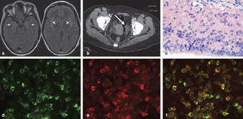
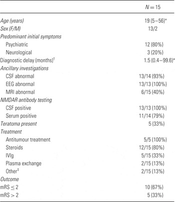
Serum and/or CSF samples from 15 patients tested anti‐NMDAR positive (Fig. 1d–f). In three patients, the anti‐NMDAR antibodies were only detectable in CSF and absent in the paired serum sample. Thirteen patients were adults (≥18 years). Clinical characteristics are summarised in Table 1.
Case vignette of a patient with NMDAR encephalitis. A 21‐year‐old female college student (Table 1; fourth row) training to become a primary school teacher was presented to the emergency room (ER) of a general hospital because of an acute confusional state. She had always been healthy and had no psychiatric or medical history. That morning while teaching, she had become agitated and confused after a request for a vacation was denied. At the ER, her behaviour apparently normalised and she was sent home with her mother after she was seen by an internist, a neurologist, as well as a psychiatrist. No medication had been given. The next day, she made statements like ‘I have to speak more softly, then the children will listen better’ and ‘I have to jump off the balcony’ and was acutely admitted to the psychiatry department. We saw an agitated patient in a catatonic state who followed commands but did not answer questions. The patient's mother in retrospect recalled that her daughter had a depressed mood for a period of about 2 weeks, prior to the admission. Furthermore, after the admission the mother visited her daughter's house and found it covered with yellow post‐it papers with unintelligible ‘to‐do reminders’. The clinical state at admission was described as ‘atypical psychosis’, and the possible underlying syndromes listed in the patient's file were stress‐induced psychotic disorder, drug‐induced psychotic disorder, schizophrenia, mood disorder/mania, ‘decompensation as part of developing personality disorder’ or ‘psycho‐organic’ (encephalitis or non‐convulsive epilepsy). Interestingly, these two latter options were explicitly written down at admission. Diagnoses were made by a senior resident after telephone consultation of the supervising psychiatrist. Initial treatment with benzodiazepines and haloperidol was started. Because the patient was unresponsive, entered other patients' rooms and showed generally disturbed behaviour, seclusion was needed from the first day on. Routine neurological examination showed no abnormalities. Routine laboratory measurements showed slightly elevated C‐reactive protein (12 mg/l, normal range 0–9 mg/l) and leucocytes (10.9 × 10⁹ cells/l, normal range 3–10 × 109 cells/l), but no other abnormalities. Anti‐NMDA receptor antibodies were not measured at the time. Over the next few days a clinical state emerged in which the patient was at times responsive (although with some slurred speech), but at other times non‐responsive, with inappropriate laughing or crying and unusual movements. Diagnosis was now: ‘catatonic psychosis’, possibly mania, and haloperidol treatment was changed to olanzapine. A neurologist was consulted who found no focal neurological deficits and no abnormalities on a CT scan of the brain. Encephalitis or non‐convulsive epilepsy was possible, but deemed very unlikely. Psychiatric examination after 6 days of admission read: in seclusion chamber; limitedly adequate and generally unresponsive; does not know where she is; somnolent; slightly slurred speech; disorientated in all modalities; disorders of formal thought: loose words and remarks, thought content: fragmented ideas; mood: slightly anxious. As the patient did not react to the medication regime, and psychoactive medication might cloud the clinical picture, olanzapine and benzodiazepine were stopped 6 days after admission. The patient continued to clinically deteriorate. Consciousness became increasingly often clouded. Agitated behaviour and responsiveness became absent. After 12 days on the psychiatry department there was no reaction to painful stimuli but she actively resisted attempts at opening her eyes. Because of decreased consciousness and general non‐responsiveness, she was transferred to the neurology ward and shortly after to the ICU where she was intubated because of central hypoventilation. Differential diagnosis now included (paraneoplastic) limbic and viral encephalitis. MRI of the brain showed medial temporal lobe (a, arrows) and insula (a, arrowheads) hyperintensities in FLAIR images. A CT scan of the abdomen showed a 2.5 × 2.3 cm left ovarian lesion (b, arrow). EEG showed a slow background pattern without epileptic phenomena. In the CSF, there was a pleocytosis with 33 WBC/µl (normal <5 WBC/µl), mainly mononuclear cells. The main differential diagnosis of the pleocytosis was viral meningoencephalitis or an immune‐mediated disorder. PCR for HSV and other neurotropic viruses was negative. The left ovary was subsequently excised and pathology showed a mature teratoma with neuronal tissue expressing the NR1 subunit of the NMDA receptor (c, red signal: AP‐labelled anti‐NR1 antibody; 400×). The patient's serum and CSF were tested for autoantibodies against the NR1 subunit of NMDAR. HEK 293 cells were transfected with an YFP‐tagged construct containing the NR1 subunit; NR1‐expressing cells become green fluorescent (d). Incubation of these transfected cells with the patient's CSF followed by incubation with Cy3‐labelled anti‐human IgG secondary antibody showed red labelling of transfected cells only (e). Colocalisation of the green and red signals to yellow was shown more clearly in (f). During her stay at the ICU, she developed mild orofacial dyskinesias and athetoid movements in the left arm. She was subsequently treated with plasma exchanges and steroids. Her condition slowly improved and she was discharged from the ICU after 4 weeks. She fully recovered over the next 6 months and started working as a primary school teacher 1 year after onset of symptoms.

Clinical characteristics in 15 patients with NMDAR encephalitis

Median (range).
Delay between onset of symptoms and final diagnosis (positive antibody testing).
Other therapies include electroconvulsive therapy and rituximab in one and mitoxantrone in another patient.
Initial presentation and referral
Table S1 shows a detailed overview of the patients. At presentation, 12 (80%) of the 15 patients had prominent psychiatric symptoms. Eight of the 13 (62%) adult patients were initially referred to a psychiatric service. Two children (5 and 8 years of age) were first seen by a paediatric neurologist. The most prominent psychiatric symptoms at initial presentation of the patients were anxiety in seven (47%), behavioural change (often bizarre) in six (40%), agitation in five (33%), delusions in four (27%) and altered mood in three (20%) (Table S1).
Early symptoms
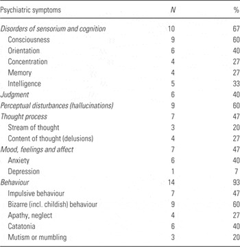
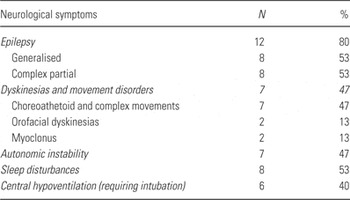
In the first 6 weeks after the onset of the syndrome, psychiatric symptoms were present in all 15 patients (100%). The most frequent psychiatric symptoms in the first 6 weeks (Table 2) were behavioural change in 14 (93%), including bizarre or childish behaviour in 9 (60%) and catatonia in 6 (40%). Perceptual disturbances (hallucinations) were present in nine patients (60%), impaired consciousness in nine (60%), anxiety or depression in seven (47%) and disorientation in six (40%). Neurological symptoms during the first 6 weeks (Table 3) included epileptic seizures in 12 (80%), choreoathetoid and complex movements in 7 (47%), sleep disturbances in 8 (53%), autonomic instability in 7 (47%) and central hypoventilation requiring admission to an ICU for mechanical ventilation in 6 (40%). The time interval between onset of symptoms and diagnosis of NMDAR encephalitis ranged between 2 weeks and 8.3 years (median 1.5 months).
Psychiatric symptoms in the first 6 weeks of NMDAR encephalitis in 15 patients

Neurological symptoms in the first 6 weeks of NMDAR encephalitis in 15 patients

Two patients were pregnant at presentation and both lost their pregnancies in the acute phase of their illness at 13 and 24 weeks of gestation.
Ancillary investigations
EEG was performed in 13 patients and showed abnormal slowing of the background pattern in all patients with additional epileptiform features in 9 of them.
MRI was abnormal in 6/15 patients (40%). MRI changes consisted of increased signal intensities in T2 and fluid‐attenuated inversion recovery (FLAIR) images. The changes were typically localised in the medial temporal lobes in four patients (Fig. 1a, arrows), with additional cortical localisations in one and post‐gadolinium enhancement in another. One patient showed subcortical changes (thalamus and putamen) and another showed changes in the periventricular white matter.
CSF was abnormal in 13/14 patients. Abnormalities included WBC (white blood cells) pleocytosis Reference Titulaer, Mccracken and Gabilondo Cuellar8, oligoclonal banding Reference Florance, Davis and Lam7 and an increased IgG index Reference Florance, Davis and Lam7, indicating intrathecal antibody synthesis. Total protein concentration was normal in all CSF samples.
Underlying tumour
In 5 of the 15 patients an ovarian teratoma was detected 1–22 months after onset of the neuropsychiatric symptoms. In two patients, the ovarian teratoma was detected and removed within a month after onset of symptoms (Fig. 1b). Immunohistochemistry revealed expression of the NR1 subunit of the NMDAR in the ovarian teratoma (Fig. 1c). In one 5‐year‐old patient multiple ovarian cysts were detected by MRI (no further follow‐up data yet available). In two patients, the teratoma was detected almost 2 years after onset of symptoms and removed to prevent relapse of the NMDAR encephalitis. In the remaining 10 patients, no teratoma or other tumours were detected.
Symptomatic treatment
Targeted management of psychiatric symptoms was mainly directed at psychotic symptoms, anxiety, agitation and mood and sleep disorders. Thirteen patients were treated with psychotropic medication including an antipsychotic drug combined with a benzodiazepine in 10 patients, antipsychotic medication only in 2 and benzodiazepine only in 1. Two patients received additional melatonin for insomnia.
In only 2 of the 13 patients, the treating physicians felt that the medication was effective in controlling the psychiatric symptoms, resulting in many medication switches. The mean number of prescribed antipsychotic drugs was 1.8 (range 1–4) and the mean number of administered benzodiazepines was 1.7 (range 1–4).
Prescribed antipsychotics included the classical antipsychotics haloperidol (in seven patients), zuclopentixol (two), levopromazine (one) and pipamperon (one) and the atypical antipsychotics risperidon (six) and olanzapine (five). The following benzodiazepines were prescribed: lorazepam (seven), diazepam (three), oxazepam (two) and to one patient each midazolam, clonazepam, alprazolam, nitrazepam, temazepam and clorazepate.
Treatment and prognosis
Two patients died 2 weeks and 5 months after onset of symptoms (mRS 6). Follow‐up in the other patients ranged from 3 months to 9 years (median 14 months). At the nadir of the disease, all patients were severely disabled and completely dependent (mRS 5). The outcome was favourable (mRS < 2) in eight patients while five patients fared less well (mRS 2–4). Twelve patients received treatment with steroids combined with intravenous immunoglobulins (IVIg) in 5, plasma exchange in 2 and rituximab in 1. Treatment was initiated 0.1–10 months after onset of symptoms (median 1.1 months). Three patients did not receive any form of immunotherapy, one died while the other two recovered with mild (mRS 1) to moderate (mRS 2) symptoms.
All five patients with a pathologically proven and removed ovarian teratoma had an excellent prognosis (mRS 0). However, in two of these patients, the teratoma was found and removed long after recovery from the NMDAR encephalitis (22 months after onset of symptoms).
In two patients, the follow‐up was shorter than 6 months and outcome was not yet evaluable. Of the evaluable patients, seven had received early treatment, within 2 months from onset. In six of these early treatment patients, the outcome was good (mRS 0–1) while it was poor (mRS ≥ 2) in one. Of six evaluable patients with no or late treatment, the outcome was poor in four and favourable in two (p = 0.053).
Autopsy in both deceased patients showed inflammatory changes in the brain, mainly in the medial temporal lobe and hippocampus (Fig. S1). Inflammatory infiltrates consisted of CD8‐positive T lymphocytes and CD20‐positive B cells in perivascular cuffs and in the neurophil. Also CD68‐positive macrophages and activated microglia were observed.
Discussion
Twelve of 15 patients (80%) with anti‐NMDAR encephalitis had prominent psychiatric symptoms at first presentation. Eight adult patients were initially admitted to a psychiatric unit. The most common diagnosis was ‘atypical psychosis’, followed by schizophrenia, depression and panic attacks. Subsequently, patients developed seizures, decline of consciousness, catatonia, abnormal movements, autonomic instability and central hypoventilation. Thirteen patients received multiple psychotropic drugs with very limited symptomatic control. Most patients were young women and in five of them pathological or radiological evidence of an underlying teratoma was found. Early aggressive treatment consisting of tumour treatment (if present) and immunotherapy, correlated with a good outcome in 8 of 13 evaluable patients.
Evaluation of the initial stages of diagnostics and patterns of referral showed that a substantial proportion of the patients initially presented with symptoms suggestive of a psychiatric disorder. At the same time, clinical records indicated that most psychiatrists were aware of an atypical presentation of the symptoms early on, leading to the suspicion of an ‘organic syndrome’, and subsequent consultation of a neurologist. Conversely, when patients were initially seen by a neurologist, an atypical presentation was also recognised, leading to psychiatric consultation. Generally, patients initially presented with little obvious biological alterations (focal neurological deficits, blood chemistry and MRI), which in many cases prompted further interpretation of symptoms as caused by a psychiatric syndrome. Recognition of the ‘neurological background’ of the syndrome usually came when either prominent EEG alterations were found, or when obvious neurological symptoms began to dominate the clinical picture (mostly seizures, and decline of consciousness). Our study and previous studies show that most of the patients with NMDAR encephalitis are initially evaluated by a psychiatrist. In the series of Dalmau et al. Reference Dalmau, Gleichman and Hughes4, 80% of the patients were initially evaluated by a psychiatrist while Irani et al. Reference Irani, Bera and Waters5 report early psychiatric symptoms in 77% of patients.
In patients of 40 years or older, with no previous history of psychiatric illness, presentation with psychiatric symptoms should always raise suspicion of an underlying neurological or other disorder. In young female patients, de novo presentation with ‘atypical’ psychosis, in particular when combined with unusual and complex behavioural changes, a diagnosis of NMDAR encephalitis should be considered Reference Kayser, Kohler and Dalmau18.
It is well‐known that expert clinical decision making greatly depends on clinical pattern recognition. It seems that the initial clinical presentation of patients with NMDAR encephalitis has elements of two apparently conflicting clinical patterns, contributing to diagnostic uncertainty. The dominant presentation of young, previously healthy women showing psychotic phenomena obviously invokes the clinical pattern of ‘psychotic disorder/schizophrenia’. However, the alternative clinical pattern (seizures, movement alterations and speech impediments in patients with good premorbid functioning without psychiatric problems) invokes in psychiatrists the clinical pattern ‘organic syndrome’. Diagnostic delay was caused by the relative absence of objective biological alteration in the early stages, in combination with unfamiliarity with this recently identified syndrome. In our series 4 out of 15 patients had been misdiagnosed as atypical psychosis or schizophrenia for months to years (range 4.2–99.6 months). It is well possible that more cases of NMDAR encephalitis with predominantly psychiatric symptoms are still misdiagnosed as primary psychiatric syndromes Reference Zandi, Irani and Lang14, Reference Barry, Hardiman and Healy19. On the other hand, a recent cross‐sectional study in 50 patients diagnosed with a psychotic disorder according to DSM IV criteria (including both first episode as well as chronic schizophrenic inpatients) did not detect any anti‐NMDAR antibodies Reference Haussleiter, Emons and Schaub20.
NMDAR encephalitis is diagnosed by detection of anti‐NMDAR antibodies in serum and/or CSF. Importantly, in three of our patients (20%) the anti‐NMDAR antibodies were exclusively detectable in CSF. Dalmau et al. Reference Dalmau, Gleichman and Hughes4 described intrathecal synthesis of anti‐NMDAR antibodies in 53 paired serum – CSF samples resulting in a higher normalised antibody concentration in CSF, using an enzyme‐linked immunosorbent assay specific for the NR1 subunit of NMDAR. Irani et al. Reference Irani, Bera and Waters5 found higher absolute anti‐NMDAR titres in serum than in CSF examining 14 matched serum – CSF sample pairs, using a cell‐based assay. One CSF sample was negative while all other samples clearly showed intrathecal synthesis of anti‐NMDAR. In our cell‐based assay, a negative serum test does not rule out NMDAR encephalitis and when suspected, CSF should be examined as well.
Negative serum titres in the presence of persistent CSF titres apparently occur in the setting of prolonged duration of the illness and after plasma exchange or other forms of immunotherapy Reference Lancaster and Dalmau12. In addition, CSF will almost always be abnormal and may show lymphocytic pleocytosis, oligoclonal banding and intrathecal IgG synthesis. EEG was abnormal in 100% of our patients and in 80–92% of patients in other series Reference Dalmau, Gleichman and Hughes4, Reference Irani, Bera and Waters5. In contrast, MRI was normal in our series, 60% similar to the series of Dalmau et al. Reference Dalmau, Gleichman and Hughes4 (45%) and Irani et al. Reference Irani, Bera and Waters5 (77%).
Antibodies from patients with NMDAR encephalitis cause a selective and reversible decrease in NDMAR surface density and synaptic localisation in hippocampal slices and in Lewis rats Reference Hughes, Peng and Gleichman21 and suppress induction of long‐term potentiation in cultured hippocampal neurons Reference Zhang, Tanaka and Sun22. In addition, the intensity of NR1 staining is strongly reduced in the hippocampus from patients with NMDAR encephalitis as compared to control brain Reference Hughes, Peng and Gleichman21. These findings indicate that the anti‐NMDAR antibodies may be pathogenic by disturbing glutamatergic neurotransmission. Indeed, the common psychotic symptoms in NMDAR encephalitis followed by amnesia Reference Finke, Kopp and Pruss23 and seizures resemble the memory impairment and psychosis caused by NMDAR antagonists such as ketamine Reference Newcomer, Farber and Jevtovic‐Todorovic24 or the recreational drug phencyclidine, also known as ‘angel dust’ Reference Paparelli, Di Forti and Morrison25, Reference Rainey and Crowder26. Moreover, there is evidence that in the schizophrenia syndrome altered activity of the NMDAR on GABA interneurons in the prefrontal cortex plays a role Reference Coyle27.
The efficacy of both classical and atypical antipsychotics and benzodiazepines alone or in combination was very limited in this patient population, as previously described Reference Chapman and Vause13, Reference Sansing, Tüzün and Ko28. As a result, the 13 patients treated with psychotropic medication received one to four different antipsychotics and one to four different benzodiazepines. The limited efficacy of psychotropic medication may be related to profound NMDAR downregulation by the antibodies. The most effective treatment of the psychiatric symptoms consisted of tumour removal (if present) and immunotherapy. Similarly, encephalitis‐related seizures are notoriously difficult to treat with antiepileptic drugs and respond much better to immunotherapy Reference Irani and Lang29, Reference Irani, Bien and Lang30. In the literature, no guidelines exist on the psychiatric treatment of autoimmune encephalitis and conclusions are difficult to draw because of the concurrent use of other treatment modalities Reference Chapman and Vause13. Generally, psychiatric and behavioural symptoms respond best when the immune response is suppressed or reversed. Amelioration of problematic symptoms depended on early aggressive immunotherapy, removal of the underlying tumour and intensive care Reference Titulaer, Mccracken and Gabilondo Cuellar8, Reference Chapman and Vause13.
In a third of our patients, the encephalitis was triggered by an underlying teratoma. In young women, ultrasound or MRI is generally preferred over computerised tomography (CT) for teratoma screening Reference Dalmau, Lancaster and Martinez‐Hernandez2. The cause of the anti‐NMDAR immune response in the patients who do not harbour an underlying tumour is not clear. Prodromal symptoms, which may indicate a triggering viral infection, were reported by 27% of our patients. In other series, prodromal symptoms were found in 27–86% Reference Peery, Day and Dunn1, Reference Dalmau, Gleichman and Hughes4, Reference Irani, Bera and Waters5, Reference Titulaer, Mccracken and Gabilondo Cuellar8.
In conclusion, autoimmune encephalitis should be considered in patients, especially young females, presenting with atypical psychiatric phenomena. The diagnosis is made by anti‐NMDAR testing in blood and/or CSF and if blood testing is negative and the condition is strongly suspected, testing for antibodies in CSF should be done. With subsequent early treatment, the prognosis of NMDAR encephalitis is relatively favourable.
Acknowledgements
The authors gratefully acknowledge the physicians who referred patients. The authors wish to thank Marcel van der Weiden for performing NMDAR immunostaining of tissue slides. P. M., E. d. G., C. H. and P. A. S. substantially contributed to conception and design of the study; P. M., E. d. G., N. M. v. B., E. H., R. M. V., K. K., M. v. D., H. H., C. H. and P. A. S. were involved in acquisition, analysis and interpretation of data; P. M., E. d. G., N. M. v. B., R. M. V., K. K., C. H. and P. A. S. were involved in drafting and/or revising the manuscript critically for important intellectual content.
Supporting Information
Figure S1. Autopsy findings in NMDAR encephalitis. A 33‐year‐old female was admitted with progressive anxiety, altered and disorganised behaviour and speech disturbances. MRI and EEG were normal. A psychiatrist was subsequently consulted and diagnosed her with an ‘organic syndrome’. She was started on haloperidol and benzodiazepine. Then she developed seizures that were controlled with phenytoin. Eleven days after admission she was found dead in bed, probably by central hypoventilation. Autopsy showed perivascular and intraparenchymal lymphocytic infiltrates in the hippocampus (a, 100×; b, in close‐up, 200×) consistent with limbic encephalitis. The perivascular infiltrates consisted of both T cells (c, CD3+) and B cells (d, CD20+). The parenchymal infiltrates consisted mainly of cytotoxic T cells (b, CD8+). Only weak IgG staining was noticed (f). Systemic autopsy did not reveal a tumour. Serum anti‐NMDAR antibodies were only demonstrated during post‐mortem.
Table S1. Initial referral and predominant initial symptoms in 15 patients with NMDAR encephalitis.
Additional Supporting information may be found in the online version of this article.
Please note: Wiley-Blackwell Publishing is not responsible for the content or functionality of any supplementary materials supplied by the authors. Any queries (other than missing material) should be directed to the corresponding author for the article.