Obesity is defined as accumulation of abnormal or excessive amounts of fat in body adipose tissues due to imbalance in energy metabolism. Many studies have shown that prevalence of childhood obesity is increasing, particularly in the last 30 years.Reference Ebbeling, Pawlak and Ludwig 1 , Reference Flegal and Troiano 2 Similarly, in our country, incidence of childhood obesity has increased from 6–7% to 15–16% in the last 20 years.Reference Tarım 3 It is known that cardiovascular disease risk increases in obese children.Reference Must, Jacques, Dallal, Bajema and Dietz 4 , Reference Franks, Hanson, Knowler, Sievers, Bennett and Looker 5 body mass index, hypertension, dyslipidaemia, insulin resistance, and type 2 diabetes mellitus have been identified as risk factors for cardiovascular diseases in children.Reference Juonala, Magnussen and Berenson 6 Although there are studies showing that obesity unfavourably affects systolic and diastolic functions of the heart, in some studies, cardiac functions have been found to be normal in obese patients.Reference Kibar, Paç, Oflaz, Ballı and Ece 7 , Reference Dayı, Tartan and Kasıkcıoglu 8 The purpose of our study was to determine the systolic and diastolic functions of the heart in obese children and evaluate the association of obesity and insulin resistance with left ventricular size and function.
Material and methods
Study population
Cases in puberty and aged 10–16 years presenting to our Paediatric Endocrinology Outpatient clinic between March, 2013 and August, 2013 and diagnosed with obesity were included in the study. Cases with syndromic obesity and also with accompanying type 2 diabetes mellitus and thyroid disease, hypertensive cases, and those with a known chronic disease were excluded from the study; 79 cases (47 girls, 32 boys) meeting these criteria were selected. The control group comprised 79 (38 girls, 41 boys) healthy and non-obese cases of similar gender and age and with no known history of drug use presenting at our Paediatric Cardiology outpatient clinic and diagnosed with non-specific chest pain or innocent murmur. As there would be differences regarding normal echocardiographic values according to age group, the patients were evaluated within their own groups after dividing them into age groups. Accordingly, patient and control groups were divided into three groups in terms of age as group 1 (10–12 years), group 2 (12–14 years), and group 3 (14–16 years).
Auxological examination
All the cases underwent detailed clinical examination by the same researcher and their age, gender, height, and weight were recorded, and their pubertal development was assessed with Tanner staging. Body weight was measured using the “Seca (Ulmer)” weighing balance, and height was measured using the “BUSSE Design Engineering ulmer stadiometer” height measurement device. Standard deviation scores of weight and height and body mass index and body mass index standard deviation scores were calculated with the auxology program using the standards determined for Turkish children. Those with body mass index percentiles above the 95th percentile adjusted for age and gender or those above +2 standard deviation according to standardised measurements adjusted for age and gender were evaluated as obese. Blood pressure was measured twice using a calibrated and suitable-sized manometer from the right arm after the patient rested for 10 minutes and their average was taken. Arterial systolic and diastolic pressures of the patients were evaluated according to the blood pressure percentile chart based on age and height, and those at the 95th percentile and above were evaluated as hypertensive. All the patients and control groups were normotensive.
Laboratory parameters
Blood glucose levels after at least 10 hours of fasting (fasting blood glucose), lipid profiles – triglyceride, total cholesterol, HDL, and LDL levels – and fasting insulin levels of the cases included in the study were analysed. Homoeostasis Model of Assessment of insulin resistance (HOMA-IR) assessment was performed using the following formula: fasting blood glucose (mmol/l)×fasting insulin (mcU/ml)/22.5.Reference Kajaia, Binder and Dittrich 9 HOMA-IR >3.16 was accepted as insulin resistance. Cases with triglyceride levels >150 mg/dl, HDL cholesterol levels <40 mg/dl, and LDL cholesterol levels >110 mg/dl were considered as having dyslipidaemia.Reference Zimmet, Alberti and Kaufman 10 , Reference Sangun, Dundar, Kosker, Pirgon and Dundar 11
Echocardiography
Echocardiography was carried out at the left decubitus position using the (2-dimension, M-mode) Vivid 5S (GE Vingmed Ultrasound AS, Horten, Norway) device and 3-MHz transducer. Each participant underwent echocardiography in accordance with the standard images and techniques presented in the American and European Echocardiography Society guidelines.Reference Edvardsen, Helle-Valle and Smiseth 12 Using the Pulse wave Doppler, the cursor of the echocardiography (ECHO) device was placed on the apex of the mitral valve leaflets, and the early diastolic E-wave on the mitral valve flow velocity curve and the late diastolic A-wave showing atrial contraction were measured. Left ventricular ejection fraction, fractional shortening, end-diastolic (LVEDD) and end-systolic (LVESD) diameters, left atrium diameter, interventricular septum (IVS) wall thickness were measured using the M-Mode ECHO in the parasternal long-axis position, and aortic valve opening was measured using the two-dimensional ECHO. All the echocardiographic parameters were indexed to age and body surface area.
Statistical evaluation
Study data were analysed using SPSS for Windows 14.0 package program. As parametric test hypotheses were met during data evaluation, significance test of the difference between two mean values and Pearson’s correlation analysis were used in independent groups. Our data were expressed as arithmetic mean±standard deviation in the tables, and error level was taken as 0.05; p<0.05 was considered to be statistically significant. Inter-observer variability (82%) was assessed by analysing all the echocardiographic parameters by two different researchers. For intra-observer variability (94%), all the echocardiographic parameters were analysed by one researcher at two different times.
Results
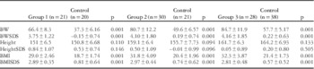
When body weight, body weight standard deviation scores, body mass index, and body mass index standard deviation scores of all the groups were compared with the control groups, the differences were statistically significant (p<0.05; Table 1).
Comparison of auxologic parameters between obese and control groups.

BMI=body mass index; BW=body weight; BWSDS=body weight standard deviation scores
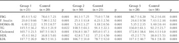
When fasting blood glucose, fasting insulin, HOMA-IR, TG, Cholesterol, HDL, and LDL levels of the cases in groups 1, 2, and 3 were compared with the control group, differences were statistically significant (p<0.05; Table 2).
Comparison of laboratory parameters between obese and control groups.

F. Insulin=fasting insulin; FBG=fasting blood glucose; HDL=high-density lipoprotein; HOMA-IR=Homoeostasis Model of Assessment-insulin resistance; LDL=low-density lipoprotein; TG=triglyceride
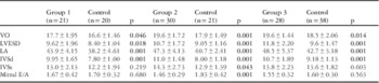
In the ECHO evaluation of the cases, valve openings, left atrium diameters, IVS diastolic thickness (IVSd), and LVESD measurements of all the three groups were statistically significantly higher when compared with their respective control groups (p<0.05). In group 2, the difference in IVS systolic wall thickness (IVSs) compared with the control group was statistically significant but at a low level. There was no statistical difference when LVEDD, ejection fraction, and fractional shortening of all three groups were compared with the control groups (p>0.05). In all patient groups, mitral valve E/A was found to be >1 (Table 3).
Comparison of indexed echocardiographic parameters between obese and control groups.

IVSd=interventricular septum diastolic wall thickness; IVSs=interventricular septum systolic wall thickness; LA=left atrium; LVESD=left ventricle end-systolic diameters; VO=valve opening
Bold values significant at p <0.05
Although a positive correlation was found between body mass index and body mass index standard deviation scores and only IVSd measurements in group 1, a positive correlation was found between body weight, body weight standard deviation scores, body mass index, and body mass index standard deviation scores and IVSd, IVSs, and mitral E measurements in group 3. A negative correlation was found between body mass index and E/A ratio in all the groups. The difference in E/A between obese and controls was statistically significant in group 2.
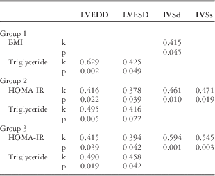
In groups 2 and 3, a positive correlation was found between fasting insulin and HOMA-IR and LVESD, LVEDD, IVSd, IVSs, and left atrium. In cases with insulin resistance, a significant positive correlation was found between insulin resistance and LVEDD, LVESD, IVSd, and IVSs (Table 4). A positive correlation was found between cholesterol levels and LVESD and LVEDD in group 3. A positive correlation was found between HDL levels and LVEDD in group 1. In all the three patient groups, there was a positive correlation between TG and LDL levels and LVESD, LVEDD, and valve openings (Table 4).
Correlation between indexed parameters.

BMI=body mass index; HOMA-IR=Homoeostasis Model of Assessment of insulin resistance; IVSd=interventricular septum diastolic wall thickness; IVSs=interventricular septum systolic wall thickness; k=Pearson’s correlation; LVEDD=left ventricle end-diastolic diameter; LVESD=left ventricle end-systolic diameter
Discussion
Obesity prevalence has a tendency to increase worldwide. Childhood obesity is an important risk factor for the development of cardiovascular diseases later in life.Reference Must, Jacques, Dallal, Bajema and Dietz 4 , Reference Franks, Hanson, Knowler, Sievers, Bennett and Looker 5 Left ventricular size has been shown to be associated with hypertension, obesity, insulin resistance, and early stage cardiovascular diseases in young adults;Reference Benjamin, D’Agostino, Belanger, Wolf and Levy 13 however, few studies have been carried out on this subject in childhood.
Obesity induces left ventricular dilation by increasing left ventricular filling pressure and volume. Dilated chamber volume increases unproportionately with stress in the left ventricular wall. Consequently, the myocardium adapts itself by increasing its contractile elements and therefore the myocardial mass; the end product of this process is ventricular hypertrophy. Left ventricular hypertrophy has been defined as the determinant risk factor for various cardiovascular diseases in obese children.Reference Hirschler, Acebo, Fernandez, de Luján Calcagno, Gonzalez and Jadzinsky 14 , Reference Juhola, Magnussen and Viikari 15 Left ventricular hypertrophy causes decrease in left ventricular compliance and impairment of diastolic relaxation pattern. Interventricular septum thickness and LVEDD are important parameters for the determinantion of left ventricular function.Reference Sasson, Rasooly, Bhesania and Rasooly 16 In the 20-case study by Berkalp et alReference Berkalp, Cesur, Corapcioglu, Erol and Baskal 17 and the 341-case study by Kono et al,Reference Kono, Yoshinaga, Oku, Nomura, Nakamura and Aihoshi 18 IVSd and LVPWd values were significantly increased in obese patients. In our study, obese and control groups were compared, and in accordance with literature data, although there was a significant difference in IVSd values, there was no significant difference in IVSs values. This was interpreted as obesity causing first an increase in diastolic wall thickness morphologically.
Significant changes secondary to volume load in morphological findings such as left atrium, LVESD, LVEDD, left ventricular mass, and preserved left ventricular systolic function have been reported in obese children in the literature.Reference Daniels, Witt, Glascock, Khoury and Kimball 19 , Reference Mehta, Holliday, Hayduk, Wiersma, Richards and Younoszai 20 Some studies have reported significantly high LVEDD in obese cases. This condition has been shown as an early determinant of left ventricular function, and a strong relationship has been established between left atrium volume index and duration and severity of diastolic dysfunction.Reference Garavaglia, Messerli, Nunez, Schmieder and Grossman 21 , Reference Sermez, Eren, Keskin and Turk 22 In another study, left atrium diameter was significantly higher in obese groups compared with the control group;Reference Kibar, Paç, Oflaz, Ballı and Ece 7 however, although there was a significant difference in left atrium and LVESD between the obese and control groups in our study, there was no difference regarding LVEDD. As in our study, left atrial enlargement was also found in obese cases without an increase in LVEDD in some studies. This condition was considered to be a physiological adaptation of the heart.Reference Sasson, Rasooly, Gupta and Rasooly 23 Again in our study, there was no statistically significant difference in ejection fraction and fractional shortening between obese cases and controls, in accordance with the literature. Left ventricular systolic functions were noted to be preserved in obese cases.
Some studies have defined early diastolic functional impairment without left ventricular systolic dysfunction.Reference Di Bello, Santini and Di Cori 24 , Reference Urheim, Edvardsen, Torp, Angelsen and Smiseth 25 Obesity is an independent determinant of diastolic dysfunction but its pathophysiology is still not clear.Reference Galinier, Pathak, Roncalli and Massabuau 26 , Reference Langenberg and Sharp 27 Endothelial dysfunction, caused by various pro-inflammatory cytokines, free fatty acids, nitric oxide, and insulin resistance, causes cardiovascular diseases in obese children.Reference Singleton, Smith, Russell and Feldman 28 Adiponectin deficiency, angiotensin 2, and leptin in obesity lead to abnormal cardiac relaxation and consequently diastolic dysfunction.Reference Zibadi, Cordova, Slack, Watson and Larson 29
The early diastolic E and late diastolic A waves on the mitral valve velocity curve and E/A ratio were used for measuring diastolic dysfunction. E/A should be 1.5 or greater in normal diastolic filling pattern. In the presence of diastolic dysfunction, E/A can be measured as <1. Diastolic dysfunction could not be shown in a study comparing mitral E/A of obese and non-obese groups.Reference Sharpe, Naylor and Jones 30 In a study by Gian et al,Reference Gian, Simone, Greco and Rosato 31 obese patients had decreased peak E velocities, mildly increased A velocities, and decreased E/A ratios compared with the controls. In a study by Erturk et al,Reference Erturk, Goksen, Ozyurek, Darcan and Coker 32 carried out in obese children, the obese group had increased A velocity and decreased E/A ratio compared with the controls. In a study by Chakko et al,Reference Alpert, Terry and Kelly 33 E/A ratio was significantly decreased in the obese group compared with the control group. Similarly, in our study, mitral E/A of all patient groups was >1 and the obese group had decreased E/A ratio compared with the control group in accordance with the literature. It can be defined as an abnormality of ventricular filling with normal cardiac function and compensatory increased atrial pressure. In this condition, the A wave upraises. Although there are studies finding a negative correlation between body mass index and E/A, there are also studies that have found no difference.Reference Cetin, Caglayan, Yıldırımer, Kızılyıldız, Deveci and Coskun 34 We also found a negative correlation between body mass index and E/A ratio in our study. Owing to the positive correlation between body mass index increase and IVSd, IVSs, and mitral E values, group 3, which has the highest body mass index and body weight values, has also been considered as having such positive correlation in terms of those parameters.
Insulin resistance can lead to structural and functional changes in the left ventricle. There are studies reporting a statistical correlation between left ventricle size and body mass index and HOMA-IR in obese children.Reference Hirschler, Acebo, Fernandez, de Luján Calcagno, Gonzalez and Jadzinsky 14 , Reference Juhola, Magnussen and Viikari 15 Although one study reported a significant correlation between insulin resistance (HOMA-IR) and left atrium and left ventricular end-diastolic volume, it has not shown a similar correlation between insulin resistance and body mass index.Reference Kibar, Paç, Oflaz, Ballı and Ece 7 In our study, a positive correlation was found between fasting insulin and HOMA-IR and LVESD, LVEDD, left atrium, IVSd, and IVSs in groups 2 and 3. Furthermore, a significant positive correlation was found between insulin resistance and LVEDD, LVESD, IVSd, and IVSs in cases with insulin resistance. Therefore, it was seen that increase in insulin resistance caused an increase in left ventricular size and wall thicknesses by increasing sympathetic autonomous activity.
Increased triglyceride and cholesterol levels in childhood are considered to be among the cardiovascular risk factors.Reference Juhola, Magnussen and Viikari 35 , Reference Morrison, Glueck, Horn, Yeramaneni and Wang 36 Similarly, in our study, a positive correlation was found between TG and LDL levels and LVESD, LVEDD, and valve opening in all three patient groups; however, a positive correlation was found between cholesterol levels and LVESD and LVEDD only in group 3. It was thought that this condition could be due to long-term exposure to hyperlipidaemia because of higher mean cholesterol levels and age.
Summary
It was shown in our study that left ventricular hypertrophy and diastolic dysfunction developed but systolic functions were preserved, and an increase in insulin resistance caused an increase in left ventricular size and wall thickness in childhood obesity. In conclusion, determination of these early cardiac changes will be important in allowing early diagnosis and treatment of cardiovascular diseases that may develop later.
Acknowledgement
None.
Conflicts of Interest
None.