


Introduction
Ammonoids are among the most widely utilized fossil groups—having been applied in nearly all disciplines of paleontological study (see Ritterbush et al. Reference Ritterbush, Hoffmann, Lukeneder and De Baets2014) from stratigraphy (e.g., Meister and Piuz Reference Meister and Piuz2013; Moreno-Bedmar et al. Reference Moreno-Bedmar, Barragán Manzo, Company Sempere and Bulot2013) to geochemistry (Moriya et al. Reference Moriya, Nishi, Kawahata, Tanabe and Takayanagi2003; Lukeneder et al. Reference Lukeneder, Harzhauser, Muelleffer and Piller2010; Kruta et al. Reference Kruta, Landman and Cochran2014), evolutionary dynamics (e.g., Monnet et al. Reference Monnet, De Baets and Klug2011; De Baets et al. Reference De Baets, Klug, Korn and Landman2012; Korn et al. Reference Korn, Hopkins and Walton2013), paleoecology (Tanabe Reference Tanabe1979, Reference Tanabe2011), functional morphology (e.g., Ward and Westermann Reference Ward and Westermann1976; Reyment Reference Reyment1980; Ritterbush and Bottjer Reference Ritterbush and Bottjer2012), and biomechanics (Daniel et al. Reference Daniel, Helmuth, Saunders and Ward1997; Hassan et al. Reference Hassan, Westermann, Hewitt and Dokainish2002). Despite this robust research history the commonly proposed primary function of the ammonite phragmocone, to serve as a hydrostatic apparatus allowing the animal to enter the water column (Teichert Reference Teichert1967; Crick Reference Crick1988), has been subject to continuing controversy. General interpretations of ammonite life habits view these animals as planktonic/nektonic (i.e., pelagic) though a few have argued for a benthic life habit (e.g., Klinger Reference Klinger1981; Shigeta Reference Shigeta1993; Ebel Reference Ebel1999; Westermann Reference Westermann2013). Ammonite hatchlings are generally considered to be planktonic largely on the basis of facies analysis of shell occurrences and comparisons with modern cephalopod hatchlings (De Baets et al. Reference De Baets, Klug, Korn and Landman2012; Ritterbush et al. Reference Ritterbush, Hoffmann, Lukeneder and De Baets2014). Prior buoyancy calculations of juvenile shells have argued for a nektobenthic lifestyle (Shigeta Reference Shigeta1993) or a benthic mode of life for hatchlings and early juveniles (Wetzel Reference Wetzel1959). In this paper we develop a novel method of buoyancy calculation utilizing quantitative computed tomography that can be used to test the function and buoyant properties of the cephalopod phragmocone and hypotheses of life habits.
Ammonoids are ectocochleate cephalopods (i.e., possessing an external shell) and thus often compared to the only extant ectocochleate cephalopod the nautilus, though they are more closely related to the coleoids: squids, octopuses, cuttlefish, and Spirula (Engeser Reference Engeser1996; Warnke and Keupp Reference Warnke and Keupp2005; Kröger et al. Reference Kröger, Vinther and Fuchs2011). The first attempt to deduce ammonite life habits based on the morphological similarity with Nautilus was done in the seventeenth century by Robert Hooke, who correctly postulated that Nautilus could alter its buoyancy by regulating the water to gas ratio in the chambers of the shell and extended this interpretation to ammonites (see Derham Reference Derham1726; Denton and Gilpin-Brown Reference Denton and Gilpin-Brown1973; Davis Reference Davis2010). Nautilus, however, should not be considered the ideal model organism to understand the mode of life of ammonites, owing to differences in reproduction style and phylogenetic distance (Jacobs and Landman Reference Jacobs and Landman1993; Ritterbush et al. Reference Ritterbush, Hoffmann, Lukeneder and De Baets2014), as well as the observation that ammonites are a morphologically diverse group whereas the modern Nautilus shows highly limited shell morphology (Jacobs and Landman Reference Jacobs and Landman1993; Warnke and Keupp Reference Warnke and Keupp2005; Mutvei and Dunca Reference Mutvei and Dunca2007; Wright Reference Wright2012). Kröger (Reference Kröger2002) showed that the buoyant properties of the Nautilus shell and an ammonite shell are quantifiably different by calculating the capacity of the shell to tolerate shell loss. It is therefore necessary to establish a reliable framework within which accurate buoyancy calculations can be performed on any variety of cephalopod shells.
Initial attempts to theoretically quantify the volume of a planispiral shell were the equations of Moseley (Reference Moseley1838), which were based on the calculation of the volume of coiled cones. Moseley never applied these equations to actual shells; this was later done by Raup and Chamberlain (Reference Raup and Chamberlain1967), who modified the equations to make them more amenable to calculations involving measurements from actual specimens. Trueman (Reference Trueman1941) avoided applying Moseley’s equations in favor of a simplified approach inspired by Thompson (Reference Thompson1917), in which the shell volume was calculated by using a modified form of the equation for the volume of a cone. Trueman (Reference Trueman1941) noted that his equation and those of Moseley assume that the cross-section of the cone changes in a uniform manner, which is a deviation from true ammonite growth patterns. Heptonstall (Reference Heptonstall1970) revised an oversight in Trueman’s equations and then calculated ammonite densities by using more accurate cephalopod shell densities (Trueman used the density of pure aragonite for the density of the shell, which led to notable error in the buoyancy calculations of both Trueman (Reference Trueman1941) and Currie (Reference Currie1957). Further quantitative study of the volumetrics of the shell through mathematical methods and implications for the life habits of extinct ammonoids and nautiloids were the topics of numerous publications in the following decades; for comprehensive discussions of this topic, see Westermann (Reference Westermann1996, Reference Westermann1999), Jacobs and Chamberlain (Reference Jacobs and Chamberlain1996), Westermann and Tsujita (Reference Westermann and Tsujita1999), and Ritterbush et al. (Reference Ritterbush, Hoffmann, Lukeneder and De Baets2014).
Ebel (Reference Ebel1983) attempted to calculate the buoyant properties of the shell by using cones with idealized geometric base areas (triangular and trapezoidal) and argued for the benthic-crawler model of ammonite life habits (Ebel Reference Ebel1990, Reference Ebel1992, Reference Ebel1999). Additional calculations by Rein (Reference Rein1999), for Ceratites, have been used to argue for negatively buoyant (i.e., benthic) life habit. Dietl (Reference Dietl1978) argued for a benthic life habit for Spiroceras on the basis of high shell variability. Delanoy et al. (Reference Delanoy, Magnin, Selebran and Selebran1991) calculated a negative buoyancy for the heteromorph ammonite Moutoniceras. These conclusions have been duly criticized, especially the calculations of Ebel (Reference Ebel1983), by Westermann (Reference Westermann1993, 1996) and Kröger (Reference Kröger2001) for unclear and incorrect calculation methodology or by claiming their results fall within the range of error of neutral buoyancy. Shigeta (Reference Shigeta1993) focused on calculating the buoyancy of Cretaceous ammonite hatchlings and attempted to correct the assumption of isometric shell growth and a circular whorl cross-section assumed in the calculations of Moseley (Reference Moseley1838) and Trueman (Reference Trueman1941) by dividing the shell into separate components and measuring the volume of these components separately. Shigeta (Reference Shigeta1993) used these calculations to argue for an initially planktonic life habit transitioning to a nektobenthic life habit for larger juveniles, and Tanabe et al. (Reference Tanabe, Shigeta and Mapes1995) also applied this method to Carboniferous ammonoid hatchlings.
Ammonites were highly abundant and widespread in Paleozoic/Mesozoic seas and therefore knowing the life habits of ammonites is vital to understanding the biology and predator-prey interactions with contemporaneous animals (e.g., Sato and Tanabe Reference Sato and Tanabe1998; Rieppel Reference Rieppel2002). Functional studies of both the hydrostatics and the hydrodynamics of the shell assume that adult ammonites lived in the water column (Chamberlain Reference Chamberlain1976; Klug and Korn Reference Klug and Korn2004; Ritterbush et al. Reference Ritterbush, Hoffmann, Lukeneder and De Baets2014). Existing buoyancy calculations have relied on simplification of shell geometry through mathematical modeling or creating casts or physical models, approaches that lose information about the internal features of the shell or fine external shell features. To achieve more accurate buoyancy calculations, we apply X-ray computed tomography (CT) to create 3-D data sets of true specimens whose physical properties, including shell and phragmocone volume, can be directly quantified. Hoffmann et al. (Reference Hoffmann, Schultz, Schellhorn, Rybacki, Keupp, Gerden, Lemanis and Zachow2014) tested this method on the extant Nautilus, which has a known nektobenthic lifestyle (Ward et al. Reference Ward, Stone, Westermann and Martin1977), and found that with sufficient resolution buoyancy can be reasonably calculated. We transfer this method to higher-resolution scans of a Cadoceras sp. and the earliest chambers of the shell of a Spirula spirula, which has been argued to be an analogue for some aspects of ammonite paleobiology (Warnke and Keupp Reference Warnke and Keupp2005), in order to test mode-of-life hypotheses and establish a methodology for calculating the buoyancy of adult ammonites.
The first application of computed tomography in paleontology dates back to the 1980s; since then, CT has been frequently applied to questions of functional morphology in the fossil record (Abel et al. Reference Abel, Laurini and Richter2012; Cunningham et al. Reference Cunningham, Rahman, Lautenschlager, Rayfield and Donoghue2014; Hoffmann et al. Reference Hoffmann, Schultz, Schellhorn, Rybacki, Keupp, Gerden, Lemanis and Zachow2014). For an extensive review of the application of CT techniques in paleontology and an introduction to a generalized methodology see Sutton et al. (Reference Sutton, Rahman and Garwood2013), and for technical and theoretical background information concerning CT see Stock (Reference Stock2008). Lehmann (Reference Lehmann1932) was the first to apply X-rays to create stereo images of the internal structures of ammonites. Recent applications of tomography to ammonites has been limited to destructive (grinding tomography), low-resolution techniques (Naglik et al. Reference Naglik, Monnet, Goetz, Kolb, De Baets, Tajika and Klug2014; Tajika et al. Reference Tajika, Naglik, Morimoto, Pascual-Cebrian, Hennhöfer and Klug2014) to study the morphometrics and function of the shell, or high-resolution computed tomography to study discrete parts of the animal such as the radula (Kruta et al. Reference Kruta, Landman, Rouget, Cecca and Tafforeau2011; Tanabe et al. Reference Tanabe, Landman and Kruta2012).
Quantitative CT-based studies of ammonites have not yet been performed, largely because it is difficult to differentiate the carbonate shell from the often-carbonate rock matrix or infill. The lack of a strong difference between the absorption properties of the shell and the surrounding material can make differentiating them nearly impossible in conventional CT data sets (Naglik et al. Reference Naglik, Monnet, Goetz, Kolb, De Baets, Tajika and Klug2014). For this reason we chose as our subject a hollow ammonite, a Cadoceras sp. hatchling, which we scanned with high-resolution synchrotron micro-tomography (SRµCT). Wetzel (Reference Wetzel1959) suggested that ammonite hatchlings would be benthic, though this claim was challenged by House (Reference House1985), who supported a planktonic life habit for hatchlings even with the protoconch as the only empty chamber. A planktonic mode of life for ammonoid hatchlings is supported by the occurrence of embryonic and juvenile shells in dysoxic sediments that are characterized by the absence of benthic fauna, as well as by comparisons to similarly sized modern cephalopod paralarvae, which are planktonic (Landman et al. Reference Landman, Tanabe and Shigeta1996a; De Baets et al. Reference De Baets, Klug, Korn and Landman2012; Ritterbush et al. Reference Ritterbush, Hoffmann, Lukeneder and De Baets2014). The hatchling phase is a distinct ontogenetic state in the development of ectocochleate cephalopods discernible by the nepionic constriction, which marks the end of the embryonic shell (ammonitella) (Fig. 1A).
Computed tomographic slice of the juvenile Cadoceras sp. (A; black arrow indicates the terminal extent of the ammonitella) and Spirula spirula (D). 3-D model of Cadoceras (B) and the first whorl of Spirula (E). Reconstructed 3-D model of Cadoceras showing a cross-section of the interior of the shell with the septa in dark red (C). Selected shell and first three chambers used in the buoyancy calculations of a potential Spirula hatchling (F).

Materials and Methods
The internal shell of Spirula spirula, collected from Fuerteventura, was scanned at the GeoForschungsZentrum (GFZ) Potsdam, using the Phoenix Nanotom-s at 70 keV. This data set is composed of 1880×1880×1900 slices with an isotropic voxel size of 8.747 µm and a file size of 13 GB. The full shell has a diameter of 17.75 mm.
The shell of a hatchling Cadoceras sp. was scanned at the Advanced Photon Source (APS) at Argonne National Laboratory, using phase contrast tomography at 27.2 keV and a sample-detector distance of 300 mm, with an isotropic voxel size of 0.74 μm, resulting in a data set with 2048×2048×1948 slices and a file size of 32 GB. The hatchling shell has a diameter of 0.98 mm. Both data sets are archived at the Ruhr Universität Bochum. CT slices, volume renderings, and segmented models are shown for Cadoceras (Fig. 1A–C) and Spirula (Fig. 1D–F).
The data sets were analyzed in Avizo Fire (version 7.0, Visualization Science Group) and Amira (Zuse Institute Berlin), whereby the shell material was isolated using the automatic threshold tool provided and, where necessary, manually corrected. Most of these manual corrections were necessary in the Cadoceras data set, owing to diagenetic alterations largely in the form of secondary calcite growth on the inner and outer surfaces of the shell and septa (Fig. 2A,B). The Spirula data set required minimal manual corrections except in regions where the shell material was very thin, or where the shell features, mainly the siphuncle, were damaged and the irregular geometry had to be manually corrected.
Two CT slices showing the original shell material of Cadoceras (A) and the labeled and reduced shell in yellow (B). The shell shows growth of secondary calcite on the shell wall and septa, which would overestimate the shell thickness (inset). We corrected for this by using the “shrink” feature in Avizo. The length of the body chamber and the number of chambers a hatchling ammonite would possess are unknown; therefore we model six scenarios. The first scenario models the protoconch as the only empty chamber and the body length (in dark brown) extends from the end of the ammonitella back to the protoconch (C). The last scenario models the protoconch and five additional chambers (D). Intermediate scenarios model the animal with the protoconch and one through four additional chambers.
We corrected for the secondary calcite in the hatchling Cadoceras by using the “shrink” feature in Avizo, which shrinks the selected region by one voxel layer while preserving the original geometry. Direct measurements of the entire hatchling shell wall of Cadoceras are not reported in the literature; however, Drushchits et al. (Reference Drushchits, Doguzhayeva and Lominadze1977) measured the thickness of the nepionic ridge of a Cadoceras to be 40 μm. After shrinking, the measured thickness of the nepionic ridge of our Cadoceras hatchling was 35–37 μm. Measurements by Landman et al. (Reference Landman, Tanabe and Shigeta1996a) of ammonitella thicknesses for the heteromorph ammonite Baculites are between 2 and 8 μm. In our specimen the original selection thickness, without corrections for the secondary calcite growth, was nearly 30 μm; reselected thickness ranges from <1 to 12 μm.
Hard Parts and Soft Tissue
The shell and chamber volumes of both specimens were segmented as separate materials. The soft-body model of Cadoceras was assumed to be equal to the entire volume of the body chamber (an overestimation; see below for a mathematical correction for this assumption); however, because it is unknown how many chambers the hatchling Cadoceras possessed and because the extent of the body chamber changes through development (Trueman Reference Trueman1941), we modeled six scenarios. The initial scenario models the hatchling with the protoconch as the only empty chamber and the rest of the volume of the ammonitella taken up completely by the soft body; this scenario is the commonly assumed condition for hatchling ammonoids (Shigeta Reference Shigeta1993; Landman et al. Reference Landman, Tanabe and Shigeta1996a). Subsequent scenarios added additional chambers, with the final scenario modeling the protoconch and five additional chambers (Fig. 2C,D). The additional chambers are modeled in order to examine the influence of the ratio of shell, soft body, and air on buoyancy. Findings of ammonitella with more than one septa are reported by Bandel (Reference Bandel1982) and Kulicki and Wierzbowski (Reference Kulicki and Wierzbowski1983). It is unclear if these actually represent ammonite hatchlings, though variation in the number of septa in Sepia hatchlings has been observed (Bandel and Boletzky Reference Bandel and Boletzky1979); see Landman et al. (1996a) for a more thorough discussion of this topic. All chambers are modeled as being completely empty (i.e., filled with gas only and no cameral fluid or gel [Landman et al. Reference Landman, Tanabe and Shigeta1996a]). The weight of the gas is presumed to have ignorable effect on the weight calculation.
Estimates of soft-body volume of Spirula are based on geometric relationships between the shell diameter and the dorsal/ventral mantle length and breadth reported in the measurements of Bruun (Reference Bruun1943), who recorded the diameter of the shell, length of the dorsal mantle, and mantle breadth as percentages of the ventral mantle length in juvenile specimens from the Indo-Pacific and Atlantic Oceans. Specimens show a range of relationships between these parameters; we use the most extreme relationships (highest and lowest percentages) to calculate minimum and maximum estimates for the soft-body volume. We present three scenarios for the hatchling Spirula: the internal shell with one, two, and three chambers. Each chamber, together with the corresponding section of the siphuncle, is extracted as a separate structure in the CT data, and dimensions of the soft body are calculated from the diameter of the single-, double-, and triple-chambered shell.
Accurate buoyancy calculations depend on many considerations in addition to the volume of the shell and soft body (Kröger Reference Kröger2002). Variation in extant cephalopod shell density forces the consideration of a range of ammonite shell densities. Measured shell density of Spirula and density values reported by Bruun (Reference Bruun1943) yield a reasonable minimum shell density of 2.5 g/cm3 and a maximum shell density of 2.7 g/cm3. Measured densities of the shell of Nautilus and reported shell densities of the externally shelled ammonites show similar variation (2.7 g/cm3 [Greenwald and Ward Reference Greenwald and Ward2010]; 2.62 g/cm3 [Okamoto Reference Okamoto1988]; 2.5958 g/cm3 [Ebel Reference Ebel1983]; 2.65 g/cm3 [Westermann Reference Westermann2013]); therefore we extract a suitable minimum and maximum value, which are the same minimum and maximum values used for Spirula.
The soft-body density is also variable as the body is composed of multiple tissue types, which have varying densities. The average soft-body density is, like the shell, represented by a minimum and maximum, of 1.05 g/cm3 and 1.07 g/cm3 respectively (1.06 g/cm3 [Greenwald and Ward Reference Greenwald and Ward2010]; 1.068 g/cm3 [Heptonstall Reference Heptonstall1970]; 1.055 g/cm3 [Longridge et al. Reference Longridge, Smith, Rawlings and Klaptocz2009]; 1.05–1.07 g/cm3 Ward and Westermann Reference Ward and Westermann1977]). Potential soft-body densities are the same for Spirula and Cadoceras.
The pellicle is a thin tissue layer lining the inner surface of the phragmocone that assists in the removal of cameral liquid (Denton and Gilpin-Brown Reference Denton and Gilpin-Brown1966). The volume of the pellicle is estimated by taking the surface area of the chamber volumes and multiplying it by the thickness, here taken as 0.5 µm. The pellicle is treated as soft tissue in terms of density.
Siphuncle
The siphuncle is a small but notable volume of soft body and, in the form of septal necks, shell volume. The septal necks of Spirula extend through an entire chamber, from septum to septum, and are well preserved, allowing easy recognition of the siphuncle volume although no soft tissue is present. The septal necks and siphuncle volume are segmented with the same method as the shell and chambers. The siphuncle, as expected, is not preserved in Cadoceras, nor does the specimen seem to possess septal necks; however, the spaces through which the siphuncle would have passed through the septa (siphuncular foramina) are preserved and allow us to trace the estimated dimensions of the siphuncle. The siphuncle of Cadoceras is therefore separated into a series of linked cylinders through each chamber. The height and radius of each cylinder is taken as the minimum and maximum extent of chamber width and siphuncular passage respectively (Supplementary Table 1).
Jaws
The jaws are a potentially significant weight especially in Cadoceras and other aptychus-bearing ammonites. Aptychi are typically composed of various layers of organic and calcareous materials (Engeser and Keupp Reference Engeser and Keupp2002); the structure and composition of the juvenile aptychus of Cadoceras is unknown, so we present a range of possible values. The volume of the aptychus is calculated in two ways: the minimum as the cross-sectional area of the aperture (Kröger Reference Kröger2002) and the maximum as equal to the volume of the final septum. According to Hewitt et al. (Reference Hewitt, Westermann and Checa1993) the density of the aptychus is between chitin (1.655 g/cm3) and calcite (2.71 g/cm3); we use these two density values as minimum and maximum estimates.
The jaws of the juvenile Spirula have an unknown volumetric relationship with the rest of the body and are therefore estimated as a volume equal to the final septal volume. The density is taken as that of chitin because Spirula lacks the calcareous plates of Nautilus jaws (Kruta et al. Reference Kruta, Landman and Cochran2014). The smallest and largest potential jaw volumes are averaged together, thereby providing a constant volume for each Spirula scenario; this has no significant effect on the buoyancy calculation.
Mantle Cavity
The assumption that 100% of the body chamber volume would have been soft body is an oversimplification and overestimation of the soft-body structure, as noted by Kröger (Reference Kröger2002). The mantle cavity is the most notable and important body chamber modification. The mantle cavity in Nautilus is 15% of the total body chamber volume and would have been filled largely with seawater, excluding the gills (Chamberlain Reference Chamberlain2010). The extent of the ammonite mantle cavity is unknown; thus Nautilus is used as a reference and 15% of the relative body chamber volume is subtracted and treated as a volume of seawater.
The size of the mantle cavity of a juvenile Spirula and its geometric relationships with other body shape parameters are unknown; however, Spirula is known to be capable of retracting its head and tentacles into the mantle cavity (Bruun Reference Bruun1943). The length and diameter of the head and tentacles in relation to the ventral mantle length are not noted by Bruun (Reference Bruun1943), so our calculations of soft-body volume assume the animal has retracted its head and tentacles into the mantle cavity, completely filling the mantle cavity volume with soft tissue.
Calculations
Buoyancy calculations traditionally involve the calculation of effective weight, the weight of the animal underwater, or density. Using the volume data derived from the CT scans and the above quoted density values, we can calculate the mass of each section of the animal (shell and soft body) as (mass=volume*density) and then convert that into weight (weight=mass*acceleration due to gravity). Buoyancy is the difference between the total weight of the animal and the buoyant force; the buoyant force is equal to the weight of displaced fluid such that the volume of fluid is equal to the total volume of the object that displaces that fluid (in this case the total volume of Cadoceras/Spirula). We use the standard density of seawater, 1.0275 g/cm3, to calculate the buoyant force. Then, dividing the total mass by the total volume, we can calculate the total density of the animal, which we can compare with the seawater density to determine buoyancy.
Swimming and Sinking
Investigations of buoyancy should take into account the ability of motile animals to generate forces to counteract potential sinking due to negative buoyancy. Many nektonic and planktonic animals exhibit slight negative buoyancy, including sharks (e.g., Wilga and Lauder Reference Wilga and Lauder2002), diatoms (e.g., Raven and Waite Reference Raven and Waite2004), and Nautilus (Ward et al. Reference Ward, Stone, Westermann and Martin1977). The swimming capability of ammonites is a subject of significant speculation (Jacobs Reference Jacobs1992; Chamberlain Reference Chamberlain1993; Parent et al. Reference Parent, Westermann and Chamberlain2014); however, Chamberlain (Reference Chamberlain1981) used the hydrodynamics of Nautilus (the only empirical hydrodynamic model for ectocochleate cephalopods) and the stability (S) of Raup (Reference Raup1967) to estimate swimming velocity:
 $$\scale94%{V=\left( {2mgSsin({\rm \rtheta })\rrho ^{{{\minus}1}} A^{{{\minus}1}} C_{D}^{{{\minus}1}} \left( {\left( {{x \over d}} \right){\minus}Ssin(\rtheta )} \right)^{{{\minus}1}} } \right)^{\wedge} {1 \over 2}$$
where V is the swimming velocity, m is the total mass of the animal, g is the gravitational acceleration, S is stability, θ is the angle of rotation (10° for Nautilus [Chamberlain Reference Chamberlain1981]), ρ is the density of seawater, A is the reference area (shell volume^[2/3] for ectocochleate cephalopods), C D is the coefficient of drag (a range of hypothetical values are used corresponding to a range of Reynolds numbers), x is the moment arm of thrust (estimated for each scenario), and d is the shell diameter.
Stability is defined as the ratio between the distance between the center of buoyancy (coincident with the center of volume) and the center of mass of the soft body to the shell diameter (Raup Reference Raup1967; Raup and Chamberlain Reference Raup and Chamberlain1967). Trueman (Reference Trueman1941) first investigated these quantities in ammonites and defined the center of buoyancy and the center of mass as the two main parameters, and considered a third parameter, which he called the center of gravity (the center of mass of the soft body and shell) too difficult to determine empirically. Later studies would follow this trend, using the center of buoyancy and the center of mass instead of the center of gravity (Okamoto Reference Okamoto1996; Westermann Reference Westermann1999; Parent et al. Reference Parent, Westermann and Chamberlain2014). With Avizo, all three parameters can be calculated. We calculate the stability of Raup (Reference Raup1967) and compare the differences between the center of mass and the center of gravity.
The swimming velocity is compared with the sinking velocity to test whether the animal would have been capable of overcoming negative buoyancy. The equation for sinking velocity (eq. 2) is a slightly modified form of the terminal velocity equation, corrected for the effect of buoyancy:
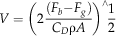 $$V=\left( {2{{(F_{b} {\minus}F_{g} )} \over {C_{D} \rrho A}}} \right)^{\wedge} {1 \over 2}$$
F g is the force of gravity (weight), and F b is the buoyant force; thus, F b−F g is the effective weight of the animal or the buoyancy. If the swimming velocity is greater than the sinking velocity then the animal would have been able to enter the water column (though potentially only for discreet periods of time) despite being negatively buoyant.
Results
Cadoceras
Volume data for the constituent parts of the ammonite are given in Supplementary Table 1. The buoyancy calculations (Fig. 3A) show a range of buoyancy values across the six potential scenarios from −2.47×10⁻⁷ N to 3.57×10⁻⁷ N (Supplementary Table 2). The Cadoceras hatchling displays a slight negative buoyancy for both minimum and maximum estimates of shell and soft-body volume, aptychi density and volume, and siphuncle volume in the first two scenarios (the protoconch and the protoconch plus one additional chamber). The formation of the third chamber is when near-neutral/slight positive buoyancy is attained under minimum estimates though maximum estimates only attain positive buoyancy with the fifth chamber. The variation of aptychus volume doesn’t make an effective difference in the buoyancy calculation (i.e., shift from negative to positive buoyancy) except in the third minimum scenario in which a near-neutral negative buoyancy becomes slightly positive with a decrease in the aptychus volume. Density calculations show the same results, near-neutral/positive buoyancy (density equal to or greater than seawater respectively) is reached between the third and fifth chamber.
Results of the buoyancy calculation for Cadoceras (A) and Spirula (B). The scenarios for Cadoceras correspond to the number of chambers/different lengths of the body chamber (see Methods and Fig. 2). The data lines correspond to minimum and maximum estimations of volumes and densities used to calculate buoyancy. Here we differentiate between the shell, soft tissue (soft body and siphuncle), and aptychus: “MinSST-MinA” corresponds to the minimum estimations of shell and soft tissue densities and volumes along with the minimum estimates for aptychus volume and density. A, B, C, D are the four negative buoyancy values used in the analysis of swimming and sinking velocities in Figure 4A. The body volume of Spirula was calculated using the ventral or dorsal mantle length. The Spirula hatchling is modeled with one, two and three chambers, each corresponding to different dimensions of the soft body.
The effect of the pellicle on the buoyancy calculation is minimal and does not shift any buoyancy result to positive or negative. The change in buoyancy is, at most, slightly less than 0.08% depending on scenario and density.
Tanabe (Reference Tanabe1975) notes that the weight of the septa ranges between 2% and 6% of the shell weight, based on measurements made by Trueman (Reference Trueman1941) of several ammonite genera. Our hatchling Cadoceras possesses a septal weight equivalent to 0.53%, 1.82%, 3.66%, 5.91%, and 9.10% of the ammonitella weight with one to five septa respectively. Shigeta (Reference Shigeta1993) uses several ratios to calculate buoyancy: shell thickness is 2% of the whorl height, the septal angle is a consistent 27°, the surface area of a septum is three times the whorl cross-section, and septum thickness is 2% of whorl height. Direct measurements of the Cadoceras hatchling reveal notable deviations from these ratios. Shell thickness is 6–9% of whorl height, expanding to 11% at the primary varix, with an average of 9%. The septal angle ranges between 20.2° and 28.9°, with an average of 22.6°. The septal surface area ranges from 11 to 20 times the whorl cross-section. Septal thickness is 2–3% of whorl height, with an average of 2.8%. Comparison of the protoconch volume using the method of Shigeta (Reference Shigeta1993), modeling the protoconch as the sum of parts of three ellipsoids, and the CT data show an error between 22.1% and 34.5%. This method consistently overestimated the volume of the protoconch. This range arises because the protoconch is not perfectly symmetrical and thus the radius for the ellipsoid changes slightly.
Raup and Chamberlain (Reference Raup and Chamberlain1967) re-derived the equations of Moseley (Reference Moseley1838) in terms of common shell parameters (eq. 3) and the equation used by Trueman (Reference Trueman1941) in terms of the same shell parameters (eq. 4).
 $$V=\left( {{2 \over 3}} \right)\rpi \left( {{{KR_{a} } \over {ln\,(W)}}} \right)\left( {1{\minus}W^{{{{{\minus}3\rtheta } \over {2\rpi }}}} } \right)$$
 $$V=\left( {{1 \over 3}} \right)(KR_{a} )\left( {\left( {\left( {{{4\rpi ^{2} } \over {ln\,(W)^{2} }}} \right){\plus}1} \right)^{\wedge} {1 \over 2}} \right)$$
W is the whorl expansion rate, K is the area of the aperture, R a is the distance between the coiling axis and the center of gravity of the aperture, and θ is the radians per revolution. Both equations give similar estimates of volume, 19431106.76 μm3 and 19571712.54 μm3, respectively (the true volume is 49354020 μm3). Both equations underestimate the true volume of shell material by about 2.5 times and have an error of 60%.
The maximum swimming speed and the potential minimum (respiratory) swimming speed are calculated using the equation of Chamberlain (Reference Chamberlain1981) and the observation by O’dor et al. (Reference O’dor, Wells and Wells1990) that the respiratory velocity of an adult Nautilus is roughly 20% of the maximum swimming speed. Under all tested C D the minimum swimming speed (respiratory velocity) is consistently lower than the sinking speed, whereas the maximum swimming speed is higher except for the most negatively buoyant result for scenario 1 (Fig. 4A).
A, The sinking and swimming velocities calculated for the negatively buoyant Cadoceras (using four negative buoyancy values, A–D) when the body chamber extends back to the protoconch (scenario 1). Sinking velocity is calculated via terminal velocity, swimming velocity is calculated via Chamberlain (Reference Chamberlain1981), and respiratory velocity is 20% of the swimming velocity (O’dor 1990). B, The change in position of Cadoceras in the water column (for scenario 1A), starting in an arbitrary position, through time based on the swimming/sinking velocities and the number of jet pulses per second. The changes in position are considered under the simplification that movement is strictly vertical. C, The relationship between increasing stability (ratio of the distance between the center of mass and the center of buoyancy to the total diameter of the shell) and velocity.
Discrepancy between the positions of the center of mass (center of the soft body) and the center of gravity (center of the shell and soft body) increase with decreasing length of the body chamber (Fig. 5A). The position of the center of gravity is less sensitive to changes in body chamber length relative to the center of mass (Supplementary Table 3). Stability for Cadoceras (calculated with the center of mass) ranges from 0.13 with the protoconch and five additional chambers (Fig. 5B) to 0.03 when the body chamber extends back to the protoconch (Fig. 5C). Nautilus has a stability around 0.09 (Parent et al. Reference Parent, Westermann and Chamberlain2014). Stability values can be used for evaluating potential swimming capability, with increasing stability favoring greater swimming ability; Figure 4C shows this in the hatchling Cadoceras, with increasing swimming speed coupled with increasing stability though not in a simple linear manner.
3-D models with the calculated centers of buoyancy, mass, and gravity. The blue diamond is the center of buoyancy, the dark blue dots are the center of mass, and the green squares are the center of gravity. B and C show the difference in life position of the hatchling depending on the length of the body chamber (Fig. 2C,D). Ammonite stability is often calculated using the center of buoyancy and the center of mass of the soft body. The center of gravity takes into account the shell and the distribution of septa. A shows the difference in position of the center of mass of the soft body and the center of gravity for three different body chamber lengths. The closer the center of gravity/mass is to the center of buoyancy, the longer the body chamber. As the length of the body chamber decreases the discrepancy between the center of mass and the center of gravity increases.
Sinking and swimming speeds can be used to model a simple scenario of a hatchling ammonite moving vertically in the water column and the number of jet cycles (pulses per second) necessary to ascend and maintain constant position in the water column (maintenance jetting). Figure 4B shows that to overcome a negative buoyancy of 0.07 µN (1 A) Cadoceras would need to jet at 1.34 pulses per second to maintain a constant position in the water column. To overcome a negative buoyancy of 0.2 µN (1C) Cadoceras would require about 28 pulses per second.
Spirula
Minimum and maximum assumptions both show an initial positive buoyancy with only the protoconch (Fig. 3B). Minimum estimates show an asymptotic decrease in buoyancy with the addition of the second and third chamber; however, maximum estimates show a near-linear decrease in buoyancy, and buoyancy becomes negative with the formation of the second chamber.
Discussion
Model Limitations
Original ammonite shell density is unknown, and could have been variable among ammonite groups as is shell density among existing cephalopods (Mutvei Reference Mutvei1983). It is reasonable then to model a range of potential densities where the range is derived from the densities of cephalopods that phylogenetically bracket ammonites (i.e., Nautilus and coleoids) (Ritterbush et al. Reference Ritterbush, Hoffmann, Lukeneder and De Baets2014). Small density differences seem to exist between the shell and septa in Nautilus, one study noted a small density difference between the outer shell and septa: 2.57 g/cm3 and 2.51 g/cm3, respectively (Hoffmann and Zachow Reference Hoffmann and Zachow2011). Our analysis makes no distinction in density between the shell wall and the septa because this difference would be minimal and the results would fall within the range covered by the minimum and maximum density calculations.
The aptychus is a significant unknown in this analysis, so we could only estimate its size and density. Moreover, nothing is known about the characteristics of the aptychus in a hatchling, and thus the sequence of development of the embryological jaws is speculative. Likewise, the relative volume of the jaws for the hatchling Cadoceras is unknown and only rough estimations could be made. The effect of the jaws relative to the shell and soft body is small, however, as can be seen in Figure 3A.
Like the shell, the density of the soft body of ammonites is unknown but can be estimated reasonably through phylogenetic bracketing. Also like the shell, the density of the constituent parts would vary (i.e., different tissues would have different densities). Therefore we model the soft body as a single density and vary that density between a minimum and maximum value. The range between minimum and maximum value should include the slight tissue density differences.
Additional aspects of the soft body also should be addressed. The volumes of the siphuncle and pellicle are small in comparison to the rest of the chamber and thus make a minimal difference to the final buoyancy calculation. The final soft-body consideration is how to account for the tentacles. The soft-body volume is taken as the volume of the body chamber, which would undoubtedly have accounted for most of the soft body. The hyponomic sinus has been documented in Devonian ammonoids at the edge of the post-embryonic shell, suggesting that the soft body extended to the edge of the aperture (House Reference House1965, Reference House1996). No modification to the calculations were made for additional material that may have been outside of the body chamber, i.e., the tentacles or the yolk (Boletzky Reference Boletzky2002). Because no preserved tentacles have been found (Klug et al. 2012; Ritterbush et al. Reference Ritterbush, Hoffmann, Lukeneder and De Baets2014), the morphology of the tentacles is unknown, and therefore it is unreasonable to speculate. Also unknown is whether the ammonite hatchlings possessed a yolk.
The volume of the soft body of Spirula is not constrained by a body chamber and estimations of the volume had to be based on the relationships reported by Bruun (Reference Bruun1943). The negative buoyancy trend seen for Spirula with maximal estimates for density and volume could represent an overestimation of the growth of the body volume relative to shell size caused by the poor understanding of the relative proportions of the hatchling animal. The animal had to be modeled as if the head had retracted into the mantle cavity because the length and volume of the head and tentacles relative to the mantle size are unknown (Chun Reference Chun1915; Bruun Reference Bruun1943). The siphuncular tube is preserved in Spirula, however, so its volume is directly quantifiable. As in Cadoceras, the jaw apparatus in Spirula is unknown and we estimated its volume in the same way as the volume of the Cadoceras aptychus.
Beyond the estimations of certain physical parameters, there are additional potential sources of error that have to be addressed. One is the state of preservation of the shell. Although the conch was “hollow,” secondary calcite crystals had formed along the inner and outer surface of the shell and septa. Owing to the high quality of the SRµCT data, we were able to identify these calcite deposits as well as the boundary between the shell and these secondary calcite deposits. Although diagenesis is corrected for (see Methods section) it is possible that further diagenetic alteration affected the geometry of the shell in imperceptible ways, though how this would affect the volume calculations is unknown but unlikely to be very appreciable. The transition from aragonite to calcite during diagenesis of the shell may also alter the shell volume by a small amount (Reyment Reference Reyment1958).
A source of error that is inevitable to some degree in quantitative tomographic work is the partial volume effect (PVE). CT images (see Fig. 1A,D) are constructed from voxels (three dimensional pixels), which are assigned a single value represented by a shade ranging from black to white depending on the material properties of the materials present in the volume of that voxel. Because each voxel is constrained to a single value if two or more materials are present in the volume represented by that voxel, the shade of the voxel will be an average of the material properties of all materials within that volume. The boundary between two materials, in this case the shell (white) and air (black), is represented by a gray gradient indicating a decreasing presence of the material of interest and an increasing volume of air present within the voxel. How this boundary condition is included in the segmentation can alter the final volume calculation. The effect of the PVE on different scan methods has been tested using the shell of Nautilus by Hoffmann et al. (Reference Hoffmann, Schultz, Schellhorn, Rybacki, Keupp, Gerden, Lemanis and Zachow2014). The PVE is minimized in our data through the use of high-resolution SRµCT and Nano-CT, in which only the first whorl of Spirula was in the field of view in order to maximize the resolution.
Life Habit
The modeling of variable body lengths in Cadoceras presents an opportunity to study the effect of body length on stability. As the distance between the center of mass and the center of buoyancy increases so too does the stability. Nautilus has a stability of about 0.09, brevidomic ammonoids have a stability of 0.05, and mesodomic ammonoids have a stability of 0.03 (Parent et al. Reference Parent, Westermann and Chamberlain2014). As the length of the body chamber decreases the stability of the animal increases, which can be seen in Cadoceras. It is interesting to note, however, that because the center of mass of ammonites is typically calculated using only the soft body and ignoring the shell, the resulting stability is artificially high for shorter body chambers, compared to ammonoids with body chambers extending back a full whorl (Fig. 5A).
The buoyancy calculation shows that the hatchling Cadoceras achieves neutral buoyancy between the third and fifth chamber depending largely on the density of the shell and the soft body, and on the volume and density of the aptychus. Depending on the number of chambers and the way chambers were added after hatching, either the hatchling possessed neutral buoyancy immediately after emerging from the egg or there was a shift in life habit early in ontogeny. The buoyancy calculation itself only models the animal floating in a still column of water and only tests whether the buoyant force generated by the animal is capable of overcoming the animal’s weight. Further calculations are necessary to integrate the potential swimming forces the animal could generate and wave motion (which would likely be more important for sub-millimeter hatchlings than for adult animals). We do not factor into these calculations any liquid that might have been in the protoconch (Landman et al. Reference Landman, Tanabe and Shigeta1996a); it is possible that the protoconch would have been emptied of liquid and filled with gas after hatching (Rouget and Neige Reference Rouget and Neige2001).
Specimens of Nautilus caught in the wild exhibit some degree of negative buoyancy that the animal can overcome and remain in the water column (Ward et al. Reference Ward, Stone, Westermann and Martin1977). It is possible some ammonites also would have exhibited this type of behavior, and therefore simply calculating a slight negative buoyancy (e.g., Ebel Reference Ebel1990) is insufficient to regulate the animal to a benthic life habit without considering the potential forces the animal could generate to counteract this negative buoyancy (i.e., swimming). To demonstrate this we compared the potential swimming speed to the potential sinking speed and found that within the possible range of swimming and sinking speeds, the juvenile could have overcome potential negative buoyancy if it had hatched with an insufficient number of chambers to allow neutral or positive buoyancy (Fig. 3A).
The calculation of the swimming speed of a hatchling Cadoceras is hypothetical, the equation of Chamberlain (Reference Chamberlain1981) was formulated and tested using an adult Nautilus, and the relationship between the swimming capabilities of an adult and a hatchling Nautilus are unknown. It should also be noted that the hatchling Cadoceras lives at low Reynolds numbers (both sinking and swimming speed were calculated at Reynolds numbers around 1), entering a realm where inertial forces begin to lose their importance, viscous forces dominate drag (Jacobs and Chamberlain Reference Jacobs and Chamberlain1996; LaBarbera Reference LaBarbera2008), and propulsive methods such as jet-driven motion become less efficient. Squid hatchlings, with a length of around 1 mm, face a similar propulsive problem (Staaf et al. Reference Staaf, Gilly and Denny2014). Squid paralarvae are negatively buoyant and compensate for this continual downward motion through periodic jet pulses, resulting in what can be referred to as hop-and-sink behavior also seen in other negatively buoyant zooplankton (Haury and Weihs Reference Haury and Weihs1976). The calculated sink/swim data (Fig. 4A) of the hatchling Cadoceras potentially fits with a similar “hopping” swimming mode of juvenile squids if the hatchling was negatively buoyant.
Squid paralarvae have been shown to swim at an average speed of 0.00268 m/s, with a range of 0.00146 m/s to 0.00484 m/s (Bartol et al. Reference Bartol, Krueger, Stewart and Thompson2009). Staaf et al. (Reference Staaf, Gilly and Denny2014) measured sinking speed of paralarvae at around 0.0046 m/s. Thus, in terms of sinking velocities the order of magnitude and range for squid hatchlings are similar to those of the hatchling Cadoceras (Fig. 4A). Figure 4B shows that Cadoceras would need roughly 1.34 pulses per second with a negative buoyancy of 0.071 µN (and 2.11 pulses per second with 0.099 µN) to maintain a constant position in the water column (maintenance jetting); the observed jet frequency of squid hatchlings during maintenance jetting is 2.13 Hz (Staaf et al. Reference Staaf, Gilly and Denny2014). Jet frequency during swimming in adult Nautilus is, on average, 1.67 pulses per second with an observed maximum of 2.67 pulses per second (Ward Reference Ward1987). Scenario 1C would force Cadoceras to maintain 28 pulses per second for maintenance jetting, a number likely far too high for the animal to accommodate and thus under this scenario Cadoceras would not be able to maintain a position in the water column. It should be noted that ammonite soft parts are rarely preserved and their physiology is unknown and therefore any reconstruction of swimming speed is speculative (Ritterbush et al. Reference Ritterbush, Hoffmann, Lukeneder and De Baets2014). Comparison to coleoid paralarvae may be more appropriate than comparison to baby Nautilus in terms of the lifestyle of the hatchling, owing to the differences in total diameter; a Nautilus hatchling has a shell diameter of around 26 mm—two orders of magnitude larger than the embryonic shell diameter of ammonites (Arnold et al. Reference Arnold, Landman and Mutvei2010; De Baets et al. Reference De Baets, Klug, Korn and Landman2012). Under the majority of estimations and tested scenarios results recover near-neutral/positive buoyancy and negative buoyancy that can be countered by motion; this indicates that hatchling Cadoceras was most plausibly planktonic, which fits well with the recovery of ammonitella in anoxic conditions, i.e., black shale (Mapes and Nützel Reference Mapes and Nützel2009) and comparisons with similarly sized cephalopod hatchlings (e.g., Calow Reference Calow1987). A planktonic mode of life would aid in rapid and global dispersion via ocean currents, which has been proposed as one mechanism for survival after mass extinctions (Ward and Bandel Reference Ward and Bandel1987; Harries et al. Reference Harries, Kauffman and Hansen1996; Tajika and Wani Reference Tajika and Wani2011; De Baets et al. Reference De Baets, Klug, Korn and Landman2012, Ritterbush et al. Reference Ritterbush, Hoffmann, Lukeneder and De Baets2014). The achievement of a planktonic life habit in the hatchling ammonite, coupled with the potential mass spawning behavior (Manger et al. Reference Manger, Stephen and Meeks1999; Walton et al. Reference Walton, Korn and Klug2010; De Baets et al. Reference De Baets, Klug, Korn and Landman2012; Ritterbush et al. Reference Ritterbush, Hoffmann, Lukeneder and De Baets2014), could lead to early and rapid dispersal. It is interesting to note that simple hydrodynamic models of current dispersal of chokka squid paralarvae show that slight negative buoyancy mitigates wave action and could allow paralarvae to stay close to their hatching location (Martins et al. Reference Martins, Roberts, Lett, Chang, Moloney, Camargo and Vidal2013). This could be beneficial if they hatch in nutrient-rich waters.
Summary
The application of very high-resolution SRµCT demonstrates the utility of computed tomography to high-precision calculations of ammonite buoyancy and the integration of buoyancy results to potential swimming velocities in order to test hypotheses of ammonite life habits. This CT method allows shell volume to be considered without loss of inherent biological variability or fine details that would necessarily be eliminated through mathematically based modeling. A Cadoceras hatchling could achieve neutral/positive buoyancy provided the ammonitella had three to five chambers (including the protoconch), whereas the Spirula hatchling had positive/neutral buoyancy with just the protoconch, though it may have hatched with two to three septa (Lukeneder et al. Reference Lukeneder, Harzhauser, Muelleffer and Piller2010). However, minimum estimations of shell and soft tissue densities indicate that the Cadoceras hatchling had sufficient swimming power to enter the water column with just the protoconch.
The high resolution of this method and flexibility of digital tomographic data enables the accounting and assessment of minute, sub-micron details; the assessment of relationships in the size, area, and volume of the shell components; and the modification of shell parameters and morphology in order to test functional hypotheses.
Some have contested, on the basis of recovery of negative buoyancy values, that ammonites were benthic organisms (Wetzel Reference Wetzel1959; Ebel Reference Ebel1990; Rein Reference Rein1999; Delanoy et al. Reference Delanoy, Magnin, Selebran and Selebran1991); however, we have demonstrated here that calculation of negative buoyancy is insufficient to deem an ammonite benthic, because swimming capacity can operate to overcome the gravitational force acting on negatively buoyant animals.
Ammonites are a very diverse and long-lived group and the heteromorph ammonites (heteromorph refers to the non-planispiral shell) show great shell disparity; there is no necessary reason that all heteromorphs must conform to the same life habit. Indeed the speculated modes of life of heteromorphs are diverse, from sessile and mobile benthic animals (Diener Reference Diener1912; Klinger Reference Klinger1981; Monks and Young Reference Monks and Young1998; Shevyrev Reference Shevyrev2005), nektobenthic organisms with persistent negative buoyancy (Higashiura and Okamoto Reference Higashiura and Okamoto2012), and burrowers (Frech Reference Frech1915), to rapid swimmers (Matsumoto and Obata Reference Matsumoto and Obata1962; Westermann Reference Westermann2013). Provided suitable specimens, this CT method can be used to determine the fundamental capacity of the ammonite phragmocone to provide sufficient buoyancy as to allow a planktonic or nektonic mode of life. The insight provided through this method expands our understanding of ammonite paleobiology—specifically, how ammonites might have interacted with their environment—as well as having implications for the interpretation of geochemical data derived from ammonite shells (Ritterbush et al. Reference Ritterbush, Hoffmann, Lukeneder and De Baets2014).
Acknowledgments
This project was funded by the Deutsche Forschungsgemeinschaft (grant number HO 4674/2-1). The authors would like to thank M. Ehlke (Zuse Institute, Berlin) for help with visualization and data management. We also thank X. Xiao (Advanced Photon Source, Argonne National Laboratory, Lemont, Illinois, U.S.A.) and E. Rybacki (Deutsches Geoforschungs Zentrum, Potsdam) for scanning assistance. We greatly appreciate the assistance of V. Mitta (Paleontological Institute of the Russian Academy of Science, Moscow) for assistance during fieldwork. We thank K. De Baets (Friedrich-Alexander Universität Erlangen Nürnberg, Germany) and D. Korn (Museum für Naturkunde, Berlin) for their helpful reviews.
Supplementary Material
Supplemental materials deposited at Dryad: doi:10.5061/dryad.fc4vf



