Aortic arch obstruction is relatively common, typically presenting in newborn infants with an incidence approaching 1:2000.Reference Hoffman, Kaplan and Liberthson1 The morphological spectrum is quite varied and accompanying a discrete coarctation shelf of ductal tissue there is not infrequently aortic arch isthmal and transverse arch tubular hypoplasia. Similar to most congenital heart defects, aortic arch obstruction occurs either in isolation or in association with other cardiac anomalies. Surgical repair of native aortic arch obstruction remains the therapy of choice in infants and young children, even though endovascular management options are increasingly being used in older children and adults.Reference Sakopoulos, Hahn, Turrentine and Brown2 Aortic angioplasty for coarctation of the aorta in infants, although possible, has been shown to be associated with a high incidence of recurrence and remains controversial.Reference Park, Lucas, Sklansky, Kashani and Rothman3 The majority of infants therefore undergo surgical repair by end-to-end anastomosis or subclavian flap through a lateral thoracotomy incision. If coexisting transverse arch hypoplasia is recognised, an extended end-to-end anastomosis and arch repair may be preferred through a median sternotomy. Surgical techniques for repair of coarctation of the aorta have advanced such that mortality is now low.Reference Sakopoulos, Hahn, Turrentine and Brown2 However, re-coarctation and failure to adequately treat transverse arch hypoplasia leaves an appreciable number of children with residual arch obstruction with its potential for severe morbidity in later life.Reference Merrill, Hoff, Stewart, Elkins, Graham and Bender4 It is in this setting that endovascular stent therapy in older children and adults could be a useful adjunct allowing further surgery to be avoided while ensuring significant residual gradients post surgery with their associated longer-term risks to be successfully managed. Endovascular bare-metal stents placed transcatheter have been used to treat re-coarctation, native coarctation, and aortic arch obstruction for over a decade.Reference Forbes, Moore and Pedra5 There remains concern, however, that bare-metal stents may be associated with aortic aneurysm formation with a longer-term risk of aortic rupture.Reference Chessa, Carrozza and Butera6 Polytetrafluoroethylene-covered stents may thus be preferred, but are relatively contraindicated if placed in such a position as to occlude head and neck vessels. Our preference is to use covered stents where possible, and here we report partially uncovering and “dove-tailing” stents to treat complex arch obstruction in two patients.
Case 1
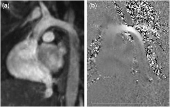
A 16-year-old, 112-kilogram boy attended for cardiac catheter assessment of his aortic arch following documented evidence of hypertension and poorly palpable femoral pulses during clinic follow-up over the preceding year. A neonatal end-to end surgical repair of coarctation had been performed, but significant residual obstruction was evident during early follow-up. Balloon angioplasty had been performed on two occasions, 12 and 3 years previously. Magnetic resonance imaging assessment before this study suggested significant aortic arch obstruction secondary to arch hypoplasia after the left common carotid artery with more discrete stenosis after the left subclavian artery (Fig 1).
Magnetic resonance images of case 1 before endovascular stent placement. (a) Whole heart steady state free precession oblique image demonstrating the change in calibre of the aortic arch after the origin of the brachiocephalic and left common carotid artery. (b) Flow still image of the aortic arch shows that flow acceleration of blood begins in the aortic arch before the origin of the left subclavian artery, confirming the suspicion of tubular arch hypoplasia seen in (a) and also demonstrated angiographically in Fig 2a.

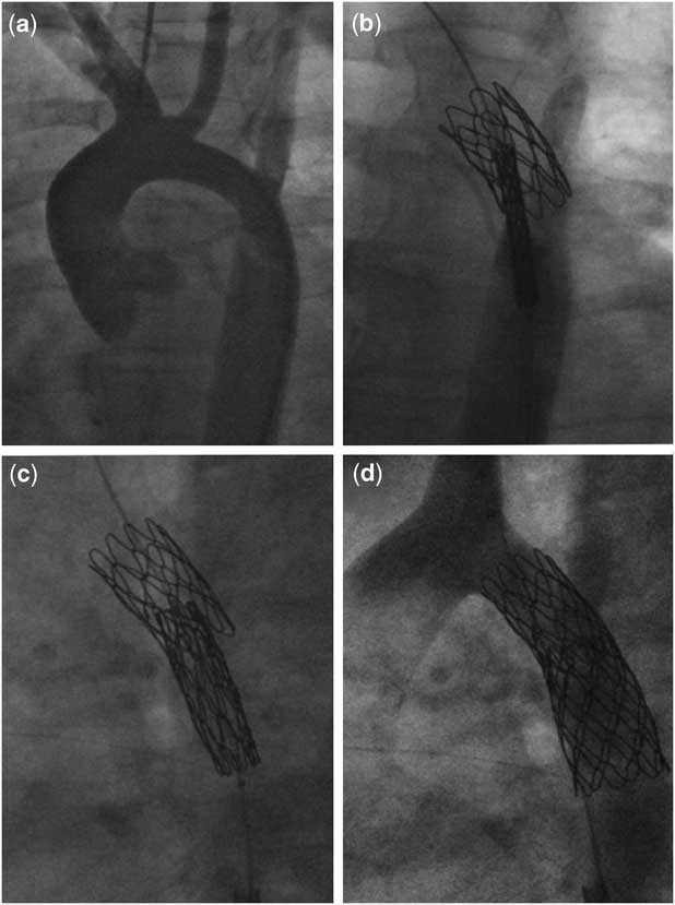
The catheter study was performed under general anaesthesia following patient consent. Access was obtained with an 8-French Terumo sheath (Terumo Corporation, Tokyo, Japan) in the right femoral artery using ultrasound guidance and a 4-French sheath in the right femoral vein. Simultaneous ascending and descending aortic pressures documented a systolic gradient around the aortic arch of 37 millimetres of mercury. A 5-French multi-purpose catheter (Cook Incorporation, Bloomington, United States of America) was used to place a 0.035-inch extra-stiff amplatz wire (Boston Scientific, Massachusetts, United States of America) in the right subclavian artery, and a 7/5 multi-track catheter (NuMed Incorporation, Ontario, Canada) was used for angiograms in a 20° left anterior oblique and straight lateral projection (Fig. 2a). The aortic arch narrowed to 9 millimetres after the origin of the left common carotid artery, with further narrowing after the left subclavian artery before increasing to 16 millimetres as the descending aorta. The overall length of obstruction exceeded 40 millimetres.
Lateral angiograms demonstrating placement of two endovascular partially uncovered covered stents to relieve significant arch hypoplasia and recoarctation. (a) The angiogram was obtained though a 5-French pigtail catheter placed retrograde from the right femoral artery. There is significant hypoplasia of the aortic arch between the left common carotid artery and the left subclavian artery. (b) Following removal of a small segment of the distal polytetrafluoroethylene covering the first stent has been carefully positioned and a second stent with a small segment of the proximal polytetrafluoroethylene covering removed is being manipulated into position. (c) The inner balloon of the BiB catheter has been inflated, allowing the second stent position to be carefully altered. (d) A satisfactory angiographic result is obtained with the two overlapping stents. Flow is seen in the left subclavian and left common carotid artery.
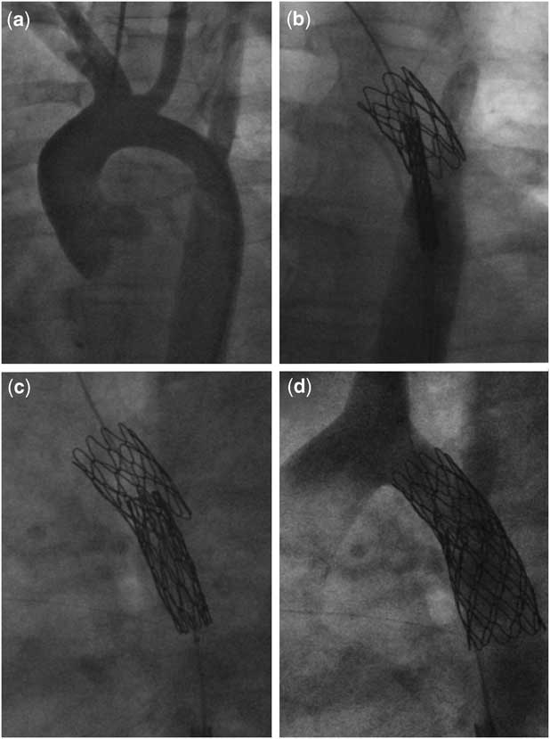
An 11-French-long Mullins sheath (Cook Incorporation, Bloomington, United States of America) was placed in the right femoral artery and a 14 millimetre × 3.5 centimetre BiB balloon (NuMed Incorporation, Ontario, Canada) was used to deliver a 28-millimetre covered Cheatham Platinum stent (NuMed Incorporation, Ontario, Canada) with the distal 10 millimetres of polytetrafluoroethylene removed. Removing polytetrafluoroethylene from the stent is straightforward using forceps, a scalpel, and fine scissors without disrupting the integrity of the remaining polytetrafluoroethylene. The stent was positioned with the leading edge adjacent to the origin of the left common carotid artery such that the uncovered part of the stent distally lay across the left subclavian artery. Repeated hand angiograms before and during inflation ensured stable and accurate positioning of the stent (Fig 2b). A second 34-millimetre Cheatham Platinum stent (NuMed Incorporation, Ontario, Canada) with the proximal 15 millimetres of polytetrafluoroethylene removed was then delivered on a 16 millimetre × 4 centimetre BIB balloon, with the leading edge of the stent lying approximately 10 millimetres inside the first stent (Fig 2c). This stent was delivered and the balloon removed. Repeat angiography demonstrated a satisfactory result without loss of flow in the left subclavian artery (Fig 2d). Pullback systolic gradient was 14 millimetres of mercury appearing midway through the two stents. Further balloon inflation of the stents will be performed in 6 months’ time.
Case 2
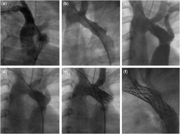
A 10-year-old, 33-kilogram boy attended for cardiac catheter assessment of his aortic arch following documented evidence of hypertension and echocardiographic evidence of residual arch obstruction during clinic follow-up in the context of multiple previous catheter procedures. Presentation was during the neonatal period with double outlet right ventricle and coarctation of the aorta. The ventricular septal defect and aortic coarctation were both repaired in early infancy with a pericardial patch. Residual aortic arch obstruction was evident in early childhood, with two angioplasty procedures performed in the second year of life (Fig 3a). This afforded some relief of obstruction, which was further assessed and treated with a bare-metal stent (EV3 Max LD intrastent, Plymouth, Minnesota, United States of America) at 5 years of age with stent re-dilatation 2 years previously (Fig 3b and c).
Left anteroposterior angiograms of a patient presenting with complex aortic obstruction following patch repair of aortic coarctation. Balloon angioplasty followed by bare-metal stent placement and lastly two partially covered stents have been placed to adequately restore the integrity of the aorta while attempting to preserve flow in the branch head and neck vessels. (a) The angiogram was obtained from the right femoral artery with a multi-track catheter (NuMed Incorporation, Ontario, Canada) over a 0.035-inch extra-stiff amplatz wire (Boston Scientific, Massachusetts, United States of America) in the right subclavian artery. Discrete stenosis is seen after the left subclavian artery, but the arch is also hypoplastic between the left common carotid artery and the left suclavian artery. (b) A bare-metal stent – Max LD Intra-stent (EV3, Plymouth, Minnesota, United States of America) has been placed, which begins to open up the arch but is too short to have adequately treated both the proximal hypoplasia and the distal discrete obstruction. No branch vessels have been crossed. (c) The stent has been further dilated at a subsequent catheter with an acceptable result, although distal obstruction remains. (d) With time, the proximal and distal obstructions have not improved and more definitive endovascular therapy is planned with placement of two partially covered Cheatham Platinum stents (NuMed Incorporation, Ontario, Canada) (e, f). (e) The proximal stent has had one zig of covering removed from its distal portion and has been positioned just distal to the left common carotid artery. (f) The second stent with partial removal of the proximal polytetrafluoroethylene covering has then been placed partially inside the first to adequately treat the distal obstruction while allowing for flow to the left subclavian artery.

The catheter study was performed under general anaesthesia following patient consent. Access was obtained with an 8-French Terumo sheath (Terumo Corporation, Tokyo, Japan) in the right femoral artery. A 5-French right coronary catheter (Cook Incorporation, Bloomington, United States of America) was used to place a 0.035-inch extra-stiff amplatz wire (Boston Scientific, Massachusetts, United States of America) in the right subclavian artery passing through the existing stent, and an 8/6 multi-track catheter (NuMed Incorporation, Ontario, Canada) was used for angiograms in an anteroposterior and straight lateral projection (Fig 3d). A systolic gradient of 13 millimetres of mercury between the transverse and descending aorta was documented. Angiography demonstrated two small aneurysms adjacent to the site of the existing stent with discrete re-coarctation just distal to the left subclavian artery.
The 8-French-short sheath was exchanged for a 12-French-long Mullins sheath (Cook Incorporation, Bloomington, United States of America) and a 39-millimetre covered Cheatham Platinum stent (NuMed Incorporation, Ontario, Canada) cut to 20 millimetres was delivered on a 14 millimetre × 4 centimetre Z-med balloon (NuMed Incorporation, Ontario, Canada) to cover both aneursyms within the existing stent (Fig 3e). A second 39-millimetre covered Cheatham Platinum stent (NuMed Incorporation, Ontario, Canada) with 5 millimetres of the proximal polytetrafluoroethylene removed was positioned on a 14 millimetre × 4.5 centimetre BiB balloon (NuMed Incorporation, Ontario, Canada), with the proximal end of the uncovered stent purposefully covering the left subclavian artery. Following stent deployment, repeat angiography demonstrated a satisfactory result with exclusion of aortic aneurysms and resolution of distal arch obstruction (Fig 3f). Flow was seen in both the left common carotid artery and left subclavian artery. At the end of the procedure, there was a pullback systolic gradient of 9 millimetres of mercury through both stents.
Discussion
The majority of surgical repairs of coarctation of the aorta are performed in early infancy on very small and occasionally complex aortic arches. Older children and adult repairs can provide an equal challenge, with concerns of spinal cord damage increasing in relation to the length of aortic cross-clamp. The preferred surgical strategy for repair of neonatal coarctation of the aorta remains controversial with a trend towards end-to-end repair in recent years.Reference Kaushal, Backer and Patel7 Patch aortoplasty is sometimes still necessary and is associated with long-term concerns of aortic integrity and aneurysm formation.Reference Botta, Russo and Oppido8 Given the formidable challenge of the variable morphological substrate, it is not surprising that the most recently published surgical series continue to report need for re-operation or re-intervention.Reference Kaushal, Backer and Patel7 Conservative management of residual gradients is not an attractive option as it is associated with long-term uncontrolled hypertension, the risk of which may be reduced to some extent by adequately treating obstructions.Reference English9 It is in this context that endovascular and repeat surgical therapies need to be evaluated.
Balloon angioplasty was initially heralded as the panacea that would address the issues of residual arch obstruction and is still the therapy of choice in small infants and children too small for endovascular stent therapy. Balloon angioplasty results in disruption of the aortic intima and media in a relatively uncontrolled manner that risks aortic dissection and aneurysm formation. By comparison, bare-metal stents result in reduced aortic damage and a more satisfactory relief of aortic obstruction.Reference Forbes, Moore and Pedra5 However, although reduced, an appreciable risk of aneurysm formation remains.Reference Forbes, Garekar and Amin10
Our preference and that of others is to use covered stents where possible.Reference Kenny, Margey, Turner, Tometzki, Walsh and Martin11–Reference Tanous, Collins, Dehghani, Benson and Horlick13 Difficulty arises when the site of aortic obstruction involves the branching vessels, as in the two cases presented here. There are several options to consider: a bare-metal stent can be used, a partially uncovered stent can be deployed as demonstrated here, surgical options can be reconsidered, the polytetrafluoroethylene can be punctured with access obtained retrograde from the branching vessel, and the opening created dilated with a balloon or the vessel can be covered.Reference Tsai, Hill and Cheatham14
There are several limitations of this technique that need to be considered. Removing part of the polytetrafluoroethylene covering removes the adhesive attachment of the covering at one end of the stent. When the stent is introduced through a sheath, there would be the potential for the polytetrafluoroethylene to crowd down to the distal adhesive covering and there would be no radiographic way of demonstrating that the integrity of the covering was intact. Cutting stents needs to be done with great care and introduces the possibility of leaving sharp edges that could disrupt the integrity of the vessel. It would have been useful to place a left radial, brachial, or axillary arterial line in both cases. This would have enabled intervention should the covering have inadvertently covered the vessel and also real-time assessment of left-arm blood pressure to assess for this possibility. This is now our routine practice for such complex arch obstruction. The follow-up of such complex cases is of equal importance to performing the procedure itself. Assessment of benefit in terms of blood pressure control necessitates occasional 24-hour blood pressure monitoring. Surveillance magnetic resonance imaging or computed tomography for stent and aortic integrity is our routine practice and is probably more informative than transthoracic echocardiography in older patients. We perform a magnetic resonance imaging study at 6 months and 1 year following complex intervention.
In summary, we propose that partial uncovering of a single or multiple covered stents is the simplest and quickest strategy to achieve a satisfactory result without incurring the risks of placing further sheaths in distal branch vessels or entrapping or rupturing balloons in the tines of the stent with attempts at perforating the polytetrafluoroethylene.