Introduction
As a highly efficient, non-toxic, and highly active photocatalyst, TiO2 has broad application prospects. It has a large forbidden bandwidth (3.28 eV), however, and its photocatalytic response is only in the ultraviolet and not in the visible region. TiO2 powder has the tendency to agglomerate and is difficult to recycle. These shortcomings greatly limit the practical applications of TiO2 powder as a photocatalyst.
An effective method to increase the photocatalytic activity of TiO2 is to dope with different elements, including metals (such as Cu, Fe, Au, and Pt) (Gomes et al., Reference Gomes, Leal, Bednarczyk, Gmurek, Stelmachowski and Zaleska-Medynska2017; Obregón et al., Reference Obregón, Muñoz-Batista, Fernández-García, Kubacka and Colón2015) and nonmetals (such as F, B, C, and N) (Asahi et al., Reference Asahi, Morikawa, Ohwaki, Aoki and Taga2001; Wang et al., Reference Wang, Huang, Dai and Whangbo2012; Yu et al., Reference Yu, Wu, Liu, Dionysiou, Yang, Wang and Liu2020). Among these elements, N-doping has been considered to be the most promising way to enhance the visible-light response of TiO2 (Jin Wang et al., Reference Jin Wang, Lewis, Hong, Manivannan, Zhi, Li and Wu2009). A previous study (Sato, Reference Sato1985) found that N-doped TiO2 exhibits visible-light activity, which presents the first example for modified TiO2 with visible-light activity. N-doped TiO2 was prepared successfully by spraying a gas mixture of N2/Ar into TiO2 (Asahi et al. (Reference Asahi, Morikawa, Ohwaki, Aoki and Taga2001). Since then, N-doped TiO2 has attracted much research attention (Asahi et al., Reference Asahi, Morikawa, Irie and Ohwaki2014; Gomes et al., Reference Gomes, Lincho, Domingues, Quinta-Ferreira and Martins2019; Huang et al., Reference Huang, Zhang, Wang, Liu and Sun2011).
Methods for the preparation of N-doped TiO2 include the sol-gel process, hydrolysis, liquid precipitation, iron implantation, vapor deposition, and templating (Joshi et al., Reference Joshi, Mangrulkar, Tijare, Padole, Parwate, Labhsetwar and Rayalu2012). These techniques use various N sources as precursors. N-doped TiO2 was prepared by Sun et al. (Reference Sun, Hu, Zheng, Zhang and Sheng2019) who detected large amounts of NO x on the TiO2 surface, which were bound via O–Ti–N bonds. Furthermore, Sun et al. (Reference Sun, Hu, Zheng, Zhang and Sheng2019) proposed that the amount of N-doping in TiO2 depended greatly on the form of N (e.g. NH3) volatilized from the composite. The effect of N-doping on the morphological structure and photocatalytic activity of TiO2 was studied systematically by Park et al. (Reference Park, Kim, Park, Tachikawa, Majima and Choi2009), revealing that Ti–O–N bonds are formed with TiO2 after N-doping, which endowed TiO2 with visible-light photocatalytic activity. Furthermore, N-doping could inhibit the growth of TiO2 grains and increase the phase transition temperature of anatase to rutile.
Many reports have been published on the microscopic mechanism of the visible-light response of TiO2 induced by N-doping, including orbital hybridization theory, independent energy level theory, and oxygen vacancy theory (Asahi et al., Reference Asahi, Morikawa, Irie and Ohwaki2014). Among these theories, oxygen vacancy theory is the one most widely recognized. According to this theory, N enters the TiO2 lattice to replace a certain number of the lattice O atoms during the roasting process, leading to visible-light absorption. According to Jansen and Letschert (Reference Jansen and Letschert2000a), O in metal oxide can be replaced by N to form a yellow oxynitride with visible-light response. Huang et al. (Reference Huang, Zhang, Wang, Liu and Sun2011) prepared an N-modified TiO2 photocatalyst by a modified sol-gel method and studied the degradation of potassium dichromate solution by this modified catalyst under visible-light irradiation. The results showed that urea decomposed to form a sensitizing cyanuramide at 400°C, resulting in a visible-light responsiveness of N-TiO2.
However, N-doping could neither solve the problem of agglomeration nor that of recycling of TiO2. The most popular solution to these problems was to incorporate TiO2 in a diatomaceous matrix (Chen & Liu, Reference Chen and Liu2016), sericite (Zhou et al., Reference Zhou, Lv, Guo, Xu, Wang, Zheng and Wu2012), zeolite (Jansson et al., Reference Jansson, Suárez, Garcia-Garcia and Sánchez2015), montmorillonite (Kun et al., Reference Kun, Mogyorosi and Dekany2006), muscovite (Yao et al., Reference Yao, Hongjuan, Tongjiang, Hao, Yating and Li2019), etc. Most of the existing studies have improved the photocatalytic performance of TiO2 by a single method. Research on the photocatalytic effect of TiO2 by employing both ion doping and muscovite loading have not been reported yet.
The objective of the present study was to improve the photocatalytic performance of TiO2 under visible light by means of incorporating nitrogen-doped TiO2 into muscovite as a nanocomposite. The hypothesis was that the influence of N substitution and muscovite loading on the photocatalytic performance of such composite materials could be verified through measurements of the structure, morphology, chemical bonding, and light absorption properties of the composite material.
EXPERIMENTAL SECTION
Sample Preparation
The muscovite used was provided by Chenxing Co., Ltd, Shijiazhuang Province, China. Urea was purchased from Chengdu Kelong Co., Ltd., Chengdu Province, China.
The composite Ti(OH)4/muscovite was prepared according to a previously published procedure (Yao et al., Reference Yao, Hongjuan, Tongjiang, Hao, Yating and Li2019), and mixed uniformly with urea at a 1:1 mass ratio. Then, the mixture was calcined in air at a selected temperature (400, 500, 600, or 700°C) for 2 h. The corresponding N-doped TiO2/muscovite nanocomposite was obtained after cooling to room temperature. The nanocomposites were labeled as N-TM-T, where N-TM stands for N-doped TiO2/muscovite nanocomposite, and T stands for the calcination temperature. A control was prepared by calcining the starting Ti(OH)4/muscovite composite in air in the absence of urea at 700°C and labeled as TM-700.
Characterization
X-ray diffraction (XRD) patterns were obtained with a PANalytical X’Pert Pro multifunctional powder diffractometer (PANalytical B.V., Eindhoven, The Netherlands) using CuKα radiation at 40 kV and 40 mA, step width of 0.02°2θ, integration time of 1 s, and scanning range from 3 to 80°2θ.
The micromorphology of the samples was observed using a Zeiss Ultra55 field emission scanning electron microscope (Carl Zeiss NTS GmbH, Oberkochen, Germany). The operating voltage was 15 kV, and the energy spectrum was resolved with a resolution larger than 127 eV (MnKα; count rate: 20,000 cps) and the peak shift was <1 eV (MnKα).
The functional groups of the nanocomposites were characterized by Fourier-transform infrared (FTIR) absorption spectrometry (Nicolet-5700 spectrometer; Thermo Fisher Scientific, Waltham, Massachusetts, USA). The samples for FTIR analysis were prepared by the KBr pellet method, and the data collection range was 4000–400 cm–1.
Optical absorption properties of the samples were analyzed using a Shimadzu Solidspec-3700 solid UV-Vis-NIR spectrophotometer (Shimadzu, Kiya-cho, Nijiyanan, Japan). The resolution was 0.1 nm, the scanning range was between 200 and 800 nm, and BaSO4 was the reference sample.
The intermediate degradation products were identified by high-performance liquid chromatography-mass spectrometry (HPLC-MS, Agilent Technologies, Inc., Santa, Clara, California, USA).
The photocatalytic performances of the nanocomposites were analyzed according to the following procedure. A 500 W xenon lamp (Beijing China Education Au-Light Co., Ltd, Beijing, China) in the visible range of 400–800 nm was used as the illumination source for photocatalytic reactions. A certain amount of photocatalytic material was dispersed in 20 mg/L RhB solution and placed on a magnetic stirrer. The light source was placed 10 cm above the solution. After stirring in the dark for 60 min, the light source was turned on and the supernatant was removed every 20 min. After the reaction, the supernatant was centrifuged to remove catalyst particles. The absorbance of the supernatant was measured on an Evolution 300 UV-Vis spectrophotometer (Shimadzu, Kiya-cho, Nijiyanan, Japan) at the maximum absorption wavelength of RhB (554 nm). The concentration change of RhB during the reaction was determined based on the absorbance at the maximum wavelength of the RhB solution, and the degradation rate of RhB was calculated from Eq. 1. The first-order kinetic equation (Eq. 2) was used to fit the photocatalytic data of the samples.


where A 0 and A t are the absorbance values of RhB at 554 nm, before and after degradation, respectively, and C 0 and C t represent the RhB concentrations before and after the degradation, respectively; k is the first-order reaction constant and t represents the reaction time.
RESULTS And DISCUSSION
Crystal Structures of N-TiO2/muscovite Nanocomposites
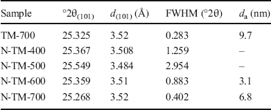
The XRD patterns of N-TiO2/muscovite nanocomposites calcined at various temperatures (Fig. 1) revealed that TiO2 was present in the form of anatase in all the samples. The anatase grain size was calculated by the Scherrer formula (Patterson, Reference Patterson1939), and the results (Table 1) indicated that, compared with the TiO2/muscovite nanocomposites, the grain sizes of the corresponding N-doped nanocomposites obtained at the same calcination temperature were reduced significantly. N-doping, therefore, inhibits the growth of TiO2 crystals. No rutile phase was present in any of the samples, indicating that N-doping retards the phase transition of TiO2 from anatase to rutile. These phenomena were consistent with previous reports (Gomes et al., Reference Gomes, Lincho, Domingues, Quinta-Ferreira and Martins2019).
XRD patterns of N-doped TiO2/muscovite nanocomposites calcined at various temperatures

Crystallographic data for anatase in the N-TiO2 /muscovite nanocomposite calcined in air at various temperatures

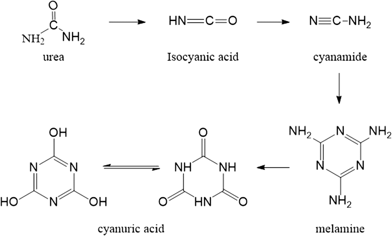
No urea diffraction peaks were detected, indicating complete decomposition of urea. However, the sample calcined at 400°C showed two main diffraction peaks at 26.2 and 29.8°2θ, which were assigned to melamine (Mitoraj & Kisch, Reference Mitoraj and Kisch2008) (pdf 00–024–1654). Hauck et al. (Reference Hauck, Jentys and Lercher2007) reported that melamine was formed by the gradual substitution of the amine group in isocyanic acid by the hydroxyl group. Furthermore, N-TM-500 exhibited a peak at 29.7°2θ, which was assigned to cyanuric acid (Jansen & Letschert, Reference Jansen and Letschert2000a, Reference Jansen and Letschert2000b). This indicated that melamine and cyanuric acid were both intermediate products of the decomposition of urea with increasing temperature. Peaks from impurities were assigned to N-containing heterocyclic organic compounds formed as side products in the pyrolysis of urea. One may conclude, therefore, that urea was decomposed and converted into the three intermediates of isocyanic acid: cyanamide, melamine, and cyanuric acid, which were then finally transformed into the N-doped TiO2/muscovite nanocomposite.
FTIR Analysis
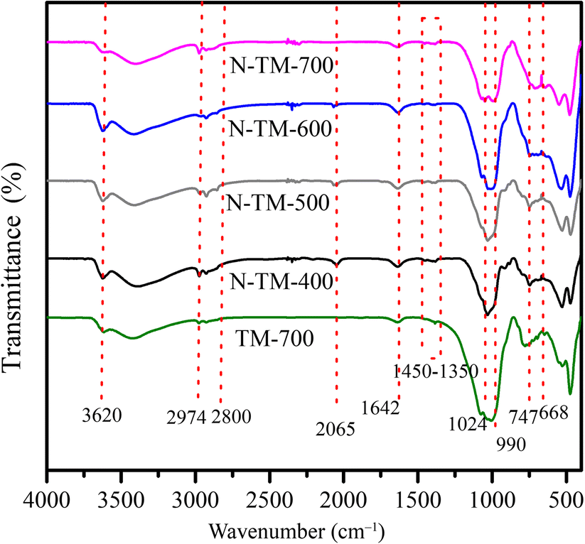
The FTIR spectra of all samples are shown in Fig. 2. The peak at 3620 cm–1 was ascribed to the structural hydroxyl groups of the 2:1 muscovite (Madejová, Reference Madejová2003). This absorption peak decreased with increasing temperature. Absorptions in the region of 3500–3400 cm–1 were assigned to the stretching vibration of the O–H bond of adsorbed water. The peak at 1024 cm–1 corresponded to the Si–O bond. Peaks in the range of 2974–2800 cm–1 corresponded to C–O, which proved the presence of cyanuric acid (Shi et al., Reference Shi, Liang, Wang, Ma and Sun2014). The peak at 2065 cm–1 belonged to HNCO (Schaber et al., Reference Schaber, Colson, Higgins, Thielen, Anspach and Brauer2004); its intensity weakened with increasing temperature and disappeared at 700°C. The peak at 1642 cm–1 was related to the C=O bond (Schaber et al., Reference Schaber, Colson, Higgins, Thielen, Anspach and Brauer2004) and was overlapped by the absorbance peak related to the flexural vibration of adsorbed water on the surface of the undoped nanocomposite. The peak at 1400 cm–1 was attributed to the stretching vibration of the triazine ring (El-Gamel et al., Reference El-Gamel, Wagler and Kroke2008; Huang et al., Reference Huang, Zhang, Wang, Liu and Sun2011; Chagas et al., Reference Chagas, Pfeifer, Rocha and Teixeira da Silva2012; Jun et al., Reference Jun, Lee, Wang, Hong, Stucky and Thomas2013) of melamine and cyanuric acid. The intensity of this 1400 cm-1 peak decreased with increasing temperature, indicating that an increasing number of isocyanic acids decomposed to form N-doped TiO2. A summary of the foregoing analysis of the decomposition of urea is shown by the reaction scheme in Fig. 3.
FTIR spectra of samples

Schematic diagram of the chemical process for the urea-decomposition reaction

Micromorphology
The SEM images (Fig. 4) of N-TM-400 and N-TM-700 showed that the microscopic morphologies of both nanocomposites were identical to those of the corresponding undoped nanocomposites. The uniformly spherical TiO2 particles formed a coating film on muscovite. In combination with the XRD patterns, the inference is that urea decomposed completely during the calcination process. Furthermore, by comparing Fig. 4d and 4f, the size of TiO2 particles in N-TM-700 was 5 nm, which is smaller than that of TM-700 (10 nm). This indicated that N-doping had a positive effect on the catalyst by inhibiting the growth/agglomeration of TiO2 (Fan et al., Reference Fan, Wu, Sun, Min, Wu and Lee2011). Furthermore, the TiO2 grains in the N-doped nanocomposites were finer and more uniformly distributed than those of the corresponding undoped sample, which improved the photocatalytic ability.
SEM images of samples a, b N-TM-400, c, d N-TM-700, e, f TM-700

Optical Absorption Characteristics
The UV-Vis absorption spectrum (Fig. 5a) showed that the adsorption of the doped nanocomposites decreased in both UV and visible regions with increasing temperature, which was caused by the formation of larger particles. In addition, the Kubelka-Munk function diagram (Fig. 5b) revealed that the absorbance of the N-doped nanocomposites in both ultraviolet and visible regions was improved. N-TM-400 exhibited a forbidden bandwidth (Eg) of 2.45 eV, while the Eg of N-TM-700 was increased to 2.74 eV. The increment of the forbidden bandwidth (Eg) with increasing heat treatment temperature was ascribed to the larger TiO2 particles (Ekimov et al., Reference Ekimov, Efros and Onushchenko1985). The lower forbidden bandwidth enhanced the visible-light absorption, as well as the visible-light catalytic efficiency, of the nanocomposites. The forbidden bandwidths of the N-doped nanocomposites were smaller than those of the undoped nanocomposites due to the doping of N atoms into TiO2, which replaced some of the lattice oxygen atoms.
a UV-Vis absorption spectra of samples and b the Kubelka-Munk function curves

Photocatalytic Degradation Activity
Adsorption ability.
The adsorption curves of N-TM-400 and N-TiO2-400 showed that when the adsorption time was >60 min, the adsorption rate remained unchanged (Fig. 6). Therefore, the photocatalytic reaction was started after the solution was stirred for 60 min in the dark. The adsorption of N-TM-400 over that time period was 10.1%. In contrast, N-TiO2 had no adsorption. This indicated that muscovite improved the adsorption ability of catalysts, which was a benefit to the photocatalyst (Laipan et al., Reference Laipan, Runliang and Hongping2016).
Adsorption curves of N-TM-400 and N-TiO2-400

Effect of heat-treatment temperature on photocatalytic performance.
The N-doped TiO2/muscovite nanocomposites had an excellent visible-light photocatalytic ability (Fig. 7) (Rangel et al., Reference Rangel, Cedeño, Espino, Bartolo-Pérez, Rodríguez-Gattorno and Alvarado-Gil2018). In contrast, the undoped nanocomposite TM-700 showed no visible-light catalytic effect. The degradation extent of the N-TM-400 after 2 h was 96%, while that of N-TM-700 decreased to 51%. The photocatalytic effect of the N-doped nanocomposites decreased gradually with increasing calcination temperature, which could be ascribed to two effects. On the one hand, the grain size of TiO2 increased with each upward increment of the heat treatment temperature. Therefore, the increasing TiO2 particle agglomeration was accompanied by a decrease in the specific surface area. On the other hand, the greater the calcination temperature, the faster the decomposition of urea and the smaller the amount of N-doping in the nanocomposites. As a result, the nanocomposites with lower calcination temperature showed a larger photocatalytic effect.
RhB visible-light photocatalytic degradation rate of the various samples (the pH of RhB solution was 5.15, the concentration of RhB solution was 20 mg/L, the amount of catalyst was 0.05 g in each case)

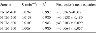
The photocatalytic degradation of RhB by the N-doped TiO2/muscovite nanocomposites followed first-order reaction kinetics (Fig. 8 and Table 2), which indicated that the degradation process followed the Eley-Rideal mechanism, and the dissolved pollutant molecules were oxidized and decomposed by the OH generated on the catalyst surface (Liang et al., Reference Liang, He, Zhong, Tan and Zhang2013). In addition, the photocatalysis of N-TM-400 was 0.0262 min–1. Moreover, the N-TiO2/muscovite composites exhibited a good photocatalytic effect under visible light, which enhances its practical applications.
Kinetic fit curves of RhB photocatalytic degradation of various samples

First-order reaction kinetics constants of RhB degradation over various samples

Cyclic performance.
After the photocatalytic reaction, the catalyst was washed ten times alternately with absolute ethanol and deionized water. Then, the catalyst was collected for the photocatalytic cycle experiment. The above steps were repeated three times. The photocatalytic degradation after the third cycle (Fig. 9) was 77%, slightly lower than the first cycle of 94%. This was due to the loss of quality in the catalyst recovery process. Nevertheless, the recyclable performance of the N-doped TiO2/muscovite nanocomposites was good. In addition, the muscovite loading made the N-TiO2 photocatalyst easy to be dispersed in and then recovered from the solution, which greatly enhanced its versatility.
Cyclic photocatalytic degradation of RhB over N-TM-400

Reaction product.
The maximum RhB absorption peak of 554 nm exhibited a significant blue shift (to lower wavelength), which was accompanied by a rapid decrease in the peak intensity over time (Fig. 10). This was due to the de-N-ethyl intermediates formed during the degradation of RhB. The main degradation products were identified by HPLC-MS (Fig. 11), revealing that five intermediates were formed in the degradation of RhB during the gradual removal of the N-ethyl group (Fig. 12).
UV-Vis absorption spectra of RhB degradation by N-TM-400 under visible light

HPLC-MS of photocatalytic degradation of RhB

Schematic diagram of the degradation of RhB

Conclusions
The N-TiO2/muscovite nanocomposites were prepared by a liquid precipitation-grinding method using muscovite as the matrix and urea as the nitrogen source. With increasing heat-treatment temperature, urea decomposed gradually into various nitrogen-containing compounds, resulting in N-doping of TiO2 due to the replacement of O atoms by N atoms. N-doping inhibited the growth/agglomeration of TiO2 nanoparticles and endowed the nanocomposite materials with a visible-light catalytic effect.
The photocatalytic performance of the N-TiO2/muscovite composite nanomaterials decreased with increasing heat-treatment temperature. The N-doped TiO2/muscovite nanocomposites that were treated at a temperature of 400°C showed the best photocatalytic performance with respect to the degradation of RhB, reaching 97% under visible-light irradiation (degradation time: 2 h; 100 mL of 20 mg/L RhB solution).
Photocatalytic degradation by N-doped TiO2/muscovite nanocomposites followed first-order reaction kinetics. Both N-doping and muscovite loading improved the photocatalytic degradation rate of TiO2.
Acknowledgments
This work was supported by the innovation team project sponsored by the Education Department of Sichuan Province, China (grant number: 14TD0012) and the Science and Technology Planning Project of Guangdong Province, China (grant number: 2017B030314175).
Funding
Funding sources are as stated in the Acknowledgments.
Declarations
Conflict of Interest
The authors declare that they have no conflict of interest.