Introduction
Free-living larvae of parasitic organisms – for example, cercariae of digeneans – usually demonstrate complicated behaviour that increases their chances of host encounter (Combes et al., Reference Combes, Fournier, Monѐ and Thѐron1994; Combes et al, Reference Combes, Bartoli, Theron, Lewis, Campbell and Sukhdeo2002; Haas, Reference Haas1994, Reference Haas2003; Rea & Irwin, Reference Rea and Irwin1994; Sukhdeo & Sukhdeo, Reference Sukhdeo and Sukhdeo2004; Prokofiev & Galaktionov, Reference Prokofiev and Galaktionov2009). Certain behavioural patterns are typical for cercariae of one species, but it was also observed that infected snails emit some cercariae with non-typical behaviour (Prokofiev, Reference Prokofiev1997, Reference Prokofiev2001, Reference Prokofiev2002, Reference Prokofiev2003; Galaktionov & Dobrovolskij, Reference Galaktionov and Dobrovolskij2003; Koehler et al., Reference Koehler, Springer, Keeney and Poulin2011; Prokofiev et al., Reference Prokofiev, Levakin, Losev, Zavirsky and Galaktionov2011). The proportion of such deviant cercariae within a species varies from 5% to 30% (Prokofiev, Reference Prokofiev2006). Variance in parasite's behaviour is commonly considered an adaptation to explore new hosts and increase transmission efficiency in the ecosystems with unstable conditions – for example, in the intertidal zone (Galaktionov & Dobrovolskij, Reference Galaktionov and Dobrovolskij2003; Prokofiev et al., Reference Prokofiev, Levakin, Losev, Zavirsky and Galaktionov2011).
The reproduction of digeneans within a first intermediate host (mollusc) is currently regarded as apomictic (diploid) parthenogenesis (Dobrovolskij & Ataev, Reference Dobrovolskij, Ataev, Combes and Jourdane2003; Galaktionov & Dobrovolskij, Reference Galaktionov and Dobrovolskij2003). A miracidium infects a mollusc and develops into a mother sporocyst. Its offspring in different groups of digeneans are either rediae or daughter sporocysts. They produce cercariae that, given parthenogenesis, are supposed to be genetically identical. The probability of infection of one mollusc individual with several (n) miracidia can be estimated as prevalence of infection (P) to the power of n – Pn (Kuris & Lafferty, Reference Kuris and Lafferty1994). So, when prevalence of mollusc infection with trematodes is low (as observed in many Arctic and subarctic communities – Galaktionov & Bustnes, Reference Galaktionov and Bustnes1999; Levakin et al., Reference Levakin, Nikolaev and Galaktionov2013a), probability of infection with several conspecific miracidia is very low. Among Littorina littorea infected with Himasthla elongata in our study region, no double infection has been detected with molecular genetic methods (Galaktionov et al., Reference Galaktionov, Podgornaya, Strelkov and Galaktionov2016). So, cercariae produced by a group of parthenitae within one snail may be considered a clone.
Studies on the behaviour of H. elongata (Mehlis, 1831) Dietz, 1909 (Echinostomatoidea: Himasthlidae) revealed a significant range of photo- and georeactions among cercariae originating from a single host individual (Prokofiev et al., Reference Prokofiev, Levakin, Losev, Zavirsky and Galaktionov2011). Unlike in the cercariae of several echinostomatid species (McCarthy, Reference McCarthy1999; Platt & Dowd, Reference Platt and Dowd2012), in H. elongata cercariae patterns of photo- and georeactions remain the same throughout their lives (Prokofiev, Reference Prokofiev2006; Prokofiev et al., Reference Prokofiev, Levakin, Losev, Zavirsky and Galaktionov2011; Nikolaev et al., Reference Nikolaev, Prokofiev, Levakin and Galaktionov2017). In addition, H. elongata larvae have strong intraclonal differences in the success of both infection of the second intermediate host (blue mussel Mytilus edulis Linnaeus, 1758), and encystment in the blue mussel haemolymph in vitro (Gorbushin & Levakin, Reference Gorbushin and Levakin2005; Levakin et al., Reference Levakin, Losev, Nikolaev and Galaktionov2013b). When comparing different ‘clones’ of H. elongata cercariae, no correlation was detected between the proportion of photo-positive cercariae and the infectivity of the cercarial ‘clone’ (Levakin & Losev, unpublished). However, it remained unknown whether behavioural features of cercariae (typical or deviant) influence their infectivity within one cercarial clone. So, the goal of our study was to find out whether there is a relationship between intraclonal variation in H. elongata cercariae behaviour – namely, their reaction to light and their infectivity for the second intermediate host. We evaluated infectivity through encystment success and used two approaches: experimental infection of the mussels, and cercariae encystment in the cell-free mix of haemolymph of the mussels.
Materials and methods
Sample collection
The research was carried out at the White Sea Biological Station of the Zoological Institute of the Russian Academy of Sciences. Periwinkles L. littorea (Linnaeus, 1758) were collected in the intertidal zone of the Chupa Inlet (Kandalaksha Bay, the White Sea) in July–August of 2017–2018. A total of 3847 L. littorea individuals were collected; snails infected with H. elongata were selected in the lab by inducing cercariae emission in the sunlight for 1 h. Uninfected blue mussels were obtained from the aquaculture facilities (Kulatchkova, Reference Kulatchkova and Lukanin1985; Nikolaev et al., Reference Nikolaev, Prokofiev, Levakin and Galaktionov2017).
Assessment of cercarial photoreaction
To group cercariae according to their reaction to light we used an approach suggested by Prokofiev (Reference Prokofiev1996, Reference Prokofiev1997). We obtained H. elongata cercariae by incubating infected periwinkles in separate transparent reservoirs with filtered seawater (50 ml) for 1 h at high light intensity (about 2500 Lx) and at a temperature of 20°С (Prokofiev et al., Reference Prokofiev, Levakin, Losev, Zavirsky and Galaktionov2011). Himasthla elongata cercariae originating from one L. littorea individual (one ‘clone’ of H. elongata) were placed into a transparent reservoir (160 × 25 × 20 mm) with seawater. The reservoir was illuminated from one side with a light bulb; for heat absorption, a flat-walled container with water (200 mm layer) was used. The total number of cercariae of each clone launched into the reservoir ranged from 150 to 800. After 15 min of exposure, we collected the cercariae that had concentrated in the most (1500 Lx, front end) and least (600 Lx, rear end) illuminated areas (5% of volume or 8 mm in front or rear end) of the reservoir. These cercariae were considered photopositive and photonegative, respectively. The light intensity was measured with a CEM DT-8809A luxmeter (CEM, Shenzhen Everbest Machinery Industry Co., Ltd, Shenzhen, China). The proportion of photopositive or photonegative cercariae was determined by their ratio to the total number of cercariae in the reservoir. We estimated variability of cercariae photoreactions within every L. littorea infected with H. elongata (a total of 311 snails).
Further use of these cercariae was guided by the size of the smaller group (commonly, the number of photopositive cercariae). If the size of the smaller group was above 60 cercariae, these cercariae could be used to determine in vitro encystment success in the cell-free haemolymph from several large mussels (see the following section).
In vitro encystment
The haemolymph was collected from the posterior adductor muscle of large mussels (shell length 70–80 mm). Encystment success has previously shown no strong dependence on the presence of mussel haemocytes (Gorbushin & Levakin, Reference Gorbushin and Levakin2005), but clumps of haemocytes could hinder the counting of cercariae and estimation of their condition. For this reason, haemocytes were precipitated by centrifugation at 1.8 krpm for 10 min. A mixture of cell-free haemolymph fractions from ten mussels was used as an in vitro encystment medium in order to level up the individual differences in mussels (Gorbushin & Levakin, Reference Gorbushin and Levakin2005; Levakin et al., Reference Levakin, Losev, Nikolaev and Galaktionov2013b).
Himasthla elongata cercariae emission from infected periwinkles, even in stable conditions, is unsteady and not simultaneous (Prokofiev, Reference Prokofiev2006), so cercariae were collected and sorted according to their photoreactions several times. The haemolymph mixture was also prepared several times. This was done to ensure that as many cercarial clones sorted in groups as possible are tested in one haemolymph mixture. If the size of the smaller group within one clone was insufficient for in vitro testing, but enough to infect one small mussel (20 cercariae), this clone was used to determine the mussel infection success (see the following section).
A total of ten H. elongata cercarial clones were tested. Water with cercariae from one group (photopositive or photonegative) was placed into 50 ml conical tubes. Cercariae were precipitated by cooling the tubes in the water with ice. After removing the excess of water, cercariae in the small water volume (10 ml) were transferred to the well of the 96-well plate with flat bottom. Photopositive and photonegative H. elongata cercariae of each clone were placed into two different wells; thus, 20 wells were used in total (ten for photopositive cercariae and ten for photonegative cercariae). Sample size per well varied from 28 to 893 cercariae; 100 µl of cell-free mussel haemolymph was added to every well. Haemolymph collection and mixing was done right before the application. After incubating at 18°С for 2 h, we counted the fully encysted cercariae. The encystment success were assessed as a ratio of the metacercariae number to the number of cercariae used for infection or placed into the haemolymph.
Mussel infection
Cercariae from one infected L. littorea individual (one ‘clone’ of H. elongata) were used to infect ten mussels: five with photopositive cercariae and five with photonegative ones. To infect small mussels, collection and sorting of cercariae was repeated from two to five times. A total of 32 mussels were infected with 20 cercariae each; all of these cercariae were either photopositive or photonegative. For infection we used small mussels with a shell length of 20–25 mm. Individual mussels were incubated together with cercariae in the plastic containers containing 100 ml (height with cap = 71.3 mm;, upper diameter = 62 mm; bottom diameter = 46 mm) of seawater for 24 h at 18°С with regular room light (about 500 Lx). To detect the resulting infection, the mussels’ soft tissues were squeezed and screened under a stereomicroscope, and the number of metacercariae was counted.
Data analysis
We calculated the prevalence of H. elongata infection (PI) in periwinkles as a fraction of infected snails in the sample. The probability of one periwinkle being infected with several miracidia was evaluated as PIn, where n is the number of miracidia.
To test the statistical significance of differences between the ratios, we used Fisher's exact test (paired) and the chi-square test (multiple). To test the statistical significance of photoreaction influence on the infection success, we used two-way analysis of variance (ANOVA) for φ-transformed ratios (factors: ‘clone’ and ‘photoreaction’). The data were checked for the homogeneity of variances (Bartlett test) and the lack of significance of factor interactions. Confidence intervals of fractions were calculated using the exact Fisher formula or obtained by the inverse transformation of the boundaries of the intervals (φ-transformation) The confidence interval was assessed for a 95% confidence limit.
Results and discussion
In the L. littorea sample, we found 311 individuals emitting H. elongata cercariae, indicating a PI of 8 ± 0.9%. Intraclonal variance in photoreactions was detected in 80% of H. elongata clones (249 L. littorea individuals). The fraction of photopositive cercariae in different clones varied from 0.2% to 60%. Only 32 out of 311 infected periwinkles emitted sufficient (more than 20 in 1 h) numbers of cercariae with non-standard photoreaction to perform the mussel infection experiment. Ten periwinkles emitted more than 60 cercariae with non-standard photoreaction in 1 h and could be used to compare encystment success of photopositive and photonegative cercariae in the mussel haemolymph in vitro.
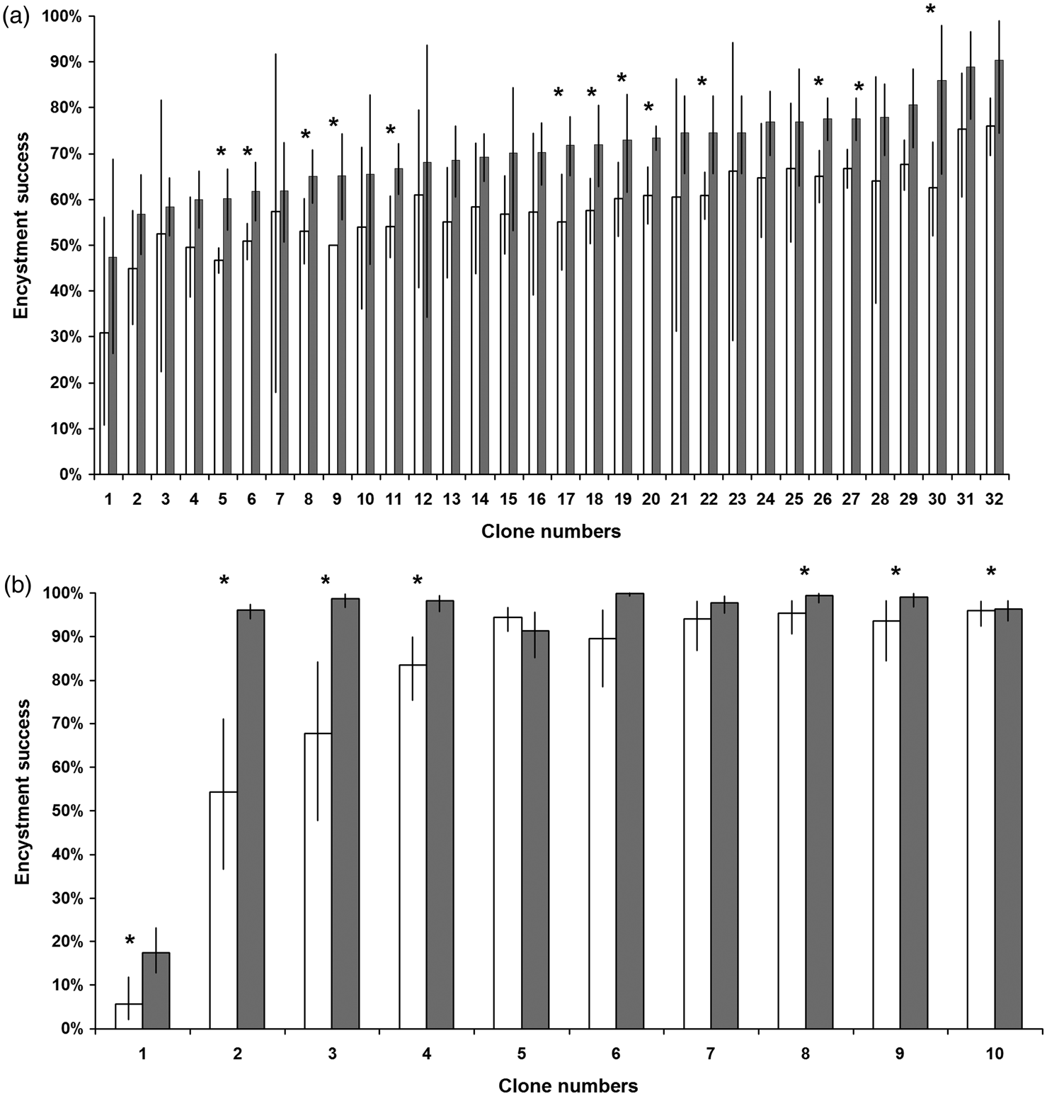
Himasthla elongata cercariae clones differed in how well they infected mussels (ANOVA: F31, 296 = 6.01, P < 0.01, η 2 = 32.4%; fig. 1a) and encysted in mussel haemolymph in vitro (chi-square test: for photopositive χ 29 = 586.76, P < 0.01; for photonegative χ 29 = 2076.87, P < 0.01; overall χ 29 = 2488.05, P < 0.01; fig. 1b). Photopositive cercariae showed lower mussel infection (ANOVA: F1, 296 = 86.9, P < 0.01, η 2 = 15%; fig. 1a) and in vitro encystment success (ANOVA: F1, 9 = 12.52, P < 0.01; fig. 1b). No interaction of factors ‘clone’ and ‘photoreaction’ on mussel infection was detected (ANOVA: F31, 296 = 0.24, P > 0.05, η 2 = 1.3%). Intraclonal differences in infection/encystment success were significant (Fisher's exact test: P < 0.05) in 13 cases out of 32 for mussel infection (fig. 1a), and in seven cases out of ten for in vitro encystment (fig. 1b).
Encystment success of Himasthla elongata cercariae with positive (empty bars) and negative (shaded bars) photoreactions: (a) average success of infecting Mytilus edulis, n = 40 for each clone (20 photopositive and 20 photonegative cercariae); (b) the proportion of cercariae that encysted successfully in a mixture of cell-free mussels’ haemolymph. Sample sizes vary from 28 to 893 cercariae(see supplementary table S1). Confidence intervals are provided for the 95% significance value. Statistical significance of intraclonal differences: *P < 0.05 (Fisher's exact test).

The probability of one periwinkle being infected by several (n) H. elongata miracidia is very low (0.08n). If we estimate the probability of infection of an individual L. littorea with H. elongata parthenitae as the infection value (0.08), then a maximum estimate of double infection can explain the variation of characters in cercaria emitted by one infected L. littorea individual for 8% of cases. So, double infection can explain variation of photoreaction in cercariae emitted by 25 infected individuals out of 311 tested individuals. We found variation of cercariae photoreaction much more frequently, so it cannot be explained by multiple infection.
Moreover, the periwinkles in the studied population were previously shown to bear single-miracidium infection. Individual cercariae amplified fragment length polymorphism (AFLP) analysis showed that larvae cluster according to their origin – that is, according to the mollusc they were extracted from (Galaktionov et al., Reference Galaktionov, Podgornaya, Strelkov and Galaktionov2016). So, in our case, multiple infection of the host by H. elongata miracidia cannot explain variation of cercarial photoreactions. Changes in the behaviour of cercariae due to their age, found in several freshwater echinostomatids (McCarthy, Reference McCarthy1999; Platt & Dowd, Reference Platt and Dowd2012), also cannot explain this variation because we used cercariae of the same age.
Several other systems with low PI and, hence, likely single miracidium infection provide evidence of genetic heterogeneity in cercariae produced within one snail individual (Khalturin et al., Reference Khalturin, Mikhaylova and Granovich2000; Semyenova et al., Reference Semyenova, Chrisanfova, Fillipova, Beer, Voronin and Ryskov2005; Solovyeva et al., Reference Solovyeva, Galaktionov and Podgornaya2013; Galaktionov et al., Reference Galaktionov, Solovyeva, Fedorov and Podgornaya2014). Intraclonal polymorphism in cercariae apparently emerges during their embryogenesis, because it is detected not just among cercariae emitted by one mollusc, but also among the offspring of one redia (Galaktionov et al., Reference Galaktionov, Podgornaya, Strelkov and Galaktionov2016) or sporocyst (Korsunenko et al., Reference Korsunenko, Chrisanfova, Arifov, Ryskov and Semyenova2013). We suppose that genetic diversity in the absence of meiosis is assumed to be guided by several possible mechanisms. One of them is the non-specific genomic rearrangements. Trematode larvae demonstrate various levels of intraclonal genetic polymorphism (Bayne & Grevelding Reference Bayne and Grevelding2003; Korsunenko et al., Reference Korsunenko, Chrisanfova, Arifov, Ryskov and Semyenova2013, Galaktionov et al., Reference Galaktionov, Solovyeva, Fedorov and Podgornaya2014). Based on the particularities of the molecular methods used, the authors suggest that ectopic recombination occurs in the larvae's genomes. On the other hand, the phenotype of developing cercariae may, for example, be influenced by epigenetic gene or non-coding DNA regulation (Slotkin & Martienssen, Reference Slotkin and Martienssen2007; Fedoroff, Reference Fedoroff2012). For example, phenotypic variation in Schistosoma mansoni is induced by direct modulation of this parasite epigenotype (Cosseau et al., Reference Cosseau, Azzi, Rognon, Boissier, Gourbière, Roger, Mitta and Grunau2010). Intraclonal genetic heterogeneity in H. elongata cercariae (Galaktionov et al., Reference Galaktionov, Podgornaya, Strelkov and Galaktionov2016) may be linked to their phenotype and may manifest as variance in both their photoreactions and infectivity. It is not unlikely that the reduced mussel infection and in vitro encystment success in deviant cercariae is due to non-specific genomic rearrangements.
Acknowledgements
We acknowledge the White Sea Biological Station of the Zoological Institute of the Russian Academy of Sciences for sampling and experiment facilities. The manuscript was translated into English by Anna Gonchar. We are grateful to Prof Kirill V. Galaktionov for his advice during manuscript preparation.
Financial support
This study was funded by Russian Foundation for Basic Research according to the research project number 17-04-02161 and state funding projects number AAAA-A19-119020690109-2 and АААА-А19-119022690122-5.
Conflicts of interest
None.
Ethical standards
The authors assert that all procedures contributing to this work comply with the ethical standards of the relevant national and institutional guides on the care and use of laboratory animals.
Supplementary material
To view supplementary material for this article, please visit https://doi.org/10.1017/S0022149X20000103