Glutathione (GSH), a tripeptide composed of glutamine, cysteine and glycine, is the most abundant endogenous antioxidant in human cells. It plays a central role in detoxification, redox homeostasis, immune modulation, DNA synthesis and protection against oxidative stress-induced cellular damage(Reference Ballatori, Krance and Notenboom1,Reference Weschawalit, Thongthip and Phutrakool2) . GSH exists in high concentrations in nearly every tissue and is essential for cellular health and recovery.
However, glutathione levels decline due to aging, poor nutrition, environmental stress and chronic diseases – conditions increasingly prevalent in modern populations(Reference Tong, Fitzmaurice and Moszczynska3). As a result, oral glutathione supplementation is gaining global attention for its therapeutic potential in managing inflammation, fatigue, skin disorders, neurodegeneration and metabolic dysfunctions(Reference Sonthalia, Jha and Lallas4,Reference Kim, Lee and Whang5) .
Despite its clinical relevance, conventional oral glutathione suffers from poor bioavailability due to rapid degradation in the gastrointestinal tract and limited intestinal transport(Reference Hou, Michiels and Kerschaver6). Several studies have confirmed that orally administered glutathione is rapidly oxidised or metabolised before reaching systemic circulation, reducing its therapeutic efficacy.
To overcome this challenge, liposomal encapsulation has emerged as a promising strategy. Liposomes – phospholipid bilayer vesicles – protect labile molecules from enzymatic degradation, enhance mucosal permeability and promote cellular uptake through membrane fusion or endocytosis(Reference Koynova and Tenchov7). Liposomal glutathione, in particular, is designed to improve stability and targeted delivery, offering sustained plasma levels and higher tissue bioavailability compared with non-encapsulated forms(Reference Sinha, Sinha and Calcagnotto8).
While pre-clinical models suggest enhanced performance of liposomal glutathione, few studies have comprehensively validated these claims through integrated in vitro and human pharmacokinetic evaluation(Reference Sinha, Sinha and Calcagnotto8). There is a need to demonstrate not just absorption but also functional outcomes at the cellular level, including uptake, proliferation and repair capacity – all of which are critical for translating antioxidant benefits into clinical practice(Reference Gandek, van der Koog and Nagelkerke9,Reference Reginald-Opara, Tang and Svirskis10) .
The present study was designed to compare the biological activity and pharmacokinetic profile of LipoDuo™ Glutathione, a liposomally encapsulated GSH formulation, with plain glutathione. We investigated cellular uptake, dose optimisation, proliferation activity and wound healing in human embryonic kidney 293T (HEK-293T) cells, followed by a human clinical trial to evaluate systemic absorption and bioavailability. This dual-model approach aims to provide mechanistic and translational evidence supporting the superior efficacy of LipoDuo™ Glutathione.
Material and methods
Reduced L-glutathione was sourced from Anhui GSH Bio Technology, China, Reduced L-glutathione United States Pharmacopeia grade, Reference Standard; Cat. No. G4251 was sourced from Sigma Aldrich, and Sunflower Lecithin was sourced from Amitex, Indore, India. Liposomal encapsulation and formulation of LipoDuo™ Glutathione was carried out at Samarth Biorigins LLP, India, using a proprietary encapsulation technology developed in-house. All cell culture reagents, including Eagle’s minimum essential medium, fetal bovine serum, antibiotics and non-essential amino acids, were procured from standard suppliers such as HiMedia (India) or Thermo Fisher Scientific (USA), unless otherwise specified.
Cell lines and culture conditions
HEK-293T cells were obtained from the National Centre for Cell Science, Pune, India. Cells were cultured in Eagle’s minimum essential medium supplemented with 10 % fetal bovine serum, 1 % non-essential amino acids, 1 % sodium pyruvate and 1 % penicillin-streptomycin. Cultures were maintained at 37°C in a humidified incubator with 5 % carbon dioxide atmosphere(Reference Karthik, Ishwara Kalyani and Krishnappa11–Reference Krishnappa, Karthik and Shantaram13).
Cell uptake studies
HEK-293T cells were seeded at a density of 0·4 × 106 cells/well in 6-well plates and incubated overnight. Cells were treated with 2 mg/ml of either plain or LipoDuo™ Glutathione. After 0, 2, 4, 6 and 8 h of incubation, cells were harvested, washed with PBS and lysed using radioimmunoprecipitation assay buffer(Reference Lykkesfeldt and Tveden-Nyborg14). Cell lysates were analysed by HPLC using a Shim-pack Solar C18 (5 um, 4·6 × 250 mm, Part Number: 227–30 600–02), with an isocratic mobile phase of 25 mM KH₂PO₄, 2·02 g/l sodium 1-heptanesulfonate, 30 ml/l methanol (pH adjusted to 3·0) and detection at 210 nm.
Dose optimisation assay
Cells were seeded at 0·18 × 106 cells/well in 24-well plates and treated with glutathione at concentrations ranging from 2 to 12 mg/well for 6 h. Sample preparation and HPLC analysis followed the same procedure as the uptake assay. Uptake was quantified as a percentage relative to baseline absorbance.
Cell proliferation assay
Cells were seeded at a density of 15 000 cells/well in 96-well plates and treated with increasing concentrations of plain and LipoDuo™ Glutathione (1–12 mg/well). After 24 h of incubation, a 3-(4,5-dimethylthiazol-2-yl)-2,5-diphenyltetrazolium bromide solution (5 mg/ml in PBS) was added and incubated for 4 h. Formazan crystals were solubilised in isopropyl alcohol, and absorbance was measured at 570 nm using a microplate reader(Reference Karthik, Ishwara Kalyani and Krishnappa11–Reference Krishnappa, Karthik and Shantaram13).
Wound healing assay (scratch assay)
HEK-293T cells were seeded in 24-well plates at 0·18 × 106 cells/well and grown to confluence. A uniform scratch was created using a sterile 10 µL pipette tip. Cells were then treated with 1 mg/well of either plain or LipoDuo™ Glutathione. Images were captured at 0, 12 and 24 h under phase-contrast microscopy. Wound closure was quantified using the software programme ImageJ developed by NIH and calculated as: This assay was conducted as per guidelines(Reference Jonkman, Cathcart and Xu15–Reference Prasad, Chaithra and Karthik18). The closure was calculated using the following formula:
 $${\text{Percent Closure}} = \frac{{{{{Migrated{\;}}cell{\rm{\;}}surface{\rm{\;}}area \times 100}}}}{{{Total{\rm{\;}}surface{\rm{\;}}area}}}$$
Human clinical bioavailability study
Study design
An open-label, randomised, parallel-group single-dose bioavailability study was conducted in twelve healthy adult volunteers (aged 24–42 years, BMI 19–30 kg/m2). Subjects were divided into two groups (n 6 each) and administered either 1 g LipoDuo™ Glutathione or 1 g plain L-glutathione (each in 2·5 g sachets under fasting conditions).
Sampling and analysis
Blood samples (5·0 ml) were collected in K₂EDTA vacutainers at fourteen time points: pre-dose and at 0·25, 0·5, 1, 1·5, 2, 3, 4, 6, 8, 12 and 24 h post-dose. Samples were stored in an ice bath and analysed using a validated LC-MS/MS method (United States Pharmacopeia compliant). Plasma concentrations of glutathione were used to derive maximum plasma concentration (Cmax), time to reach maximum concentration (Tmax), AUC and plasma retention profiles.
This study was conducted according to the guidelines laid down in the Declaration of Helsinki, and all procedures involving human subjects were approved by the Clinical Trials Registry of India (CTRI). Written informed consent was obtained from all subjects. The study protocol was approved by an institutional ethics committee and registered under the CTRI registration identification no. CTRI/2024/12/078004.
The human clinical bioavailability study was reviewed and approved by the Independent Institutional Ethics Committee of MedStar Specialty Hospital, Bangalore, Karnataka, India (approval date: 23 November 2024). The protocol was prospectively registered with the CTRI (CTRI/2024/12/078004) on 12 December 2024. All procedures involving human participants were conducted in accordance with the Indian Council of Medical Research guidelines, the Declaration of Helsinki (2013 revision) and Good Clinical Practice standards. Written informed consent was obtained from all participants before enrolment.
Statistical analysis
All analyses were performed in GraphPad Prism v9.0. Normality was verified by the Kolmogorov–Smirnov test. For factorial designs in cell-based assays, we used two-way ANOVA (treatment × time for uptake and wound healing; treatment × dose for proliferation) with Tukey’s HSD post hoc test when the ANOVA was significant (α = 0·05). For single-factor dose–response within a formulation, we used one-way ANOVA with Tukey’s post hoc test. The human pharmacokinetic comparison was analysed with two-way repeated-measures ANOVA (between-subjects factor: formulation; within-subjects factor: time), followed by Bonferroni adjustment for planned pairwise contrasts across time. Linear associations were quantified with Pearson’s r, where parametric assumptions held. We report mean (sd), two-sided 95 % CI and P-values for all main effects and interactions.
Plasma concentration–time data were analysed by two-way repeated-measures ANOVA (between-subjects: formulation; within-subjects: time), with Bonferroni correction for planned pairwise comparisons between formulations at each time point.
Results and discussion
The objective of this assay was to evaluate and compare the intracellular uptake efficiency of liposomal glutathione (LipoDuo™) and plain glutathione in HEK-293T cells over time. Liposomal encapsulation is hypothesised to enhance membrane permeability and intracellular delivery by protecting glutathione from extracellular degradation.
Bioavailability studies
HEK-293T cells were treated with 2 mg/ml of either LipoDuo™ or plain glutathione, and intracellular levels were measured using HPLC at 0, 2, 4, 6 and 8 h.
Peak uptake
LipoDuo™ Glutathione achieved maximum uptake at 6 h (∼45%), nearly 2× that of plain glutathione at its peak (∼23% at 2 h). Sustained Uptake: While plain glutathione exhibited a rapid uptake followed by a steep decline, LipoDuo™ Glutathione maintained elevated intracellular levels for up to 8 h. Mechanism Insight: The delayed peak and sustained uptake suggest a controlled release mechanism and enhanced cellular permeation facilitated by the phospholipid bilayer. Nutritional Relevance: These results indicate that LipoDuo™ Glutathione can deliver higher and more prolonged intracellular glutathione, potentially offering superior antioxidant defense at the cellular level compared with standard oral glutathione. It also demonstrated that proliposomal glutathione formulations significantly improve uptake and stability in epithelial cell models, highlighting the advantage of liposomal encapsulation in intracellular delivery as represented in Figure 1. Intracellular glutathione uptake data were analysed using two-way ANOVA (treatment × time) followed by Tukey’s post hoc test in GraphPad Prism v9.0, after confirming data normality with the Kolmogorov–Smirnov test. Results are presented as mean (sd) (% of control) with 95 % CI = 21·76, 40·19. A strong positive correlation was observed between incubation time and glutathione uptake (r = 0·9560, P = 0·0009), indicating a significant time-dependent intracellular accumulation influenced by both treatment and duration.
Time-course of intracellular glutathione uptake in human embryonic kidney 293T (HEK-293T) cells (x-axis – time in h; y-axis – % glutathione uptake). Time-course of intracellular glutathione uptake in HEK-293T cells. HEK-293T cells were treated with 2 mg/ml of either LipoDuo™ or plain glutathione, and intracellular glutathione levels were quantified by reverse-phase HPLC at 0, 2, 4, 6 and 8 h. Data represent mean (sd) from three independent replicates, with 95 % CI (lower and upper limits = 21·76, 40·19) calculated by two-way ANOVA (treatment × time) followed by Tukey’s post hoc test (P < 0·01). A strong positive correlation between uptake and incubation time (Pearson’s r = 0·9560, P = 0·0009) confirms time-dependent intracellular accumulation of LipoDuo™ relative to plain glutathione.

Dose optimisation
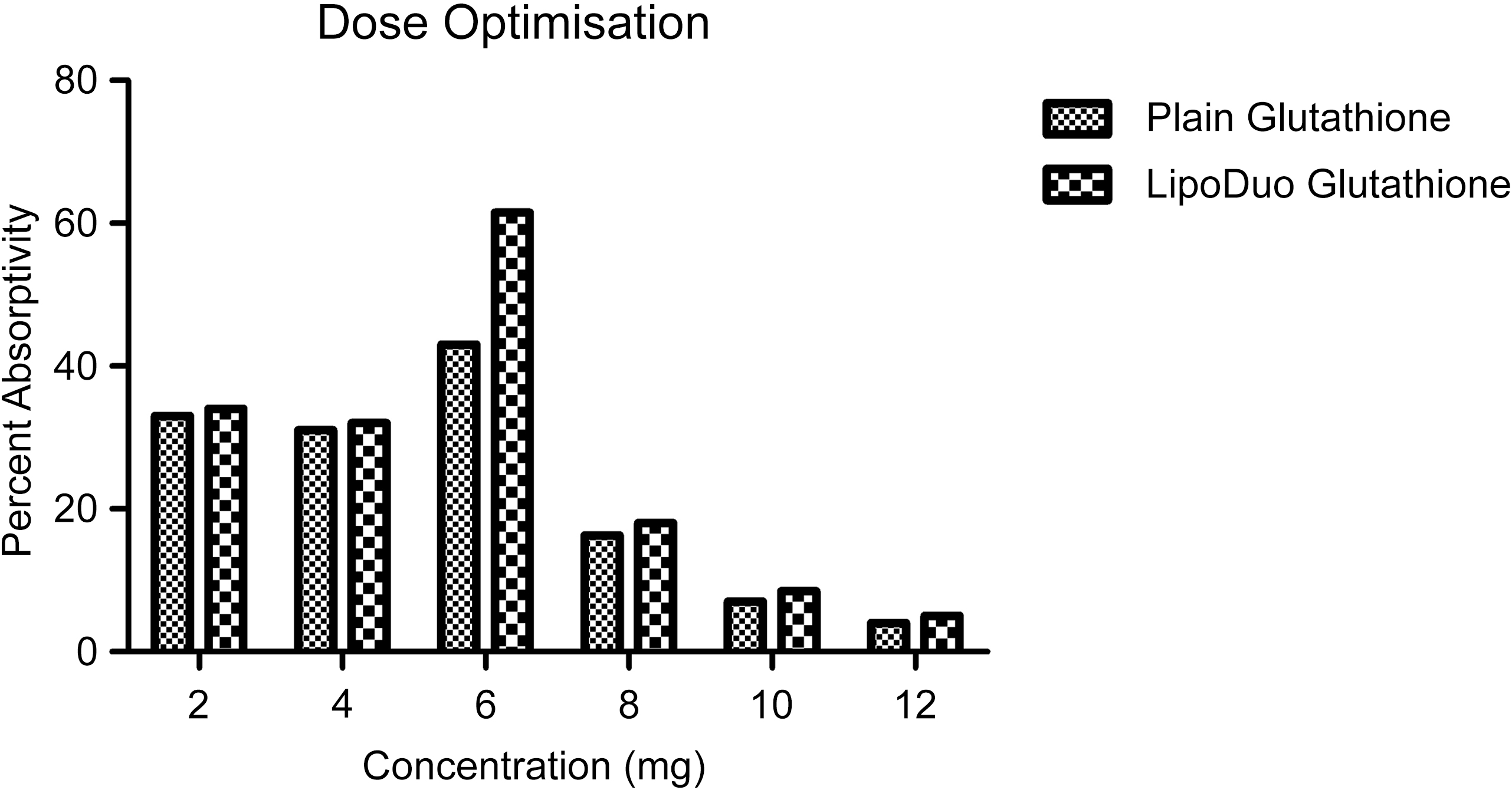
To identify the most effective dose for intracellular delivery, HEK-293T cells were treated with a concentration range of 2–12 mg/well of either LipoDuo™ Glutathione or plain glutathione. Intracellular glutathione levels were measured at 6 h post-treatment using HPLC analysis, as represented in Figure 2.
Dose optimisation studies for plain and LipoDuo™ Glutathione using human embryonic kidney 293T (HEK-293T) cells. Dose-dependent intracellular glutathione uptake in HEK-293T cells treated with plain or LipoDuo™ Glutathione. HEK-293T cells were exposed for 6 h to increasing concentrations (2–12 mg/well) of either formulation, and intracellular glutathione was quantified by reverse-phase HPLC. Data are expressed as mean (sd) (% uptake relative to untreated control) from three independent replicates. Statistical significance across doses within each treatment was determined by one-way ANOVA followed by Tukey’s multiple-comparison test (P = 0·0009 for overall effect of dose in the LipoDuo™ group). A strong positive correlation between dose and intracellular uptake was confirmed (Pearson’s r = 0·9560, P = 0·0009), indicating a linear increase in cellular glutathione content up to 6 mg/well, followed by a plateau suggesting saturation of the transport mechanism.

Dose-dependent uptake of LipoDuo™ and plain glutathione in HEK-293T cells was measured at 6 h. Uptake peaked at 6 mg/well for LipoDuo™ and plateaued thereafter, indicating saturation kinetics.
LipoDuo™ Glutathione demonstrated a clear dose-dependent increase in cellular uptake up to 6 mg/well, reaching a maximum of 61·5 %. Beyond this point, uptake plateaued, indicating saturation. Plain glutathione showed significantly lower uptake at all doses, peaking at 43 %.
The results indicate that LipoDuo™ Glutathione achieves optimal intracellular delivery at a concentration of 6 mg/well. The plateau beyond this point suggests a saturation of cellular transport mechanisms, which may include passive diffusion or receptor-mediated uptake enhanced by the liposomal bilayer. This dose optimisation is critical for achieving therapeutic intracellular levels while avoiding unnecessary overdosage. Dose–response data were analysed in GraphPad Prism v9.0 using one-way ANOVA within each treatment group, followed by Tukey’s post hoc test, after confirming normality with the Kolmogorov–Smirnov test. A significant dose effect was observed for LipoDuo™ Glutathione (P = 0·0009), supported by a strong positive correlation (r = 0·9560, P = 0·0009), indicating a concentration-dependent increase in intracellular uptake. Uptake rose progressively up to 6 mg/well, after which a plateau suggested saturation of the transport mechanism.
These findings align with earlier work by Byeon et al. (2019)(Reference Byeon, Lee and Kim19), who demonstrated that proliposomal glutathione achieved maximal uptake at specific concentration thresholds, beyond which no significant gain in intracellular levels was observed. Similarly, AlGhamdi et al. (2020)(Reference AlGhamdi, Kumar and AL-Rikabi20) reported that increasing concentrations of plain glutathione reduced proliferation and uptake in melanocytes, further supporting the importance of dose precision in glutathione therapy(Reference Ballatori, Krance and Notenboom1,Reference Sercombe, Veerati and Moheimani21) .
Wound healing assay
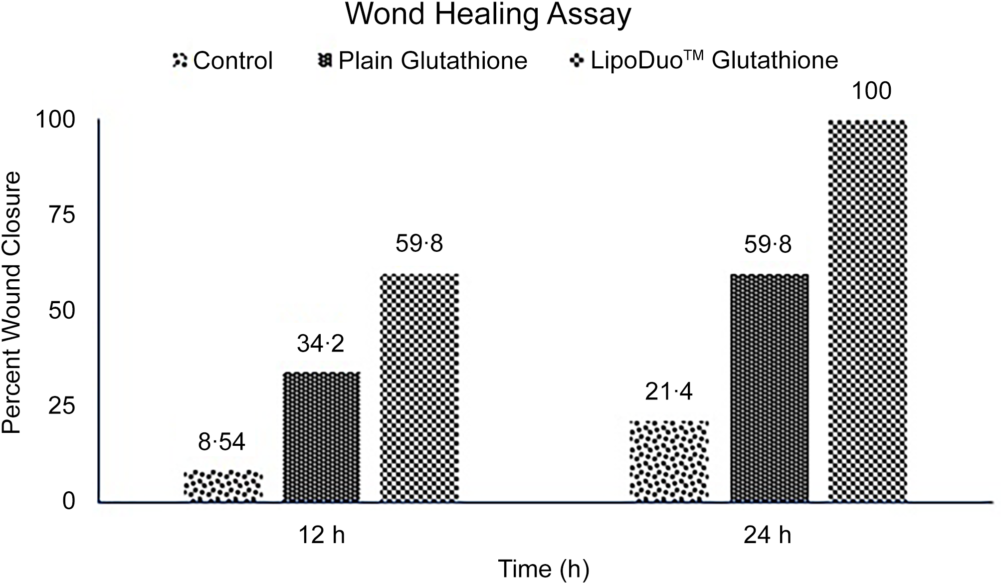
A scratch assay was conducted to evaluate the effect of LipoDuo™ Glutathione on cell migration and wound healing. Confluent HEK-293T cell monolayers were scratched to create a uniform wound and treated with 1 mg/well of either LipoDuo™ or plain glutathione. Wound closure was monitored at 0, 12 and 24 h using phase-contrast microscopy, as shown in Figure 3.
Wound closure over time in human embryonic kidney 293T (HEK-293T) cells (x-axis: time in h; y-axis: % wound closure). Wound closure kinetics in HEK-293T cells treated with LipoDuo™ or plain glutathione. Confluent monolayers were scratched and exposed to 1 mg/well of either formulation; closure was monitored at 0, 12 and 24 h under phase-contrast microscopy. Data represent mean (sd) (% wound closure) from three independent experiments. Statistical differences were evaluated by two-way ANOVA (treatment × time) followed by Tukey’s post hoc multiple-comparisons test. A significant overall treatment effect was observed (P = 0·0425), and a strong positive correlation between time and wound closure was confirmed (r 2 = 0·9575; r ≈ 0·979), indicating time-dependent cell migration and enhanced re-epithelialisation in the LipoDuo™ group.

LipoDuo™ Glutathione significantly enhanced wound closure in HEK-293T cells, achieving 100 % closure at 24 h. In contrast, plain glutathione and control treatments resulted in 59·8 and 21·4 % closure, respectively (Figure 4). These results indicate a superior effect of the liposomal formulation on promoting cell migration and wound healing.
Captured microscopic images of the wound healing process in human embryonic kidney 293T (HEK-293T) cells treated with plain and LipoDuo™ Glutathione, respectively, at 12 and 24 h. Representative phase-contrast micrographs showing the wound-healing process in confluent HEK-293T cell monolayers treated with LipoDuo™ (liposomal glutathione) or plain glutathione at 1 mg/well. Images were captured at 0, 12 and 24 h after scratching to monitor cell migration and wound closure. LipoDuo™-treated cells exhibited faster wound edge migration and nearly complete closure by 24 h, whereas plain glutathione and control groups showed partial healing (≈ 59·8 and 21·4 %, respectively). Data represent mean (sd) (% wound closure) from three independent experiments. Statistical analysis was performed using two-way ANOVA (treatment × time) followed by Tukey’s post hoc multiple comparisons. A significant overall treatment effect was observed (P = 0·0425), and a strong positive correlation between time and wound closure was confirmed (r 2 = 0·9575; r ≈ 0·979), indicating a time-dependent re-epithelialisation and enhanced migratory potential under LipoDuo™ treatment.

The enhanced wound healing observed with LipoDuo™ Glutathione may be attributed to its improved cellular uptake and sustained intracellular glutathione levels, which are critical for cellular processes such as proliferation and migration. Liposomal encapsulation facilitates better membrane permeability, leading to more effective delivery of glutathione into cells. Wound-closure data at 0, 12 and 24 h were analysed using a two-way ANOVA (treatment × time) in GraphPad Prism v9.0, with Tukey’s post hoc test applied where appropriate. A significant treatment effect was observed (P = 0·0425), indicating greater wound closure with LipoDuo™. A strong time-dependent trend was confirmed by linear fitting (r 2 = 0·9575; r ≈ 0·979). Results are reported as mean (sd) (% wound closure).
This finding aligns with previous studies demonstrating that liposomal formulations can enhance the therapeutic efficacy of bioactive compounds by improving their bioavailability and cellular uptake. For instance, Zhai et al. (2023)(Reference Zhai, Fu and Zhang22) reported that liposomal quercetin improved cellular uptake and reduced cytotoxicity in macrophages, highlighting the potential of liposomal delivery systems in enhancing cellular functions.
Cell proliferation activity
The cell proliferation assay is a key indicator saying that liposomal glutathione is more suitable for cell metabolic activity, their viability and duplication than plain glutathione, as evidenced and shown in Figure 5. The cell proliferation activity of LipoDuo™ Glutathione showed ≈3-fold higher at lower concentrations than plain glutathione up to 0·192 mMol/well, whereas at > 0·224 mMol/well, LipoDuo™ showed ≤ 2-fold cell proliferation activity than plain glutathione. This further necessitates pharmacokinetic and pharmacodynamic studies to understand the potential of the novel liposomal form. Cell proliferation (% of control) after 24 h was analysed using a two-way ANOVA (treatment × dose) in GraphPad Prism v9.0 with Tukey’s post hoc test. The overall model was significant (P < 0·0001), with LipoDuo™ showing greater proliferation than plain glutathione at lower concentrations and a plateau at higher doses. A strong dose–response relationship was confirmed (r = 0·9203; r 2 = 0·7916; 95 % CI: 30·82, 65·72), consistent with a saturable proliferation pattern.
The cell proliferation assay on distinct forms of glutathione (LipoDuo™ and plain) using human embryonic kidney 293T (HEK-293T) cells. Cell proliferation response of HEK-293T cells to LipoDuo™ and plain glutathione. Cells were exposed for 24 h to increasing concentrations (0·096–0·256 mMol/well) of each formulation, and viability/proliferation was measured by the 3-(4,5-dimethylthiazol-2-yl)-2,5-diphenyltetrazolium bromide assay. Data represent mean (sd) (% of control) from three independent experiments. Statistical analysis was performed using two-way ANOVA (treatment × dose) followed by Tukey’s post hoc test. The omnibus model was significant (P < 0·0001), confirming that LipoDuo™ produced greater proliferative activity across doses. A strong positive dose–response correlation was observed (r = 0·9203; r 2 = 0·7916; 95 % CI = 30·82, 65·72), indicating concentration-dependent stimulation up to ≈ 0·192 mMol/well and plateauing thereafter, consistent with saturation of cellular growth mechanisms.

LipoDuo™ Glutathione exhibits superior plasma bioavailability in human subjects
A randomised, open-label, single-dose, parallel-group study was conducted in twelve healthy volunteers (n 6 per group) under fasting conditions. Participants received either LipoDuo™ Glutathione (1 g) or plain glutathione (1 g) orally. Blood samples were collected at fourteen time points over 24 h. Plasma glutathione concentrations were quantified using a validated LC-MS/MS method(Reference Ballatori, Krance and Marchan23).
Cmax: LipoDuo™ Glutathione reached a peak plasma concentration of ∼1800 ng/ml at 2 h (Tmax), 6× higher than plain glutathione (∼300 ng/ml at 1–1·5 h). Sustained levels: LipoDuo™ maintained plasma concentrations > 500 ng/ml at 24 h, while plain glutathione declined to near baseline (< 100 ng/ml) after 6 h, as represented in Figure 6. AUC: Significantly greater for LipoDuo™, reflecting superior overall exposure. Bimodal distribution – an initial absorption peak at 2 h and a secondary shoulder around 8–12 h. This pattern was absent in the plain glutathione group, which showed a single, transient peak followed by a rapid decline. Plasma glutathione kinetics and statistical evaluation: Plasma glutathione concentrations were analysed using a two-way repeated-measures ANOVA (formulation × time) in GraphPad Prism v9.0, with Bonferroni post hoc comparisons. Significant formulation and time effects were observed (P < 0·0005), and the interaction indicated consistently higher levels with LipoDuo™ (P < 0·01). A biphasic time–concentration relationship was confirmed (r 2 = 0·6462; 95 % CI: 586·3, 200·1). LipoDuo™ produced a higher Cmax (∼1800 ng/ml), delayed Tmax (2 h) and sustained plasma levels (> 500 ng/ml at 24 h), demonstrating superior systemic bioavailability compared with plain glutathione.
Plasma concentration–time profile of glutathione in human subjects (x-axis: time in h; y-axis: glutathione concentration in ng/ml). Plasma concentration–time profile of glutathione in human subjects following single-dose administration of LipoDuo™ and plain formulations. Participants (n 12; 6 per group) received 1 g oral glutathione under fasting conditions, and plasma samples were collected over 24 h. Glutathione concentrations were quantified by LC–MS/MS and expressed as mean (sd) (ng/ml). Statistical analysis was performed by two-way repeated-measures ANOVA (formulation × time) followed by Bonferroni’s multiple-comparison test (P < 0·0005 for formulation effect). A positive time–concentration correlation (r 2 = 0·6462; r = 0·2369) and a biphasic absorption pattern were observed, with Cmax ≈ 1800 ng/ml at 2 h and sustained levels > 500 ng/ml at 24 h in the LipoDuo™ group, confirming prolonged systemic retention compared with plain glutathione.

The pharmacokinetic profile of LipoDuo™ Glutathione demonstrates improved oral absorption, delayed clearance and prolonged systemic availability. These effects are consistent with the protective role of liposomal carriers, which shield glutathione from gastric degradation and facilitate lymphatic or enterocyte-based absorption.
The bimodal plasma profile observed (initial and secondary peaks) may indicate enterohepatic recirculation or sustained tissue release – a well-recognised pattern in liposomal nutraceuticals. This sustained delivery is crucial for maintaining antioxidant capacity and therapeutic plasma levels.
Comparable findings were reported by Sinha et al. (2018)(Reference Sinha, Sinha and Calcagnotto8), where liposomal glutathione supplementation led to sustained increases in blood glutathione and improvements in immune function. Bruggeman et al. (2019) also noted that non-liposomal nano-glutathione had inferior plasma retention, underscoring the value of advanced delivery formats(Reference Bruggeman, Storo and Fair24).
Discussion
The present study evaluated the comparative effectiveness of a novel liposomal glutathione formulation, LipoDuo™ Glutathione, against plain glutathione using a comprehensive in vitro and human clinical model. Our findings demonstrate that liposomal encapsulation significantly enhances glutathione’s cellular uptake, proliferation-promoting activity, tissue repair potential and systemic bioavailability – features critical for improving therapeutic and nutritional outcomes in oxidative stress-related conditions.
Enhanced cellular uptake and functional activity
The superior uptake of LipoDuo™ Glutathione in HEK-293T cells confirms the advantages of liposomal encapsulation in promoting intracellular delivery. The phospholipid bilayer likely facilitates membrane fusion or passive diffusion, leading to higher intracellular concentrations. This enhanced uptake translated into significantly increased cell proliferation and accelerated wound closure, indicating that intracellular glutathione levels are a key determinant of cellular regeneration and metabolic health.
These in vitro findings are consistent with previous reports showing that liposomal antioxidants exhibit improved biological activity compared with non-encapsulated forms. For instance, Byeon et al. (2019) demonstrated that proliposomal glutathione achieved higher intracellular levels and antioxidant capacity(Reference Byeon, Lee and Kim19). Similarly, AlGhamdi et al. (2020) found that optimal glutathione concentration was essential for maintaining melanocyte viability and proliferation(Reference AlGhamdi, Kumar and AL-Rikabi20).
Pharmacokinetic superiority and bimodal absorption
The human bioavailability study revealed a 6-fold higher Cmax for LipoDuo™ Glutathione compared with plain glutathione, with sustained plasma concentrations up to 24 h. Notably, the bimodal absorption pattern observed in the LipoDuo™ group (peaks at 2 h and ∼8 h) points to a secondary release mechanism, potentially involving enterohepatic recirculation or tissue re-diffusion.
This profile aligns with the behaviour of advanced liposomal systems, which are known to shield bioactives from gastric degradation, enhance lymphatic absorption and reduce renal clearance. Sinha et al. (2018) similarly reported that liposomal glutathione maintained elevated plasma and immune markers over time(Reference Sinha, Sinha and Calcagnotto8), and Mantripragada (2002) described comparable biphasic pharmacokinetics in lipid depot formulations(Reference Mantripragada25).
Proliferation study, reported using human melanocyte cells, the concentration range 100 µg/well to 10 mg/well where lower concentrations showed higher cell proliferative activity but at higher concentrations showed lower proliferation activity using plain glutathione(Reference AlGhamdi, Kumar and AL-Rikabi20).
A study reported plain glutathione at different concentrations on two ovarian cancer cell lines, namely, A2780 and IGROV-1 cells, used to assess the cell anti-proliferative activity. The cell proliferation of tumour cells is completely reliant on γ-glutamyl transpeptidase levels, whereas GSH exerts an inhibitory effect on the activity of γ-glutamyl transpeptidase, a key factor for retarding the growth of cancer cells. For this reason, GSH is used in combination with cisplatin at high doses for therapy of advanced ovarian cancers(Reference Perego, Paolicchi and Tongiani26,Reference Traverso, Ricciarelli and Nitti27) .
This indicates, glutathione is required in Recommended Dietary Intake (RDI) levels to maintain regular cell metabolic activity, altered levels leads to severe health conditions. The liposomal glutathione composed of active ingredient surrounded lipid has better absorptivity. Replacing plain GSH with liposomal GSH might possess higher efficacy in a drug delivery system, as it is known that water and lipid soluble component will have more absorption in cells mediated through the cell membrane.
Clinical implications
Together, the data support the use of LipoDuo™ Glutathione as a clinically relevant, bioavailable and functionally superior alternative to plain glutathione. Its sustained plasma presence and regenerative cellular effects make it particularly suited for applications in nutritional support for oxidative stress and inflammation, skin health and wound recovery, immune modulation and detoxification and adjunctive therapy in chronic or degenerative diseases. Given its oral convenience and safety profile, LipoDuo™ may serve as a foundational antioxidant intervention in a wide range of populations, especially where glutathione depletion is common – such as aging, stress, chronic illness and exposure to toxins.
While the present findings provide strong evidence for the enhanced intracellular delivery, wound-healing potential and bioavailability of LipoDuo™ Glutathione, it is important to note that these results are based on in vitro assays and a single-dose pharmacokinetic evaluation. The clinical efficacy, safety profile and long-term functional outcomes of such formulations have not yet been directly tested in controlled human trials. Therefore, the potential applications and benefits – including antioxidant support, cellular regeneration and systemic redox balance – should be considered hypothetical until verified through dedicated in vivo and clinical studies. Future work should aim to translate these preliminary findings into mechanistic, pre-clinical and ultimately randomised clinical validation studies.
Limitations and future directions
The present study, though promising, has certain limitations. The clinical trial had a small sample size (n 12) and a short observation window (24 h). Future work should include larger randomised trials with multiple doses; studies in diseased or oxidative stress models; evaluation of immunological, dermatological or neuroprotective endpoints; and mechanistic studies on the pathways of cellular uptake and redistribution. LipoDuo™ Glutathione presents a compelling advancement in glutathione delivery, with both biological efficacy and pharmacokinetic robustness, validating its role as a next-generation antioxidant supplement for therapeutic and preventive use.
Conclusion
The present study provides comprehensive evidence that LipoDuo™ Glutathione, a liposomal formulation of GSH, significantly improves cellular uptake, proliferation and wound-healing potential in HEK-293T cells and enhances systemic bioavailability in humans compared with the plain formulation. Statistical analyses across all experimental models confirmed consistent and significant treatment effects, validating the robustness of the observed responses. The liposomal encapsulation likely facilitates membrane fusion and sustained intracellular delivery, resulting in prolonged glutathione availability and improved redox stability.
The dose-dependent and time-dependent trends observed in this study establish a mechanistic basis for the superior performance of LipoDuo™ over non-encapsulated glutathione. These findings suggest that liposomal carriers can overcome inherent limitations in glutathione absorption and metabolic turnover, offering a promising strategy for antioxidant supplementation and cellular protection. Nevertheless, the current work represents pre-clinical and single-dose pharmacokinetic evidence. The broader therapeutic and nutritional applications discussed herein remain hypothesis-driven and require validation through in vivo studies and controlled clinical trials to confirm efficacy, safety and long-term benefits. Future investigations should explore mechanistic pathways underlying cellular transport, evaluate chronic administration outcomes and assess clinical endpoints in populations under oxidative or metabolic stress. Overall, this study underscores the potential of liposomal encapsulation technology to enhance glutathione’s biological utility and sets a foundation for its rational development as a next-generation nutraceutical and therapeutic antioxidant.
Acknowledgements
The authors sincerely acknowledge Samarth Biorigins LLP, Tumkur, Karnataka, India, for providing laboratory infrastructure, formulation facilities and analytical support necessary to conduct this study. The authors also thank the independent clinical research team and the Institutional Ethics Committee of MedStar Specialty Hospital, Bangalore, for their oversight and approval of the human pharmacokinetic study. The authors are grateful to all healthy volunteers who participated in the clinical investigation. Technical assistance provided during cell culture experiments, analytical method development and LC–MS/MS analysis is also duly acknowledged. The authors appreciate the constructive discussions and internal scientific reviews that contributed to improving the quality and clarity of the manuscript.
This research received no external funding from governmental, commercial or not-for-profit agencies. The work was supported through internal institutional resources provided by Samarth Biorigins LLP, which offered laboratory infrastructure and analytical facilities. The company had no role in data interpretation, statistical analysis or the preparation of the manuscript.
K. N. P. and C. C. conceived and supervised the study. Y. K., G. V. G. and S. P. performed the in vitro and pharmacokinetic experiments, processed data and conducted statistical analyses. Y. K. prepared the initial manuscript draft. C. C. and K. N. P. critically reviewed and refined the final manuscript. All authors contributed to data interpretation, approved the final version of the paper and are jointly accountable for the integrity and accuracy of the work.
All authors are employees of Samarth Biorigins LLP (Tumkur, India), which developed and manufactures the LipoDuo™ Glutathione formulation investigated in this study. The in vitro experiments, formulation work and analytical development were conducted using the company’s internal R&D facilities.
To ensure independence, the human pharmacokinetic study (dosing, sample collection, clinical oversight and safety monitoring) was performed at an independent clinical research organisation under the approval of an external Institutional Ethics Committee (MedStar Specialty Hospital), as documented in the CTRI registration (CTRI/2024/12/078004).
Although the company provided infrastructure and access to laboratory resources, Samarth Biorigins LLP had no role in statistical analysis, data interpretation or the decision to submit the manuscript for publication. All analyses were conducted by the authors following a pre-specified analytical plan, and the authors alone are responsible for the content, interpretation and presentation of the results.
The datasets generated and analysed during the present study are available from the corresponding author on reasonable request. Additional supporting materials related to this research may be shared with qualified investigators for academic and non-commercial purposes upon approval by Samarth Biorigins LLP.