Implications
MicroRNAs (miRNAs) are predicted to regulate the expression of 20% to 30% of the genes in a genome. Studies in laboratory animals have revealed that miRNAs play critical roles in many aspects of animal development and physiology. Hundreds of miRNAs have been also identified in various farm animal species. Understanding the functions of miRNAs in farm animals is essential to know how the various biological processes are regulated and to developing novel strategies to improve production efficiency in farm animals.
Introduction
MicroRNAs (miRNAs) are a class of small (∼22 nucleotides) single-stranded noncoding RNAs that inhibit gene expression post-transcriptionally (Bartel, Reference Bartel2004). The first miRNA was discovered in Caenorhabditis elegans, and was called lin-4 because it represses the expression of protein-coding gene lin-14 (Lee et al., Reference Lee, Feinbaum and Ambros1993). Since then hundreds of miRNAs have been identified in various animal and plant species, and miRNAs have been shown to play roles in many biological processes, including cell proliferation, differentiation, apoptosis and development (Bartel, Reference Bartel2004; Wienholds and Plasterk, Reference Wienholds and Plasterk2005). This review will focus on the identification, expression, function and regulation of miRNAs in major farm animals.
Biogenesis of miRNAs
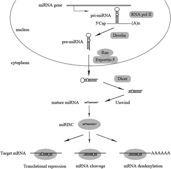
In animals miRNAs are generated from miRNA genes located in intergenic regions or introns of protein-coding genes (Bartel, Reference Bartel2004) (Figure 1). Non-protein-coding miRNA genes are transcribed by RNA polymerase II to generate primary miRNAs (pri-miRNAs). In the nucleus, pri-miRNAs are processed by the RNase III Drosha to form ∼70-nucleotide (nt) precursor miRNAs (pre-miRNAs), which are characterized by the stem-loop structure (Lee et al., Reference Lee, Ahn, Han, Choi, Kim, Yim, Lee, Provost, Radmark, Kim and Kim2003; Kim, Reference Kim2005). Pre-miRNAs are transported from the nucleus to the cytoplasm by the shuttle protein Exportin-5 with the assistance of the G-protein Ran. In the cytoplasm, pre-miRNAs are recognized and cleaved by the RNase III Dicer to form ∼22 nt double-stranded RNAs (Hutvagner et al., Reference Hutvagner, McLachlan, Pasquinelli, Balint, Tuschl and Zamore2001; Lund et al., Reference Lund, Guttinger, Calado, Dahlberg and Kutay2004). When the double strands unwind, one strand enters the RNA-induced silencing complex (RISC), and this strand becomes mature miRNA and can exercise its gene silencing function (Khvorova et al., Reference Khvorova, Reynolds and Jayasena2003). Generation of miRNAs from introns of protein-coding genes, termed mirtrons, follows a slightly different procedure. When pri-miRNAs are transcribed from mirtrons, they accurately imitate the structures of pre-miRNAs without involving Drosha (Carthew and Sontheimer, Reference Carthew and Sontheimer2009).
Biogenesis and mechanism of action of miRNAs. Primary miRNA (pri-miRNA), precursor miRNA (pre-miRNA), Drosha and Dicer are nuclear and cytoplasmic RNase III, respectively; miRISC, miRNA containing RNA-induced silencing complex. See text for details modified from references (Hutvagner et al., Reference Hutvagner, McLachlan, Pasquinelli, Balint, Tuschl and Zamore2001; Khvorova et al., Reference Khvorova, Reynolds and Jayasena2003; Lee et al., Reference Lee, Ahn, Han, Choi, Kim, Yim, Lee, Provost, Radmark, Kim and Kim2003; Lund et al., Reference Lund, Guttinger, Calado, Dahlberg and Kutay2004; Kim, Reference Kim2005) and (Lee et al., Reference Lee, Feinbaum and Ambros1993; Bartel, Reference Bartel2004; Guo et al., Reference Guo, Xie, Fei and Chua2005; Giraldez et al., Reference Giraldez, Mishima, Rihel, Grocock, Van Dongen, Inoue, Enright and Schier2006; Wu et al., Reference Wu, Fan and Belasco2006).

Like mRNAs, miRNA expression is controlled at the transcriptional level by gene-specific transcription factors (Li et al., Reference Li, Xi, Zhu, Zeng, Zhang, Guo, Hao, Liu, Feng, Chen, Chen, Lv, Liu and Liang2011d; Wang et al., Reference Wang, Lin, Li, Xu, Yao, Zhu, Xie, Shen, Sze, Li, Lu, Chan, Poon, Kung and Lin2011). miRNA expression can also be regulated by epigenetic mechanisms. Many miRNA genes contain CpG islands in the promoters, and methylation of these islands represses the transcription of the miRNA genes (Bueno et al., Reference Bueno, Perez de Castro, Gomez de Cedron, Santos, Calin, Cigudosa, Croce, Fernandez-Piqueras and Malumbres2008). miRNA expression can be also induced or inhibited by histone acetylation and/or methylation (Chen et al., Reference Chen, Liao, Hsieh, Wang, Hu and Juo2012b; Saito et al., Reference Saito, Suzuki, Taya, Nishizawa, Tsugawa, Matsuzaki, Hirata, Saito and Hibi2012). Some miRNA genes are regulated by CpG island methylation as well as histone modification (Saito and Saito, Reference Saito and Saito2012). miRNA biogenesis can be also regulated at the post-transcriptional level (Siomi and Siomi, Reference Siomi and Siomi2010).
Mechanism of miRNA action
A miRNA regulates the expression of its target mRNA in three manners (Figure 1), depending on the degree of complementarity between them. An imperfect complementarity leads to translational repression whereas a perfect or nearly perfect complementarity leads to cleavage of the target mRNA. Animal miRNAs are often imperfectly complementary to their target sites; so translational repression is the predominant mechanism by which miRNAs inhibit mRNA expression in animal cells (Lee et al., Reference Lee, Feinbaum and Ambros1993). Plant miRNAs in general have perfect or nearly perfect complementarity to their target sites. As such, cleavage of target mRNAs is the major consequence of miRNA–mRNA binding in plants (Guo et al., Reference Guo, Xie, Fei and Chua2005). Some animal miRNAs can cause the cleavage of their target mRNAs despite imperfect complementarity between them. This is how miR-196 regulates the expression of homeobox protein HOXB8 in mouse embryos (Yekta et al., Reference Yekta, Shih and Bartel2004). The third manner by which miRNA regulates mRNA expression involves mRNA deadenylation. When binds to the imperfectly complementary target site of mRNA, miRNA accelerates the poly(A) tail to be deadenylated. This manner seems to be exclusive to animal miRNAs, and this is how zebrafish miR-430 accelerates the degradation of maternal mRNAs (Giraldez et al., Reference Giraldez, Mishima, Rihel, Grocock, Van Dongen, Inoue, Enright and Schier2006), and mammalian miR-125b and let-7 regulate the expression of their target mRNAs (Wu et al., Reference Wu, Fan and Belasco2006). Although the complementarity between a miRNA and its target mRNA does not have to be perfect, nucleotides 2 to 8 from the 5′ end of the miRNA, also called seed sequence, must be perfectly complementary to the target mRNA (Bartel, Reference Bartel2004).
miRNA target prediction and validation
miRNA target genes can be predicted by computational methods. These methods include miRanda and TargetScan, which are mainly based on sequence complementarity (Enright et al., Reference Enright, John, Gaul, Tuschl, Sander and Marks2003; Lewis et al., Reference Lewis, Burge and Bartel2005), and PicTar, DIANA-microT, RNAhybrid, microInspector, RNA22 and SVMicrO, which use different algorithms (Liu et al., Reference Liu, Yue, Chen, Gao and Huang2010). It is difficult to rate the accuracy of these programs, but a target predicted by multiple programs has greater potential to be a true miRNA target than a target predicted by one program. miRNA target genes predicted by computational methods need to be validated by experiments. A common experiment to validate miRNA targets involves overexpression or knockdown of the miRNA and subsequent detection of the predicted target mRNAs using qRT-PCR or luciferase reporter assays. miRNA knockdown is usually achieved by antisense oligonucleotides (ASO), which bind to the complementary miRNAs so that the latter can not bind to their target mRNAs. 2′-O-methyl antisense oligonucleotides (AMO) and locked nucleic acids (LNA) are two kinds of chemically modified ASO. Compared with AMO, LNA have a higher inhibition efficiency (Naguibneva et al., Reference Naguibneva, Ameyar-Zazoua, Nonne, Polesskaya, Ait-Si-Ali, Groisman, Souidi, Pritchard and Harel-Bellan2006b). Another kind of ASO is miRNA sponges. miRNA sponges are RNA sequences transcribed from expression vectors, containing multiple tandem binding sites to miRNAs. miRNA sponges competitively bind to miRNAs, and thereby prevent them from binding to their targets (Ebert et al., Reference Ebert, Neilson and Sharp2007; Ebert and Sharp, Reference Ebert and Sharp2010). Large-scale miRNA target validation can be achieved through proteomic approaches. For example, using selected reaction monitoring (SRM) and isotope-coded affinity tag quantification, hundreds of potential let-7 target proteins extracted from wild-type and let-7 mutant C. elegans were screened. One hundred and sixty-one proteins can be quantified, and of these, 29 showed significant changes in abundance between wild-type and let-7 mutant. The results of SRM were similar to those of independent experimental downstream analyses such as genetic interaction, as well as polysomal profiling and luciferase assays, indicating that SRM-based validation of miRNA targets is effective and efficient (Jovanovic et al., Reference Jovanovic, Reiter, Picotti, Lange, Bogan, Hurschler, Blenkiron, Lehrbach, Ding, Weiss, Schrimpf, Miska, Grosshans, Aebersold and Hengartner2010).
miRNAs in cattle
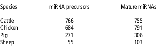
Extensive work has been conducted to identify bovine miRNAs, and a total of 755 mature miRNAs processed from 766 pre-miRNAs have been identified in cattle by various labs (Table 1; (Kozomara and Griffiths-Jones, Reference Kozomara and Griffiths-Jones2011)). By cloning small RNAs from bovine embryo, thymus, small intestine and lymph node, Coutinho and colleagues identified 129 bovine miRNAs (Coutinho et al., Reference Coutinho, Matukumalli, Sonstegard, Van Tassell, Gasbarre, Capuco and Smith2007). Using a similar approach, Gu et al. identified 59 miRNAs from bovine adipose tissue and mammary gland (Gu et al., 2007), and Jin et al. discovered 101 bovine miRNAs from 11 bovine tissues (Jin et al., Reference Jin, Grant, Stothard, Moore and Guan2009). Using computational methods, a total of 496 miRNA genes were predicted from bovine genome (Bovine Genome Sequencing and Analysis Consortium, 2009). More recently, deep sequencing was used to detect miRNAs in primary bovine retinal microvascular endothelial cells, resulting in the identification of 255 known bovine miRNAs. Interestingly, there were multiple new members of the miR-2284/2285 family detected, suggesting this family of miRNAs may play important roles in angiogenesis (Guduric-Fuchs et al., Reference Guduric-Fuchs, O'Connor, Cullen, Harwood, Medina, O'Neill, Stitt, Curtis and Simpson2012).
Numbers of identified miRNAs in four farm animal species*
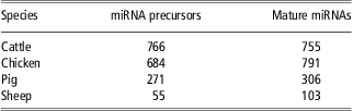
*Based on miRBase 19.
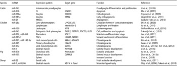
Functional studies of bovine miRNAs have concentrated on the roles of miRNAs in adipose, skeletal muscle, oocyte and early embryonic developments (Table 2). Jin et al. compared the expression of 89 bovine miRNAs in adipose tissues between three crossbreeds differing in backfat thickness, and found that 42 miRNAs were differentially expressed, of which miR-378 was most differentiated (Jin et al., Reference Jin, Dodson, Moore, Basarab and Guan2010). miR-378 has been shown to play a role in lipogenesis in mice (Gerin et al., Reference Gerin, Bommer, McCoin, Sousa, Krishnan and MacDougald2010). Thus, miR-378 might participate in the control of subcutaneous growth in cattle. Expression of miR-143 is upregulated during differentiation of bovine intramuscular preadipocytes into mature adipocytes. Transfection of miR-143 antisense oligonucleotide represses the differentiation of bovine intramuscular preadipocytes (Li et al., Reference Li, Zhang, Zhou, Wang, Wang and Han2011b). These observations further support a role of miR-143 in intramuscular fat development in cattle. Romao and colleagues reported that adipose tissue expression of 8 miRNAs, including miR-19a, -92a, -92b, -101, -103, -106, -142-5p and -296, was responsive to high-fat diet feeding (Romao et al., Reference Romao, Jin, He, McAllister and Guan le2012). Thus, these miRNAs might be parts of the gene regulatory network that mediates high-fat diet-induced adipogenesis in cattle.
miRNAs with different functions in farm animals

miR-1 and miR-206 are two muscle-specific miRNAs that play an important role in muscle cell differentiation (Townley-Tilson et al., Reference Townley-Tilson, Callis and Wang2010). Miretti and colleagues analyzed the expression of miR-1 and miR-206 in skeletal muscle of Piedmontese and Friesian cattle, of which the former are ‘double-muscled’ because of a point mutation in the myostatin gene (Berry et al., Reference Berry, Thomas, Langley, Sharma and Kambadur2002). Expression of miR-1 was not different between the two breeds or sexes, whereas miR-206 was expressed significantly higher in female Piedmontese than in female Friesian cattle (Miretti et al., Reference Miretti, Martignani, Taulli, Bersani, Accornero and Baratta2011), suggesting that upregulated expression of miR-206 might contribute to muscle hypertrophy in female Piedmontese cattle. miR-181b is known to be involved in myoblast differentiation (Naguibneva et al., Reference Naguibneva, Ameyar-Zazoua, Polesskaya, Ait-Si-Ali, Groisman, Souidi, Cuvellier and Harel-Bellan2006a). In Angus cattle, after acute stress (anesthetization and placement of a rumen catheter), miR-181b was upregulated in the muscle, implying it may play a role in myogenesis in Angus cattle (Zhao et al., Reference Zhao, Tian, Yu, Liu, Zan, Updike and Song2012a).
Six miRNAs enriched in oocytes, including miR-205, -150, -122, -96, -146a and -146b-5p, were found to decrease dramatically in expression from 0 to 22 h of maturation (Abd El Naby et al., Reference Abd El Naby, Hagos, Hossain, Salilew-Wondim, Gad, Rings, Cinar, Tholen, Looft, Schellander, Hoelker and Tesfaye2011). These miRNAs might therefore play a role in bovine oocyte maturation. Expression of miR-106a was greater in bovine oocytes than in cumulus–oocyte complexes (COCs), whereas expression of the miR-106a target gene WEE1 homolog (Schizosaccharomyces pombe) (WEE1A) was less in oocytes compared with COCs (Miles et al., Reference Miles, McDaneld, Wiedmann, Cushman, Echternkamp, Vallet and Smith2012). These differences suggest that miR-106a might play a role in bovine oocyte development by downregulating the WEE1A gene. Similarly, miR-196a and miR-181a may regulate bovine oocyte development by targeting the newborn ovary homeobox gene (NOBOX) and the nucleoplasmin 2 (NPM2) gene, respectively, which are essential for folliculogenesis and nuclear and nucleolar organization, respectively (Lingenfelter et al., Reference Lingenfelter, Tripurani, Tejomurtula, Smith and Yao2011; Tripurani et al., Reference Tripurani, Lee, Wee, Smith and Yao2011). Expression of miR-21 and miR-130a in bovine embryos was increased from the 1-cell to 8-cell stage. These two miRNAs might be involved in early bovine embryonic development (Mondou et al., Reference Mondou, Dufort, Gohin, Fournier and Sirard2012).
miRNAs in chickens
The first study of chicken miRNAs was conducted in 2006, which identified 25 miRNAs from chicken embryos and adult chickens through small RNA cloning and sequencing (Xu et al., Reference Xu, Wang, Du and Li2006). Using the same cloning strategy, Wang et al. identified 47 miRNAs from chicken adipose tissue and skeletal muscle (Wang et al., Reference Wang, Yu, Zhang, Gong and Gu2012). Using deep sequencing, 123 miRNAs were discovered from embryonic chick livers at embryonic days 15 (E15) and E20 (Hicks et al., Reference Hicks, Trakooljul and Liu2010), 121 known chicken miRNAs, 449 new chicken miRNAs and 39 mirtrons were identified from E5, E7 and E9 chicken embryos (Glazov et al., Reference Glazov, Cottee, Barris, Moore, Dalrymple and Tizard2008), and 63 and 33 novel chicken miRNAs were identified from chicken preadipocytes and skeletal muscle, respectively (Li et al., Reference Li, Wu, Zhang and Zhu2011c; Yao et al., Reference Yao, Wang, Wang, Wang and Li2011).
Microarrays were used to profile the expression of chicken miRNAs at different stages of gonadal sex differentiation. The study showed that miR-202*, a sexually dimorphic miRNA, was upregulated in male gonads, and this result was validated by in situ hybridization and Northern blot, indicating a potential role of miR-202* in male gonad development (Bannister et al., Reference Bannister, Tizard, Doran, Sinclair and Smith2009). miR-363 was identified to be another sexually dimorphic miRNA, whose expression in chicken gonads was different between sexes at E4.5 and E6.5 (Huang et al., Reference Huang, Gong, Peng, Li, Yang and Feng2010). Using in situ hybridization, 111 miRNAs were profiled in chicken embryos and 75 of them were differentially expressed during embryonic development, suggesting these miRNAs might play roles in chicken embryonic development (Darnell et al., Reference Darnell, Kaur, Stanislaw, Konieczka, Yatskievych and Antin2006). MiR-302 and miR-456 maintain the undifferentiated stage of blastoderm by targeting the sex-determining region Y box 11 (SOX11). MiR-181a* inhibits the somatic differentiation of primordial germ cells (PGCs) by targeting homeobox A1 and prevents PGCs from entering meiosis by inhibiting the nuclear receptor subfamily 6, group A, member 1 (NR6A1) gene (Lee et al., Reference Lee, Lee, Hwang, Lee, Rengaraj, Song, Park and Han2011). Also in PGCs the gene controlling DNA methylation DNA (cytosine-5-)-methyltransferase 3 beta (DNMT3B) is regulated by miR-15c, -29b, -383 and -222 (Rengaraj et al., Reference Rengaraj, Lee, Lee, Seo and Han2011).
Wang et al. found that 10 miRNAs including miR-1a, -133a and -122 were differentially expressed in chicken adipose tissue or skeletal muscle at different developmental stages, suggesting a potential role of these miRNAs in chicken adipose tissue and skeletal muscle development (Wang et al., Reference Wang, Yu, Zhang, Gong and Gu2012). Li and colleagues further identified the activin receptor 2B (ACVR2B) gene as a miR-1 target in chicken muscle (Li et al., Reference Li, Wu, Zhang and Zhu2011c).
Several chicken miRNAs were reported to be involved in chondrogenesis. In chicken limb mesenchymal cells, miR-221 inhibits slug degradation by targeting mdm2 to regulate chondrogenic differentiation (Kim et al., Reference Kim, Song and Jin2010), and miR-142-3p regulates chondrogenesis by directly modulating the disintegrin and metalloprotease 9 (ADAM9) gene (Kim et al., Reference Kim, Song, Kim, Kang and Jin2011b). In primary chicken chondrocytes, miR-365 is a mechanically responsive miRNA that stimulates chondrocyte differentiation by targeting histone deacetylase 4 (HDAC4), an inhibitor of chondrocyte hypertrophy (Guan et al., Reference Guan, Yang, Wei and Chen2011). miR-34a negatively regulates chondrogenesis by targeting EphA5 and suppressing the migration of chondroblasts (Kim et al., Reference Kim, Song, Kim, Chun and Jin2011a) or the reorganization of the actin cytoskeleton, which is essential for chondrocyte differentiation (Kim et al., Reference Kim, Song, Kim, Park, Chun, Sonn and Jin2012).
In chicken cone photoreceptors, the expression of miR-26a is diurnally rhythmic; miR-26a regulates the expression of L-type voltage-gated calcium channel α1C subunit (L-VGCCα1C) according to its own diurnal rhythm to allow retinal photoreceptors to exercise rhythmic function (Shi et al., Reference Shi, Ko and Ko2009). As a tumor suppressor, miR-26a targets interleukin-2, and the downregulation of miR-26a contributes to proliferation of lymphoma cells (Xu et al., Reference Xu, Yao, Smith and Nair2010). Interestingly, a few miRNAs identified in avian herpes viruses including Marek's disease virus might also have regulatory functions (immune evasion, anti-apoptosis and proliferation) in chickens (Burnside and Morgan, Reference Burnside and Morgan2011).
miRNAs in pigs
The first set of porcine miRNAs identified included miR-17, miR-18a, miR-19a, miR-20a, miR-19b and miR-92a, and they all belong to the miR17-92 cluster and were identified based on sequence homology search with known human miRNAs (Sawera et al., Reference Sawera, Gorodkin, Cirera and Fredholm2005). Using small RNA cloning, Sharbati-Tehrani et al. identified 11 miRNAs from various pig tissues (Sharbati-Tehrani et al., Reference Sharbati-Tehrani, Kutz-Lohroff, Scholven and Einspanier2008). Combining deep sequencing and computational analysis, 777 unique miRNAs were identified from six prenatal and four postnatal pigs, including 30 tissue-specific miRNAs (Li et al., Reference Li, Xia, Gu, Zhang, Lang, Chen, Guan, Luo, Chen, Li, Li, Li, Jiang, Shuai, Wang, Zhu, Zhou and Gao2010). Using similar approaches, 15 novel pig-specific miRNAs and 56 novel candidate miRNAs were identified from pig testis (Lian et al., Reference Lian, Sun, Niu, Yang, Liu, Lu, Meng, Qiu, Zhang and Zhao2012); 57 miRNAs were identified from pig skeletal muscle (Xie et al., Reference Xie, Huang, Shen, Li, Zhang, Zhu, Qin and Zhao2010); 293 miRNAs were identified from pig adipose tissue (Li et al., Reference Li, Li, Li, Ning, Li and Yang2011a); 215 miRNAs were identified from the backfat of Large White and Chinese Meishan pigs (Chen et al., Reference Chen, Deng, Qiao, Zheng, Chai, Ding, Peng and Jiang2012a); and 89 miRNAs were identified from pig skeletal muscle and adipose tissue (Cho et al., Reference Cho, Kim, Seo, Lim do, Hong, Park, Park, Hong, Whang and Lee2010).
Expression profiles of porcine miRNAs were mostly analyzed by miRNA microarrays. Using microarrays based on known porcine and human miRNAs, 332 miRNAs, 201 of which were potentially novel porcine miRNAs, were found to be expressed in pig intestine (Sharbati et al., Reference Sharbati, Friedlander, Sharbati, Hoeke, Chen, Keller, Stahler, Rajewsky and Einspanier2010). Between the sexually immature and mature porcine testes, 129 miRNAs are differentially expressed. In the mature testes, 51 miRNAs are upregulated and 78 are downregulated, suggesting that a large number of miRNAs participate in spermatogenesis (Luo et al., Reference Luo, Ye, Liu, Shao, Zheng, Ren, Zuo, Xu, Lei, Jiang, Deng, Xiong and Li2010). The expression levels of let-7a, let-7d, let-7e and miR-22 were higher whereas that of miR-15b lower in abnormal sperm compared with normal sperm (Curry et al., Reference Curry, Safranski and Pratt2011); thus, these miRNAs might be involved in sperm development. MiR-122 is an important regulator of lipid metabolism. The expression level of miR-122 is lower in minipigs fed a high-cholesterol diet than those fed a standard diet, implying a potential role of miR-122 in obesity (Cirera et al., Reference Cirera, Birck, Busk and Fredholm2010). Using deep sequencing, Chen et al. found that the expression of miR-215, -135, -224 and -146b is higher, whereas the expression of miR-1a, -133a, -122, -204 and -183 is lower in the Large White pigs than in the Meishan pigs (Chen et al., Reference Chen, Deng, Qiao, Zheng, Chai, Ding, Peng and Jiang2012a). The Meishan pigs are known to have more fat than the Large White pigs. Differential expression of these miRNAs suggests the potential involvement of these miRNAs in adipose tissue development and growth in pigs.
Functions of porcine miRNAs are beginning to be revealed. MiR-148b potentially targets integrin alpha 5 (ITGA5), which is related to splay leg in piglets. The miR-148b gene has two SNPs, one of which alters a potential miRNA core promoter sequence. This SNP has different frequencies between different breeds of pig, but is not associated with splay leg condition (Maak et al., Reference Maak, Boettcher, Komolka, Tetens, Wimmers, Reinsch, Swalve and Thaller2010). One T/C SNP in miR-27a gene, detected in Large White and Dam line of Chinese lean-type new lines (DIV) pigs, is associated with litter size (Lei et al., Reference Lei, Gao, Luo, Xia, Jiang, Deng, Xiong and Li2011). Between different genotypes, there is a significant difference of litter size in DIV but no significant difference of litter size in Large White pigs, implying the specific role of miR-27a in litter size in DIV pigs (Lei et al., Reference Lei, Gao, Luo, Xia, Jiang, Deng, Xiong and Li2011). In the ovarian granulosa cells, miR-378 downregulates aromatase expression and estradiol production by targeting the 3′-UTR of the aromatase mRNA (Xu et al., Reference Xu, Linher-Melville, Yang, Wu and Li2011). Thus, miRNA-378 might play a role in ovarian follicular development (Xu et al., Reference Xu, Linher-Melville, Yang, Wu and Li2011). Zhao et al. analyzed the expression of Olfactomedin-like 3 (OLFML3) in developmental skeletal muscle and found it was regulated by miR-155 (Zhao et al., Reference Zhao, Zhang, Hou, Zan, Wang, Tang and Li2012b). MiR-155 was also highly expressed in spleen and was upregulated after lipopolysaccharide and poly (I:C) stimulation, and it positively regulated TLR3/TLR4 signaling pathways. These imply a role of miR-155 in regulating porcine immunity (Li et al., Reference Li, He, Zhu, Zhao and Li2013).
miRNAs in sheep
MiR-431, -433, -127, -434, -432 and -136 were first sheep miRNAs identified through sequence homology search from the Callipyge sheep, which display the muscle hypertrophy phenotype (Davis et al., Reference Davis, Caiment, Tordoir, Cavaille, Ferguson-Smith, Cockett, Georges and Charlier2005). From the same sheep, 747 miRNAs representing 472 miRNA precursors were identified from the skeletal muscle of callipyge sheep through deep sequencing (Caiment et al., Reference Caiment, Charlier, Hadfield, Cockett, Georges and Baurain2010). Using small RNA cloning, Sheng and colleagues reported the identification of 31 sheep miRNAs (Sheng et al., Reference Sheng, Song, Yu, Niu, Li, Li, Wei, Liu, Zhang and Du2011). More recently, McBride et al. reported the identification of 212 sheep miRNAs by sequencing small RNA libraries from sheep ovarian follicles and corpus lutea at various reproductive stages (McBride et al., Reference McBride, Carre, Sontakke, Hogg, Law, Donadeu and Clinton2012).
Using miRNA microarrays, Wenguang et al. compared the expression of 159 miRNAs in body skin and ear skin from sheep, finding that 19 miRNAs were specifically expressed or greatly enriched in body side skin (Wenguang et al., Reference Wenguang, Jianghong, Jinquan and Yashizawa2007). This raises the possibility that these miRNAs play a role in hair growth in sheep. Torley et al. compared the expression of 128 miRNAs in sheep ovaries and testes at gestational day (GD) 42 and GD75 by real-time qRT-PCR, finding that 24 miRNAs were differentially expressed between ovaries and testes at GD42 while 43 miRNAs at GD75. Using in situ hybridization, Torley et al. revealed that miR-22 was located in Sertoli cells and proposed that this miRNA might play a role in suppressing the estrogen-signaling pathway during fetal testicular development (Torley et al., Reference Torley, da Silveira, Smith, Anthony, Veeramachaneni, Winger and Bouma2011). Prenatal testosterone treatment leads to ovarian pathology. In the fetal ovaries from gestational ewes treated with testosterone, miR-497 and miR-15b were upregulated, suggesting that these two miRNAs might have a role in testosterone-induced ovarian pathology (Luense et al., Reference Luense, Veiga-Lopez, Padmanabhan and Christenson2011). Clop et al. found that the myostatin (MSTN) gene of Texel sheep had a G to A mutation in the 3′-UTR, and they further found that this mutation created a target site for miR-1 and miR-206, and thereby repressed the expression of MSTN protein and resulted in muscular hypertrophy in Texel sheep (Clop et al., Reference Clop, Marcq, Takeda, Pirottin, Tordoir, Bibe, Bouix, Caiment, Elsen, Eychenne, Larzul, Laville, Meish, Milenkovic, Tobin, Charlier and Georges2006).
Conclusions and perspectives
MicroRNAs are important regulators in biology. Hundreds of miRNAs have been identified in cattle, chickens, pigs and sheep. Many of these miRNAs are found to be expressed in a spatiotemporal-specific manner, which indicates that miRNAs play roles in specific tissue types or at specific developmental stages. Although limited functional studies of farm animal miRNAs have been conducted, these studies indicate that miRNAs play important roles in muscle development and hypertrophy, adipose tissue growth, oocyte maturation and early embryonic development in farm animals. The key to understanding the functions of miRNAs is to identify their mRNA targets. Therefore, future studies of miRNAs in farm animals should focus on the identification of mRNAs targeted by miRNAs and the physiological processes where the mRNA–miRNA interactions take place. Recent genome-wide association studies have shown that complex traits are often strongly associated with noncoding SNPs (Easton et al., Reference Easton, Pooley, Dunning, Pharoah, Thompson, Ballinger, Struewing, Morrison, Field, Luben, Wareham, Ahmed, Healey, Bowman, Meyer, Haiman, Kolonel, Henderson, Le Marchand, Brennan, Sangrajrang, Gaborieau, Odefrey, Shen, Wu, Wang, Eccles, Evans, Peto, Fletcher, Johnson, Seal, Stratton, Rahman, Chenevix-Trench, Bojesen, Nordestgaard, Axelsson, Garcia-Closas, Brinton, Chanock, Lissowska, Peplonska, Nevanlinna, Fagerholm, Eerola, Kang, Yoo, Noh, Ahn, Hunter, Hankinson, Cox, Hall, Wedren, Liu, Low, Bogdanova, Schurmann, Dork, Tollenaar, Jacobi, Devilee, Klijn, Sigurdson, Doody, Alexander, Zhang, Cox, Brock, MacPherson, Reed, Couch, Goode, Olson, Meijers-Heijboer, van den Ouweland, Uitterlinden, Rivadeneira, Milne, Ribas, Gonzalez-Neira, Benitez, Hopper, McCredie, Southey, Giles, Schroen, Justenhoven, Brauch, Hamann, Ko, Spurdle, Beesley, Chen, Mannermaa, Kosma, Kataja, Hartikainen, Day, Cox and Ponder2007; Greenman et al., Reference Greenman, Stephens, Smith, Dalgliesh, Hunter, Bignell, Davies, Teague, Butler, Stevens, Edkins, O'Meara, Vastrik, Schmidt, Avis, Barthorpe, Bhamra, Buck, Choudhury, Clements, Cole, Dicks, Forbes, Gray, Halliday, Harrison, Hills, Hinton, Jenkinson, Jones, Menzies, Mironenko, Perry, Raine, Richardson, Shepherd, Small, Tofts, Varian, Webb, West, Widaa, Yates, Cahill, Louis, Goldstraw, Nicholson, Brasseur, Looijenga, Weber, Chiew, DeFazio, Greaves, Green, Campbell, Birney, Easton, Chenevix-Trench, Tan, Khoo, Teh, Yuen, Leung, Wooster, Futreal and Stratton2007; Saxena et al., Reference Saxena, Voight, Lyssenko, Burtt, de Bakker, Chen, Roix, Kathiresan, Hirschhorn, Daly, Hughes, Groop, Altshuler, Almgren, Florez, Meyer, Ardlie, Bengtsson Bostrom, Isomaa, Lettre, Lindblad, Lyon, Melander, Newton-Cheh, Nilsson, Orho-Melander, Rastam, Speliotes, Taskinen, Tuomi, Guiducci, Berglund, Carlson, Gianniny, Hackett, Hall, Holmkvist, Laurila, Sjogren, Sterner, Surti, Svensson, Tewhey, Blumenstiel, Parkin, Defelice, Barry, Brodeur, Camarata, Chia, Fava, Gibbons, Handsaker, Healy, Nguyen, Gates, Sougnez, Gage, Nizzari, Gabriel, Chirn, Ma, Parikh, Richardson, Ricke and Purcell2007; Scott et al., Reference Scott, Mohlke, Bonnycastle, Willer, Li, Duren, Erdos, Stringham, Chines, Jackson, Prokunina-Olsson, Ding, Swift, Narisu, Hu, Pruim, Xiao, Li, Conneely, Riebow, Sprau, Tong, White, Hetrick, Barnhart, Bark, Goldstein, Watkins, Xiang, Saramies, Buchanan, Watanabe, Valle, Kinnunen, Abecasis, Pugh, Doheny, Bergman, Tuomilehto, Collins and Boehnke2007; Yeager et al., Reference Yeager, Orr, Hayes, Jacobs, Kraft, Wacholder, Minichiello, Fearnhead, Yu, Chatterjee, Wang, Welch, Staats, Calle, Feigelson, Thun, Rodriguez, Albanes, Virtamo, Weinstein, Schumacher, Giovannucci, Willett, Cancel-Tassin, Cussenot, Valeri, Andriole, Gelmann, Tucker, Gerhard, Fraumeni, Hoover, Hunter, Chanock and Thomas2007; Stefansson et al., Reference Stefansson, Ophoff, Steinberg, Andreassen, Cichon, Rujescu, Werge, Pietilainen, Mors, Mortensen, Sigurdsson, Gustafsson, Nyegaard, Tuulio-Henriksson, Ingason, Hansen, Suvisaari, Lonnqvist, Paunio, Borglum, Hartmann, Fink-Jensen, Nordentoft, Hougaard, Norgaard-Pedersen, Bottcher, Olesen, Breuer, Moller, Giegling, Rasmussen, Timm, Mattheisen, Bitter, Rethelyi, Magnusdottir, Sigmundsson, Olason, Masson, Gulcher, Haraldsson, Fossdal, Thorgeirsson, Thorsteinsdottir, Ruggeri, Tosato, Franke, Strengman, Kiemeney, Melle, Djurovic, Abramova, Kaleda, Sanjuan, de Frutos, Bramon, Vassos, Fraser, Ettinger, Picchioni, Walker, Toulopoulou, Need, Ge, Yoon, Shianna, Freimer, Cantor, Murray, Kong, Golimbet, Carracedo, Arango, Costas, Jonsson, Terenius, Agartz, Petursson, Nothen, Rietschel, Matthews, Muglia, Peltonen, St Clair, Goldstein, Stefansson and Collier2009; Ellinor et al., Reference Ellinor, Lunetta, Glazer, Pfeufer, Alonso, Chung, Sinner, de Bakker, Mueller, Lubitz, Fox, Darbar, Smith, Smith, Schnabel, Soliman, Rice, Van Wagoner, Beckmann, van Noord, Wang, Ehret, Rotter, Hazen, Steinbeck, Smith, Launer, Harris, Makino, Nelis, Milan, Perz, Esko, Kottgen, Moebus, Newton-Cheh, Li, Mohlenkamp, Wang, Kao, Vasan, Nothen, MacRae, Stricker, Hofman, Uitterlinden, Levy, Boerwinkle, Metspalu, Topol, Chakravarti, Gudnason, Psaty, Roden, Meitinger, Wichmann, Witteman, Barnard, Arking, Benjamin, Heckbert and Kaab2010). Given the role of miRNAs in gene expression, one possible explanation for such trait-noncoding SNP associations is that the associated SNPs are parts of miRNA binding sites or miRNA sequences. Therefore, another potential direction of future miRNA studies in farm animals is to identify SNPs that overlap with miRNA binding sites or miRNA sequences and are associated with production or health traits in farm animals. Such studies could have immediate implications for selecting farm animals with superior production or health traits and for improving genetics in farm animals.
Acknowledgments
This project is supported by National Natural Science Foundation of China (30871789, 31072025, 31272438) and Innovation Project of Jiangsu Graduate Research (CXZZ11_0988).