Introduction
Since the original description of Aspersentis megarhynchus by von Linstow (Reference von Linstow1892) from Antarctic fishes in South Georgia as Echinorhynchus megarhynchus, the identity of this acanthocephalan has been riddled with many controversies, diverse interpretations and disagreements. We suspect that von Linstow (Reference von Linstow1892) used the name ‘megarhynchus’ in reference to the large ventral hooks compared to the small dorsal hooks on the proboscis (von Linstow, Reference von Linstow1892, Fig. 34) even though he did not literally describe the dorso-ventral differentiation of hook size. In 1929, two major developments took place: Baylis (Reference Baylis1929) described the same species as Rhadinorhynchus wheeleri Baylis, 1929 from Notothenia rossii Richardson, Parachaenichthys georgianus Fischer and Trematomus hansoni Boulenger in South Georgia Island in the West Antarctic with one figure of a proboscis; and Van Cleave (Reference Van Cleave1929) created and diagnosed the genus Aspersentis in the family Rhadinorhynchidae Lühe, 1912 and described the same acanthocephalan species as Aspersentis austrinus Van Cleave, Reference Van Cleave1929 from Trematomus sp. or Notothenia sp. in the South Shetland Islands based on three specimens in the Baylis collection in the British Museum that have been tentatively labelled Echinorhynchus campbelli, with three figures of three proboscides. Footnote no. 1 in Baylis (Reference Baylis1929, p. 557) stated that
Since this report was sent to press, an account has been published by Van Cleave (Ann. Mag. Nat. Hist. (10) IV, p. 229, August, 1929) of a from Trematomus or Notothenia from the South Shetlands, which he names Aspersentis austrinus, gen. et sp. n. This form seems to approach very closely to R. hadinorhynchus wheeleri … .
Chandler (Reference Chandler1934) assigned R. wheeleri to the genus Aspersentis. Meyer (Reference Meyer1933) provided an abbreviated version of Van Cleave's (Reference Van Cleave1929) description of A. austrinus using his figures of three proboscides and a separate version of Baylis (Reference Baylis1929) of R. wheeleri under the genus Rhadinorhynchus. Joyeux & Baer (Reference Joyeux and Baer1954) described a dozen largely contracted specimens of A. megarhynchus from N. rossii in the Kerguelen Islands. Edmonds (Reference Edmonds1955) described 30 mostly female specimens of A. austrinus from Notothenia coriiceps Richardson and Notothenia cyanobrancha Richardson in Heard Island and later, Edmonds (Reference Edmonds1957) reported over 300 specimens of A. austrinus from five species of fish in the T.H. Johnston's collection of the British Australian and New Zealand Antarctic Expedition of 1929–1931 in Crozet and Kerguelen Islands. Petrochenko (Reference Petrochenko1956) included the genus Aspersentis in the family Arhythmacanthidae Yamaguti, 1935 and reported A. wheeleri and A. austrinus as distinct species differing only in the proboscis armature, the only distinguishing character; he erroneously reported the armature of A. wheeleri, which created a false disparity between two otherwise similar species. Golvan (Reference Golvan1960) created the family Aspersentidae and appropriately established A. megarhynchus (von Linstow, Reference von Linstow1892) Golvan, Reference Golvan1960 (=E. megarhynchus von Linstow, Reference von Linstow1892 = R. wheeleri Baylis, Reference Baylis1929 = A. austrinus Van Cleave, Reference Van Cleave1929) as the type species. Golvan (Reference Golvan1969) retained his arrangement of 1960 as did Amin (Reference Amin, Crompton and Nickol1985, Reference Amin2013), and later Golvan (Reference Golvan1969) provided a description of A. megarhynchus purely based on that of Baylis (Reference Baylis1929) verbatim and using Van Cleave's figures of three proboscides. Golvan (Reference Golvan1994), however, reversed his position and listed A. austrinus, A. megarhynchus and A. wheeleri as separate species, much like what Petrochenko (Reference Petrochenko1956) did, without any justification except to mention that A. wheeleri may be a synonym of A. megarhynchus. In its most comprehensive taxonomic treatment, Zdzitowiecki (Reference Zdzitowiecki1981) redescribed A. austrinus from 30 fishes of five species collected in two sites of West Antarctica, the South Shetlands and South Georgia, and provided ten detailed figures and measurements. Zdzitowiecki (Reference Zdzitowiecki1981) synonymized A. austrinus with A. wheeleri and A. megarhynchus. In his synopsis of Antarctic Acanthocephala and in all his subsequent publications, Zdzitowiecki (Reference Zdzitowiecki, Wägele and Sieg1991) recognized A. megarhynchus as the primary name, with A. austrinus Van Cleave, Reference Van Cleave1929, R. wheeleri Baylis, Reference Baylis1929 and Heteracanthocephalus hureaui Dollfus, 1965 as synonyms. Pichelin et al. (Reference Pichelin, Smales and Bray2002) questioned the validity of A. megarhynchus (von Linstow, Reference von Linstow1892) Golvan, Reference Golvan1960 and considered it a species inquirenda based on statements in the description of E. megarhynchus by von Linstow (Reference von Linstow1892) that could be interpreted a number of ways or on the incomplete description of some of the characters as may have been more appropriate in 1892. We disagree with the extreme assessment of Pichelin et al. (Reference Pichelin, Smales and Bray2002) and we find more reason to support the legitimacy of von Linstow's (Reference von Linstow1892) description and validity of A. megarhynchus as proposed by Golvan (Reference Golvan1960) and as were concurred with later by Amin (Reference Amin, Crompton and Nickol1985, Reference Amin2013) and in this presentation.
In the present work we provide new morphological observations of a new collection of specimens from a new location in Antarctica, which is noteworthy considering the number of descriptions made of this species to date. We also include new morphometric data, SEM and microscopic images of newly reported characters, chemical analysis of hooks, and confirm the distinctiveness of this population. Finally, we provide a molecular characterization of this material based on the small subunit SSU 18S DNA gene, which represents the first genetic data for the genus Aspersentis and also for the family Heteracanthocephalidae, and explore phylogenetic affinities with other paleacanthocephalan genera and families.
Materials and methods
Collections
Collections of helminths were made in April 2014–February 2015 during the 19th Ukrainian Antarctic expedition to the Ukrainian Antarctic Station (UAS) ‘Akademik Vernadsky’ on the Galindez Island, Argentine Islands (65°15′S, 64°16′W). A total of 106 individuals of N. coriiceps were caught using a fishing rod off the shore of the Galindez Island at depths from 10 to 30 m (see Kuzmina et al., Reference Kuzmina, Salganskij, Lisitsyna and Korol2020). All fishes collected were immediately transported to the laboratory, measured and examined using the standard parasitological techniques (see Zdzitowiecki & Laskowski, Reference Zdzitowiecki and Laskowski2004; Weber & Govett, Reference Weber and Govett2009). Acanthocephalans were collected manually, kept in tap water for 30 min to 3 h for proboscis evagination prior to their fixation in 70% ethanol; they were counted and stored separately. Tentative identification of the parasites was performed in temporary total mounts of the Berlese medium according to Zdzitowiecki (Reference Zdzitowiecki1983, Reference Zdzitowiecki1984a, Reference Zdzitowieckib, Reference Zdzitowiecki1987) in the laboratory of the Department of Parasitology, I.I. Schmalhausen Institute of Zoology in Kyiv, Ukraine, where specimens were deposited (see Kuzmina et al., Reference Kuzmina, Salganskij, Lisitsyna and Korol2020).
About 45 specimens were made available to study at the Scottsdale, Arizona lab. Thirteen specimens (12 females and one male) were processed for microscopical examination and measured; sex could not be determined before processing. Twelve specimens including three males with everted bursa were used for scanning electron microscopy (SEM) and energy-dispersive X-ray analysis (EDXA), four specimens for molecular analysis and the remaining specimens are stored in ethanol in the Amin collection.
Microscopical methods and images
Freshly collected acanthocephalans were extended in water until proboscides were everted then fixed in 70% ethanol for transport to our Institute of Parasitic Diseases in Arizona, USA, for processing and further studies. Worms were punctured with a fine needle and subsequently stained in Mayer's acid carmine, de-stained in 4% hydrochloric acid in 70% ethanol, dehydrated in ascending concentrations of ethanol reaching 100% (24 h each) and cleared in 100% xylene then in 50% Canada balsam and 50% xylene (24 h each). Whole worms were then mounted in Canada balsam. Measurements are in micrometres, unless otherwise noted; the range is followed by the mean values between parentheses. Width measurements represent maximum width. Trunk length does not include proboscis, neck or bursa.
Microscope images were created using 10× and 40× objective lenses of a BH2 light Olympus microscope (Olympus Optical Co, Osachi-shibamiya, Okaya, Nagano, Japan) attached to an AmScope 1000 video camera (United Scope LLC, dba AmScope, Irvine, California, USA), linked to an ASUS lab top equipped with HDMI high-definition multimedia interface system (Taiwan-USA, Fremont, California, USA). Images from the microscope are transferred from the lab top to a USB and stored for subsequent processing on a computer.
Line drawings were created by using a Ken-A-Vision micro-projector (Ward's Biological Supply Co, Rochester, New York, USA), which uses cool quartz iodine 150W illumination with 10×, 20× and 43× objective lenses. Images of stained whole-mounted specimens were projected vertically on 300 series Bristol draft paper (Starthmore, Westfield, Massachusetts, USA), then traced and inked with India ink. Projected images were identical to the actual specimens being projected.
Specimens were deposited in the University of Nebraska's State Museum's Harold W. Manter Laboratory collection in Lincoln, Nebraska, USA.
SEM
About 15 specimens that had been fixed and stored in 70% ethanol were processed for SEM following standard methods (Lee, Reference Lee1992). These included critical-point drying in sample baskets and mounting on SEM sample mounts (stubs) using conductive double-sided carbon tape. Samples were coated with gold and palladium for 3 min using a Polaron #3500 sputter coater (Quorum (Q150 TES), www.quorumtech.com) establishing an approximate thickness of 20 nm. Samples were placed and observed in an FEI Helios Dual Beam Nanolab 600 (FEI, Hillsboro, Oregon, USA) Scanning Electron Microscope with digital images obtained in the Nanolab software system (FEI, Hillsboro, Oregon, USA) and then stored on a USB for future reference. Samples were received under low vacuum conditions using 10 KV, spot size 2, 0.7 Torr using a Gaseous Secondary Electron Detector.
EDXA
Standard methods were used for preparation similar to the SEM procedure. Specimens were examined and positioned with the above SEM instrument, which was equipped with a Phoenix energy-dispersive X-ray analyser (FEI, Hillsboro, Oregon, USA). X-ray spot analysis and live scan analysis were performed at 16 Kv with a spot size of 5 and results were recorded on charts and stored with digital imaging software attached to a computer. The TEAM (Texture and Elemental Analytical Microscopy) software system (FEI, Hillsboro, Oregon, USA) was used. Data were stored on a USB. The data included weight percent and atom percent of the detected elements following correction factors and were stored on a USB. All figures on the USB can be viewed by contacting the second author. The hooks were cut and scanned at two positions (tip and middle) with a gallium (Ga) beam liquid ion metal source (LIMS) using a dual-beam scanning electron microscope. The alignment of the hook previous to cutting generated a cross-section of the area.
Ion sectioning of hooks
A dual-beam SEM with Ga ion source (GIS) is used for the LIMS part of the process. The Ga beam (LIMS) is a gas injection magnetron sputtering technique whereby the rate of cutting can be regulated. The hooks of the acanthocephalans were centred on the SEM stage and cross-sectioned using a probe current between 0.2 and 2.1 nA according to the rate at which the area is cut. The time of cutting is based on the nature and sensitivity of the tissue. Following the initial cut, the sample also goes through a milling process to obtain a smooth surface. The cut was then analysed with an X-ray at the tip, middle and base of hooks for chemical ions with an electron beam (Tungsten) to obtain an X-ray spectrum. Results were stored with the attached imaging software then transferred to a USB for future use. The intensity of the GIS was variable according to the nature of the material being cut.
Molecular methods
Total genomic DNA was extracted from two specimens of A. megarhynchus preserved in ethanol 70% using Qiagen™ (Valencia, California, USA) DNeasy® Tissue Kit following manufacturer's instructions. Partial nuclear small subunit ribosomal DNA (18S rDNA) gene was amplified (50 μl total volume) using ExcelTaq™ SMOBIO® PCR Master Mix (Taiwan). Primer pairs and amplification conditions used were as described in Amin et al. (Reference Amin, Heckmann, Dallarés, Constenla and Ha2019b). In every polymerase chain reaction (PCR) run, a negative and positive control were included in order to detect any potential contamination and to have a reliable sample to compare with, respectively. PCR amplicons were sequenced directly for both strands using the same PCR primers.
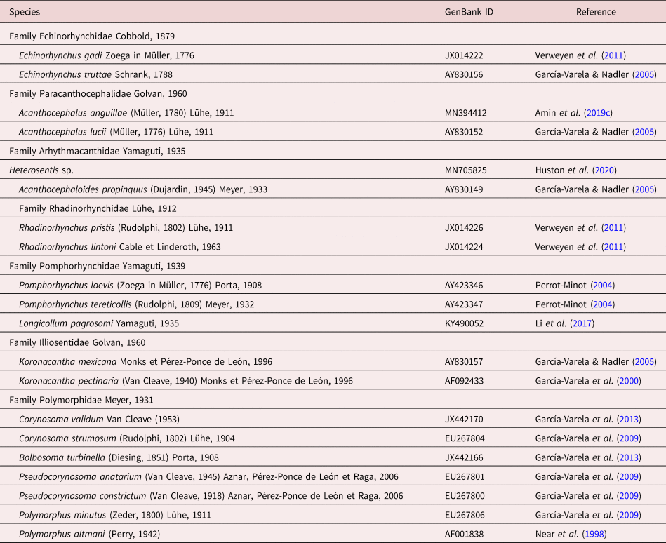
Sequences were assembled and edited using ContigExpress implemented in the software Vector NTI Advance® v10.3.0 and submitted to GenBank under accession numbers MW916820 and MW916821. Sequences were aligned using Muscle as implemented in MEGA v6 (Tamura et al., Reference Tamura, Stecher, Peterson, Filipski and Kumar2013) alongside representatives of main paleacanthocephalan families. Detailed data on the sequences included in the alignment and subsequent phylogenies can be found in table 1. Rotaria rotatoria (Pallas, 1776) (DQ089736) was selected as an outgroup. Alignment reliability was assessed with the web-based program GUIDANCE (Penn et al., Reference Penn, Privman, Landan, Graur and Pupko2010). The aligned sequences (864 nt positions, of which 113 were excluded prior to analysis) were used for comparative sequence analysis and phylogenetic reconstructions.
Sequences belonging to six paleacanthocephalan families retrieved from GenBank and included in the 18S rDNA alignment and subsequent phylogenetic analyses.

Gblocks 0.91b implemented on the Phylogeny.fr site (Dereeper et al., Reference Dereeper, Guignon and Blanc2008) was used to select blocks of evolutionarily conserved sites. Maximum likelihood (ML) and Bayesian inference (BI) algorithms were used for phylogenetic tree reconstruction after determination of the best-fit model of nucleotide substitution with jModelTest v2.1.4 (Darriba et al., Reference Darriba, Taboada, Doallo and Posada2012) using the Akaike Information Criterion and the Bayesian Information Criterion, respectively. The best-fitting model selected for the ML algorithm was the GTR + I + G model (nst = 6, rates = gamma, ngammacat = 4), while for BI it was TrN + I+G (nst = 6, rates = gamma, ngammacat = 4). ML analyses were performed in PhyML v3.0 (Guindon et al., Reference Guindon, Dufayard, Lefort, Anisimova, Hordijk and Gascuel2010) with a non-parametric bootstrap of 100 replicates. BI analyses were carried out with MrBayes v3.2.6 (Ronquist et al., Reference Ronquist, Teslenko and Van Der Mark2012) implemented on the CIPRES Science Gateway v3.3 (Miller et al., Reference Miller, Pfeiffer and Schwartz2010). Log likelihoods were estimated over 10,000,000 generations using Markov Chain Monte Carlo searches on two simultaneous runs of four chains, sampling trees every 1000 generations. The first 25% of the sampled trees were discarded as ‘burn-in’ and a consensus topology and nodal support estimated as posterior probability values (Huelsenbeck et al., Reference Huelsenbeck, Ronquist, Nielsen and Bollback2001) were calculated from the remaining trees. Pairwise genetic distance matrices were calculated using the ‘uncorrected P-distance’ model implemented in MEGA v6.
Results
The hosts
All fishes, N. coriiceps, examined (100%) were found infected with one or more of 25 species of helminths near the UAS ‘Akademik Vernadsky’ in 2014–2015 including eight species of acanthocephalans. Acanthocephalans were found in 93.4% of N. coriiceps with intensity from 1 to 106 (mean intensity 24.7; median intensity 17) specimens per fish with a mean abundance of 23.04. The prevalence and mean intensity of specimens of A. megarhynchus found in the 106 individuals of N. coriiceps examined were 13.2% and 4.0, respectively (Kuzmina et al., Reference Kuzmina, Salganskij, Lisitsyna and Korol2020). Prevalence (%) and mean abundance of A. megarhynchus infections of N. coriiceps in the waters surrounding the UAS ‘Akademik Vernadsky’ in 2014–2015 were 13.2% and 0.53, respectively (Kuzmina et al., Reference Kuzmina, Salganskij, Lisitsyna and Korol2020) compared to 2.0% and 0.16 in 2002 (Zdzitowiecki & Laskowski, Reference Zdzitowiecki and Laskowski2004).
The parasite
The examination of new material and relevant controversial literature on A. megarhynchus made it possible to have a fresh look at the generic diagnosis of the genus Aspersentis, which is here amended and updated.
Emended generic diagnosis
Aspersentis Van Cleave, Reference Van Cleave1929 [syn. Heteracanthocephalus Petrochenko, Reference Petrochenko1956; Echinorhynchus von Linstow, Reference von Linstow1892; Rhadinorhynchus Baylis, Reference Baylis1929; Heterosentis Van Cleave, 1931]
Generic diagnosis (newly observed features are bolded). Small cylindroid to fusiform worms with trunk spines over entire trunk including bursa but especially conspicuous anteriorly and ventrally but much smaller to barely visible or almost missing dorsally and posteriorly. Sexual dimorphism evident in shared structures. Proboscis, bent ventrad, relatively short, cylindrical, widest anteriorly, with longitudinal rows of hooks much larger ventrally and anteriorly than dorsally and posteriorly. All ventral hooks with prominent roots markedly shorter than blades; posterior two or three hooks with vestigial roots having conspicuous anterior manubria. Apical ventral hook slender with slightly manubriated root anteriorly. Second or third anterior ventral hooks largest and widest at base. All dorsal hooks with smaller and anteriorly prominently manubriated roots. Lateral hooks intermediate in shape and size between dorsal and ventral hooks. Proboscis receptacle double-walled, considerably thicker dorsally than ventrally and with cross spiral striations of outer wall. Cerebral ganglion large, drop-shaped, near posterior end of receptacle. Lemnisci flattened, slightly to markedly longer than receptacle, may extend to level of anterior testis. Male reproductive system in posterior two-thirds of trunk. Testes ovoid, often contiguous, in tandem to diagonal. Cement glands six, pyriform to rounded, compacted, each with its own duct joining posterior end of Saefftigen's pouch independently. Male gonopore may be dorsal. Vagina and uterus well-developed and uterine bell with many glands. Egg fusiform with polar prolongation of fertilization membrane. In marine fish.
Description
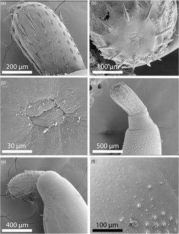
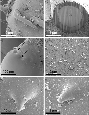
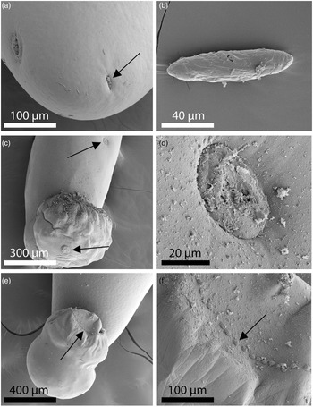
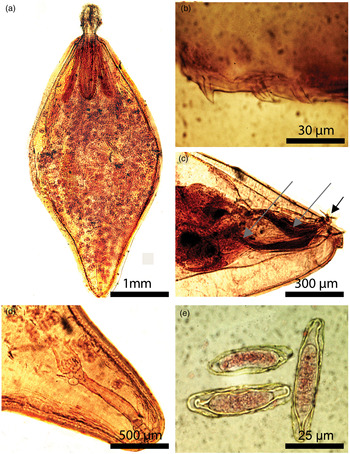
Most of the descriptions of A. megarhynchus were based on those of Van Cleave (Reference Van Cleave1929) who provided line drawings of three proboscides, one with anterior trunk, and Baylis (Reference Baylis1929) who included one line drawing with anterior trunk spines. Actually, the much earlier description of von Linstow (Reference von Linstow1892) provided coloured line drawings of anterior and basal hooks with roots, an egg and a section at the anterior trunk. All other descriptions, including line drawings, were outdone by the most comprehensive description of Zdzitowiecki (Reference Zdzitowiecki1981) that included ten figures of the most complete coverage of structures. Zdzitowiecki (Reference Zdzitowiecki, Wägele and Sieg1991, p. 32) also provided good line drawings of a whole male, proboscis and posterior end of a female, and Golvan (Reference Golvan1969, p. 155) provided many more. We opted not to be repetitious in light of the availability of these line drawings and, instead, we decided to use microscope and SEM images of actual specimens depicting the natural appearance of structures being reported (Figs. 1–5).
SEM images of specimens of Aspersentis megarhynchus from Notothenia coriiceps in the Western Antarctica off Galindez Island. (a) Lateral view of the proboscis. Note the larger ventral hooks on the ventral side (left). (b) The apical end of a proboscis showing the double-lobed orifice of the apical organ. (c) A higher magnification of the apical organ orifice. (d) Variations in the distribution of the larger trunk spines. Large trunk spines almost encircling the whole anterior trunk; note the large neck. (e) Variations in the distribution of the larger trunk spines. Large trunk spines limited to a small ventral portion of the anterior trunk of another specimen. (f) Transition between the larger and smaller spines in the anterior trunk of a specimen.

SEM images of specimens of Aspersentis megarhynchus from Notothenia coriiceps in the Western Antarctica off Galindez Island. (a) A large anterior hook of a specimen; note its angular curvature. (b) A large Gallium-cut hook. Note its thick cortical layer probably containing a high level of sulphur. (c) Two of a number of sensory pits on the anterior neck of a specimen. (d) Micropores on the cuticular surface of the mid-trunk. (e) A high magnification of a large antero-ventral spine; note its conical shape. (f) A high magnification of one of the smaller and more slender trunk spines. Note the extension of micropores on the spine.

SEM images of specimens of Aspersentis megarhynchus from Notothenia coriiceps in the Western Antarctica off Galindez Island. (a) Variations in the orientation of the female gonopore. The slit-shaped gonopore is vertical or occasionally horizontal. Note the large sensory plate (black arrow). (b) A ripe egg revealing the sub-surface fibrillar coat. (c) The posterior portion of a male specimen showing the ventrally curved bursa and two sensory plates (arrows) similar to those seen in females (fig. 3a). (d) High magnification of a sensory plate like those in fig. 3c. (e) Different perspectives and magnifications of the bursa showing the one circle of the bead-like sensory receptors characteristic of this species. (f) Different perspectives and magnifications of the bursa showing the one circle of the bead-like sensory receptors characteristic of this species

Microscope images of specimens of Aspersentis megarhynchus from Notothenia coriiceps in the Western Antarctica off Galindez Island. (a) A ventral view of a whole mounted female specimen showing the typical inflation at mid-trunk in our material and the relatively longer lemnisci than the proboscis receptacle. (b) Lateral view of three anterior ventral spines. Note the single rod core per spine. (c) The posterior part of the male reproductive system showing the dark cement gland ducts (three on each side, grey arrows) surrounding Saefftigen's pouch and the dorsal gonopore (short black arrow) a distance from the papilla-like posterior end of the trunk. The bursa was cut off. (d) The conically shaped posterior end of the female showing the whole reproductive system. (e) Three eggs. The smaller top egg is not as ripe compared to the two other larger eggs featuring well-developed polar prolongation of fertilization membrane.

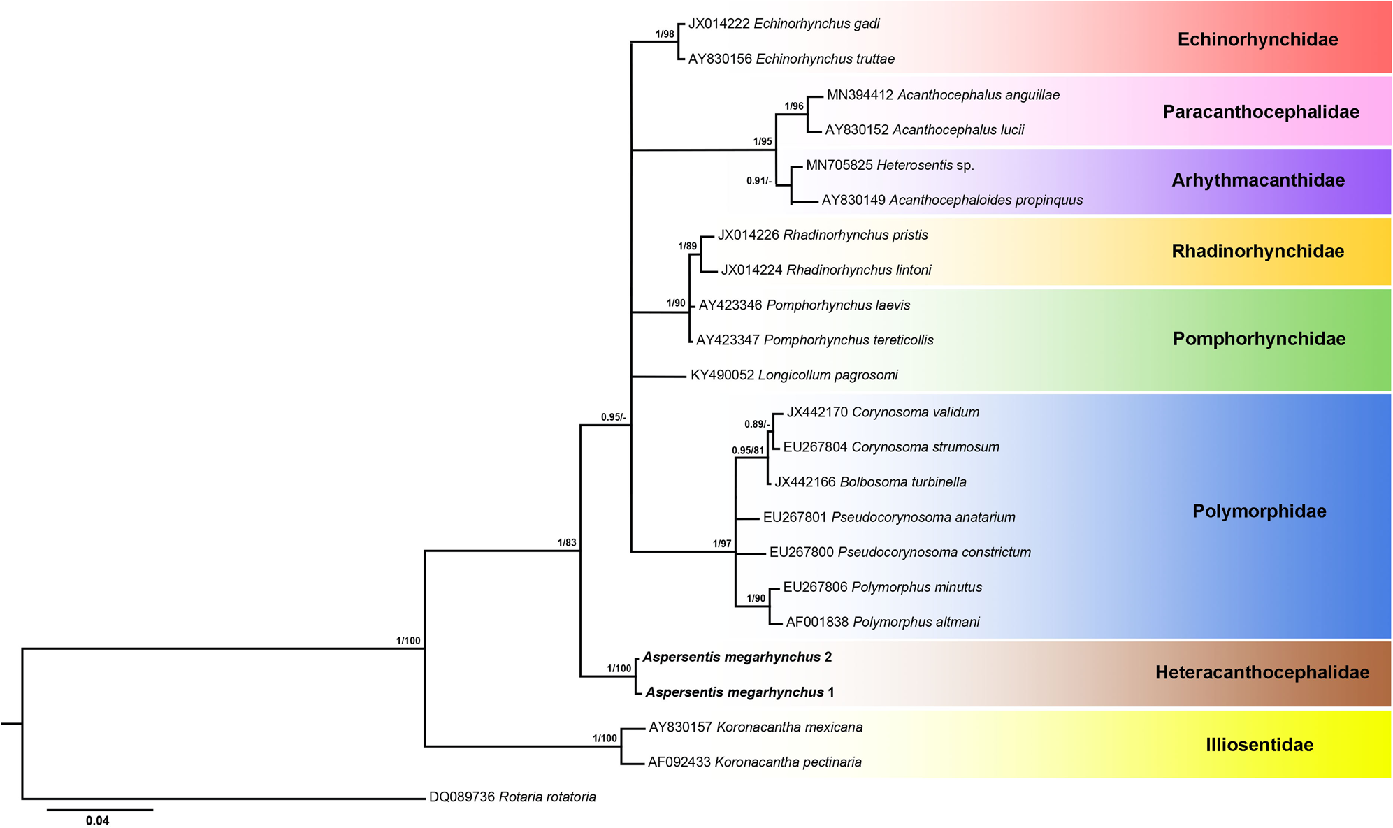
ML phylogram reconstructed using newly generated 18S rDNA sequences for Aspersentis megarhynchus (in bold) and retrieved sequences from GenBank for representatives of the paleacanthocephalan families (highlighted in different colours). Outgroup: Rotaria rotatoria. Nodal supports for ML and BI analyses are indicated as BI/ML. Posterior probability values lower than 0.9 and bootstrap values lower than 70 are omitted, and nodes associated to such low values are displayed collapsed as polytomies. The scale bar indicates the expected number of substitutions per site.

Aspersentis megarhynchus (von Linstow, Reference von Linstow1892) Golvan, 1960
General. With characters of the genus Aspersentis as emended above. Shared structures larger in females than in males. Trunk, extremely fusiform tapering at both ends (fig. 4a) with electron-dense micropores (fig. 2d) that also cover spines (fig. 3b) and randomly scattered sensory discs in both males and females (fig. 3a, c, d). Trunk with large spines (fig. 2e), with single-core rod support (fig. 4b) of variable anterior distribution (fig. 1d–f) and smaller spines (fig. 2f) that may extend to posterior end of trunk. Proboscis bent ventrally with two-lobed apical organ and 14–16 rows of 8–10 hooks each. Hooks longer anteriorly (fig. 2a) and ventrally than dorsally (fig. 1a, d), with prominent roots about as long as blades, and thick cortical layer with high level of sulphur (fig. 2b). Neck prominent (fig. 1d, e) with sensory pores (fig. 2c). Proboscis receptacle double-walled, about twice as long as proboscis, with an outer layer of spirally arranged muscle fibres and large drop-shaped cerebral ganglion near its posterior end. Lemnisci longer than proboscis receptacle (fig. 4a).
Male (based on one measured whole mounted mature specimen and four specimens used for SEM). Trunk extremely fusiform, 3.95 mm long by 1.45 mm wide (width 37% of length) with a prominent anterior zone of 25 long ventral spines with one central shaft and faint 15 long limited dorsal spines. Proboscis 561 long by 239 wide anteriorly with 15 rows of nine hooks each. Second anterior ventral hook 105 long; opposite dorsal hook 42 long. Neck 229 long dorsally by 364 at interface with trunk. Proboscis receptacle 1.35 mm long by 0.23 mm wide. Lemnisci equal, longer than receptacle, 1.51 mm long by 0.14 mm wide. Reproductive system in posterior two thirds of trunk. Testes rounded, relatively large, post equatorial, contiguous; anterior testis 675 long by 750 wide; posterior testis 625 long by 675 wide. Cement glands six in three pairs, 420 long by 312 wide, spheroid to pyriform, clustered, overlapping posterior testis, each with independent cement gland duct surrounding prominent drop-shaped Saefftigen's pouch (fig. 4c), 572 long by 260 wide, before running into ejaculatory duct and penis. Male gonopore dorsal, a short distance anterior to pointed posterior trunk tip (fig. 4c). Bursa pinched medially with one circle of bead-like sensory papillae (fig. 3e, f). Gonopore near terminal on dorsal side (fig. 4c).
Females (based on 12 measured gravid specimens and five more specimens used for SEM). Trunk extremely fusiform (fig. 4a) reaching 7.70 mm in length and 3.25 mm in width; width 28–54 (39%) of length. See measurements and counts in table 2. Lemnisci longer than proboscis receptacle (fig. 4a). Reproductive system with well-developed vagina, moderate uterus and base of uterine bell with many glands (fig. 4d). Gonopore terminal to near terminal on dorsal side, slit-like vertically or horizontally (fig. 3a). Egg fusiform-elongate with polar prolongation of fertilization membrane and a fibrillar coat (figs 3b and 4e).
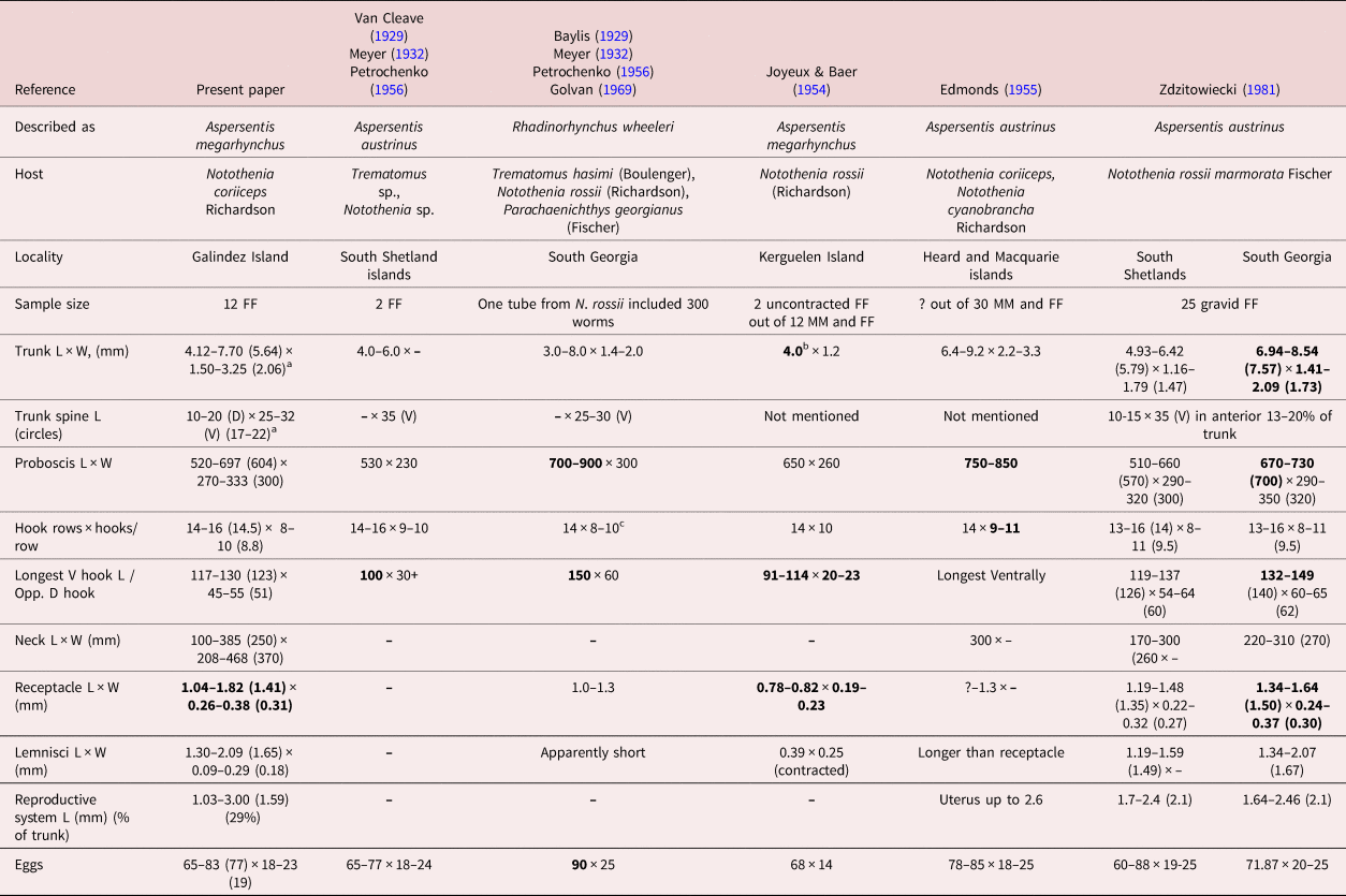
Morphometric comparisons among populations of Aspersentis megarhynchus.

FF, females; MM, males; L, length; W, width; V, ventral; D, dorsal.
a Range (mean) in micrometres unless otherwise noted.
b Bolded figures of some characters are markedly larger or smaller than others of the same characters.
c Petrochenko erroneously transcribed the number of hook rows as 12 instead of 14, as noted in Baylis (Reference Baylis1929).
Taxonomic summary
-
Type species. Aspersentis megarhynchus (von Linstow, Reference von Linstow1892) Golvan, Reference Golvan1960.
-
Type host. Notothenia coriiceps Richardson (Nototheniidae).
-
Other hosts. See references in table 2, among others.
-
Type locality. South Georgia, Antarctica (54°15′S, 36°45′W).
-
Other localities in Antarctica. See table 2.
-
Site of infection. Intestine and some in body cavity.
-
Etymology. Von Linstow (Reference von Linstow1892) may have named the species ‘megarhynchus’ because of the extremely large ventral proboscis hooks compared to dorsal hooks; he did not elaborate.
Micropores
Micropores covered the whole trunk of male and female specimens of A. megarhynchus (fig. 2d), including the epicuticle of trunk spines (fig. 2f) as is commonly observed in other species of acanthocephalans.
EDXA
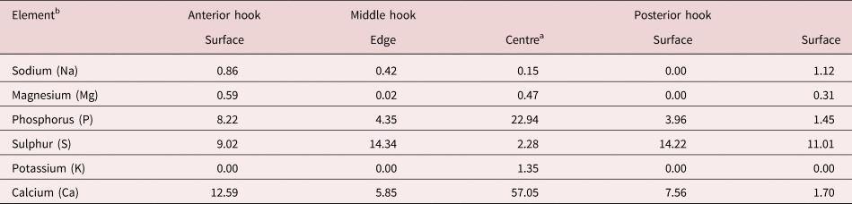
The results of the EDXA of anterior hooks of A. megarhynchus show the anterior hooks to have the highest levels of calcium and phosphorus at the centre hooks (57.05 and 22.94%, respectively) but the highest levels of sulphur at the surface and edge of all hooks (table 3). Similar analyses have not been conducted for other species of Aspersentis; thus, comparisons of the chemical profile of hooks could not be made. A baseline for future comparisons is, however, established.
Chemical composition of hooks of adult Aspersentis megarhynchus at three levels. One hook (anterior hook) cut with a gallium beam for internal X-ray.

a There was a small amount (2.53) of zinc (Zn) in the centre.
b Common protoplasmic elements (C, N, O) and processing elements (Au, Pd, Ga) omitted. Reported in WT%.
Molecular results
Two partial 18S rDNA sequences (852 and 863 nt length) differing by 2 nt (0.24%) were generated from two adult specimens of A. megarhynchus. Regarding phylogenetic reconstructions, ML and BI algorithms produced trees with identical topologies. Therefore, a consensus tree based on the ML phylogram is displayed in fig. 5.
The phylogram built from the 18S rDNA alignment displays the family Heteracanthocephalidae represented by present sequences, as a separate clade from other paleacanthocephalan families with high nodal support. Overall, phylogenetic reconstructions showed high support in most nodes although low bootstrap and posterior probability values occurred in nodes separating representatives of the families Echinorhynchidae, Arhythmacanthidae, Rhadinorhynchidae, Pomphorhynchidae and Polymorphidae, which remained as unresolved relations. Present newly generated sequences were more closely related to representatives of Pomphorhynchidae (52–56 nt, 6.21–6.68% difference), the echinorhynchid genus Echinorhynchus (54–55 nt, 6.44–6.57% difference), Rhadinorhynchidae (60–62 nt, 7.16–7.42% difference) and Polymorphidae (71–76 nt, 8.52–9.10% difference). Intermediate genetic distances were observed with respect to Arhythmacanthidae (82–91 nt, 9.84–10.92% difference) and the echinorhynchid genus Acanthocephalus (85–87 nt, 10.19–10.43% difference). Finally, the greatest genetic divergence was observed with respect to Illiosentidae (125–127 nt, 14.97–15.21% difference).
Discussion
Types
Our designation of the type host and type locality of A. megarhynchus is based on its original description of von Linstow (Reference von Linstow1892) as E. megarhynchus, which was later relegated to the genus Aspersentis Van Cleave, 1929 as listed and accepted by Amin (Reference Amin2013) and other observers (table 2). The specimens of von Linstow (Reference von Linstow1892) were obtained from the Natural History Museum in Hamburg and collected from the German Station, South Georgia in 1882–1883. Golvan (Reference Golvan1960) designated A. megarhynchus as the type species with A. austrinus Van Cleave, 1929 and R. wheeleri Baylis, 1929 as synonyms, diagnosed the genus Aspersentis based on Van Cleave (Reference Van Cleave1929), and mentioned that it is a parasite of Nototheniidae in the Antarctic Ocean and the South Pacific. Neither von Linstow (Reference von Linstow1892) nor Golvan (Reference Golvan1960) designated the type host or the type locality.
Variability
The many taxonomic accounts of A. megarhynchus, summarized in the introductory remarks, were in part prompted by its variability expressed in various populations from different host species and geographical localities. Variability in selected structures of females from seven different locations and a number of host species are shown in table 2. Specimens with the smallest trunk, proboscis hooks and proboscis receptacle were reported from N. rossii off Kerguelen Island by Joyeux & Baer (Reference Joyeux and Baer1954). In contrast, specimens from South Georgia and South Shetlands, West Antarctica, reported from the same host species by Zdzitowiecki (Reference Zdzitowiecki1981) manifested the largest larger size of the same structures, as well as the size of proboscis (table 2). Zdzitowiecki's (Reference Zdzitowiecki1981) own specimens from N. rossii were considerably larger from South Georgia than from South Shetlands pointing to the effect of geographical variability. Similarly, the specimens reported by Edmonds (Reference Edmonds1955) from N. coriiceps and N. cyanobrancha from Heard and Macquari Islands had considerably larger proboscis with more hooks per row than our specimens from N. coriiceps in Galdinez Island. Like Zdzitowiecki's (Reference Zdzitowiecki1981) specimens from South Georgia, those described by Baylis (Reference Baylis1929) also from South Georgia had the largest proboscis (700–900 long) and the longest ventral hooks (150 long) (table 2). Structures of specimens from South Georgia appear to attain the largest size compared to the same structures from other localities. The relationship between geographical factors and size of morphometric characters has been noted in other species of acanthocephalans. For example, populations of Mediorhynchus papillosus Van Cleave, 1916 have a wide distribution in North and South America, Eastern Europe, Asia and many former Soviet republics, east to China and Taiwan. Morphometric comparisons of specimens of M. papillosus from Maryland, Colorado, Taiwan, Trans-Baikal, Lower Yenesei River, Volga, Ukraine and Bulgaria exhibited distinctive morphological variability in certain key characters, ‘with specimens from Colorado and Taiwan being at the opposite ends of the variability spectrum’ (Amin & Dailey, Reference Amin and Dailey1998). Populations of A. megarhynchus may have a disjunct distribution similar to that demonstrated in M. papillosus that was attributed to ‘geographical restriction of intermediate (and adult hosts) in the apparent absence or scarcity of paratenic/reservoir hosts. Disjunct populations would promote distinct geographical diversity with consistent morphological difference from each other’ (Amin & Dailey, Reference Amin and Dailey1998).
Validity and conflicts
While the taxonomy of A. megarhynchus has been marred by its many names and relegations, it has always been recognizable as one valid entity not confused with related sympatric forms. Only Pichelin et al. (Reference Pichelin, Smales and Bray2002) discredited the original description by von Linstow (Reference von Linstow1892) by declaring his species Echinorhynchus megarynchus a species inquirenda even though they acknowledged Zdzitowiecki's (Reference Zdzitowiecki1983) statement that ‘the hooks of E. megarhynchus are different from those of M. dalmori, Echinorhynchus petrotschenkoi, Heterosentis heteracthus’. Pichelin et al. (Reference Pichelin, Smales and Bray2002) stated that
Linstow (Reference von Linstow1892, p. 70) described the worms as lacking spines, as having 18 rows of six hooks running from smaller at the base to larger at the apex of the proboscis, and gave measurements and figures for the smallest and largest hooks … the presence or absence of trunk spines is problematical … .
We have observed considerable variation in the expression of trunk spines (fig. 1d, e) varying between evident only anteriorly (larger ventrally than dorsally) and barely visible, if present, especially posteriorly.
Zdzitowiecki believed that
Linstow (Reference von Linstow1892, p. 70) may have counted acanthocephalan hooks differently from the method presently used making Linstow's formula for the hooks of E. megarhynchus (i.e. 18 rows of 6 hooks) could be interpreted as 12 rows of nine hooks which would be nearer to the hook formula of A. autrinus.
He also believed that ‘the hooks' in Linstow's figures (Linstow, Reference von Linstow1892, Figs 34 and 35) are identical to with those of A. austrinus (Pichelin et al., Reference Pichelin, Smales and Bray2002, pers. comm.). We agree with Zdzitowiecki's assessment. These variable interpretations are easily resolved when seen in context. We have addressed a similar issue regarding the counting of proboscis hooks of Bolbosoma capitatum Linstow (1880) Porta, 1908 and we quote below from Amin & Margolis (Reference Amin and Margolis1998):
PROBOSCIS ARMATURE: Porta (Reference Porta1906, p. 186) described 12–18 transverse rows of hooks on the proboscis of B. capitatum. In his 2 later papers (Porta, Reference Porta1908, Reference Porta1909), he omitted the word ‘transverse’ and referred only to 12–18 rows of hooks’. Meyer (Reference Meyer1932), Petrochenko (Reference Petrochenko1958), and Yamaguti (Reference Yamaguti1963) incorrectly interpreted Porta's descriptions as 12–18 longitudinal rows of hooks, without data on the number of hooks per ‘longitudinal’ row. Actually, neither the number of longitudinal rows of hooks nor the number of hooks in each transverse row was given by Porta in any of his papers. However, the 12–18 transverse rows of hooks translates into 6–9 hooks per longitudinal row, because of the alternating ‘transverse’ arrangement of hooks in adjacent longitudinal rows. Delyamure (Reference Delyamure1955) correctly cited the number of transverse, i.e., circular, rows of hooks as 12–18; his description of 18–20 longitudinal rows may have come from Baylis (Reference Baylis1929), who stated ‘18 (−20?).’ We cannot determine the source of the Baylis (Reference Baylis1929) number. Edmonds (Reference Edmonds1957) gave a formula of 14–16 longitudinal rows of 8 hooks each. Yamaguti (Reference Yamaguti1963) cited these numbers also, attributing them to Edmonds (Reference Edmonds1957), who later (Edmonds, Reference Edmonds1987) reported 15–17 longitudinal hook rows from an additional collection. Kikuchi & Nakajima ‘s (Reference Kikuchi and Nakajima1991) description of 10–20 longitudinal rows must be considered a typographical error for 16–20 rows, which is the number given in Kikuchi & Nakajima‘s (Reference Kikuchi and Nakajima1993) description (16–20 in males, 18–20 in females). In their 1991 abstract, Kikuchi and Nakajima report 12–16 circular (transverse) rows of hooks on the proboscis. In their 1993 paper, the number of transverse rows was given as 12–18 in males and 15–18 in females. This translates into 6–9 hooks per longitudinal row in males and 7–9 in females.
Pichelin et al. (Reference Pichelin, Smales and Bray2002) further stated that Linstow (Reference von Linstow1892) ‘made no mention of the dorso-ventral asymmetry of the hooks which is a key feature of the family.’ We agree with Zdzitowiecki (Reference Zdzitowiecki1981) who, however, ‘considered it unlikely that Linstow would have missed such an obvious character.’ We actually believe that von Linstow's (Reference von Linstow1892) choice of the specific name ‘megarhynchus’ was in reference to the large ventral hooks as opposed to the smaller dorsal hooks, but cannot substantiate. Pichelin et al. (Reference Pichelin, Smales and Bray2002) concluded that Linstow's species E. megarhynchus ‘cannot be properly identified’ given the information available at the time but not in light of currently available knowledge using freshly collected material as presented here. Pichelin et al. (Reference Pichelin, Smales and Bray2002) also stated that the species is ‘not a heteracanthocephalid’, which would negate the Golvan's (Reference Golvan1960) currently accepted designation. We reject the notions of Pichelin et al. (Reference Pichelin, Smales and Bray2002) and accept the validity of A. megarhynchus as designated by Golvan (Reference Golvan1960) and accepted by Zdzitowiecki (Reference Zdzitowiecki, Wägele and Sieg1991), Amin (Reference Amin, Crompton and Nickol1985, Reference Amin2013), among others.
Micropores
Micropores are present throughout epidermal surface of the trunk of A. megarhynchus, like those reported in other species of the Acanthocephala. They are associated with internal crypts and vary in diameter and distribution in different trunk regions corresponding with differential absorption of nutrients. We have documented this phenomenon in 16 species of acanthocephalans (Heckmann et al., Reference Heckmann, Amin and El-Naggar2013) and a few more since. The functional aspects of micropores in a few other acanthocephalan species including Rhadinorhynchus ornatus Van Cleave, 1918, Polymorphus minutus (Goeze, 1782) Lühe, 1911, Moniliformis moniliformis (Bremser, 1811) Travassos (1915), Macracanthorhynchus hirudinaceus (Pallas, 1781) Travassos (1916, 1917) and Sclerocollum rubrimaris Schmidt & Paperna, 1978 were reviewed earlier by Amin et al. (Reference Amin, Heckmann, Radwan, Mantuano and Alcivar2009). Wright & Lumsden (Reference Wright and Lumsden1969) and Byram & Fisher (Reference Byram and Fisher1973) reported that the peripheral canals of the micropores are continuous with canalicular crypts. These crypts appear to ‘constitute a huge increase in external surface area. implicated in nutrient up take.’ Whitfield (Reference Whitfield1979) estimated a 44-fold increase at a surface density of 15 invaginations per 1 μm² of M. moniliformis (Bremser, 1811) Travassos, 1915 tegumental surface. The micropores and the peripheral canal connections to the canaliculi of the inner layer of the tegument were demonstrated by transmission electron micrographs in Corynosoma strumosum (Rudolphi, 1802) Lühe, 1904 from the Caspian seal Pusa caspica (Gmelin) in the Caspian Sea (Amin et al., Reference Amin, Heckmann, Halajian and El-Naggar2011, Figs 19, 20) and in Neoechinorhynchus personatus Tkach, Sarabeev & Shvetsova, 2014 from Mugil cephalus Linn. in Tunisia (Amin et al., Reference Amin, Sharifdini, Heckmann, Rubtsova and Chine2020, Figs 26, 29, 30).
EDXA
Our studies of acanthocephalan worms have usually involved X-ray scans (EDXA) of Ga cut hooks and other worm structures (Heckmann et al., Reference Heckmann, Amin and Standing2007, Reference Heckmann, Amin, Radwan, Standing, Eggett and El Naggar2012; Standing & Heckmann, Reference Standing and Heckmann2014). Hooks are evaluated for chemical ions with sulphur, calcium and phosphorus being the prominent elements. Sulphur is usually seen at the outer edge of large hooks and calcium and phosphorus are major ions in the base and middle of hooks (table 3). Large hooks play a major role for host tissue attachment. In Cavisoma magnum (Southwell, 1927) Van Cleave, 1931 from M. cephalus in the Arabian Sea, unusually high levels of sulphur in hook tips (43.51 wt. %) and edges (27.46 wt. %) were found. This element (sulphur) is part of the prominent outer layer of most acanthocephalan hooks and is a major contributor of the hardening process of this attachment structure. Our results are comparable to those of mammalian teeth enamel. The centre and base of hooks of the same worms had negligible sulphur levels and contained mostly phosphorus and calcium, the two other essential elements for hook structure (Amin et al., Reference Amin, Heckmann and Bannai2018). These findings are comparable to those of A. megarhynchus in this study (table 3). Like fingerprints, the EDXA pattern appears to be species-specific and have significant diagnostic value in acanthocephalan systematics (Amin et al., Reference Amin, Heckmann, Sharifdini and Albayati2019a). For example, Moniliformis cryptosaudi Amin, Heckmann, Sharifdini & Albayati, 2019 was erected based primarily on its EDXA pattern (Amin et al., Reference Amin, Heckmann, Sharifdini and Albayati2019a). The present findings will provide a baseline for future comparisons with other species of Aspersentis. Our methodology for the detection of the chemical profile of hooks in the Acanthocephala has also been used in other parasitic groups including the Monogenea (Rubtsova et al., Reference Rubtsova, Heckmann, Smit, Luus-Powell, Halajian and Roux2018, Rubtsova & Heckmann Reference Rubtsova and Heckmann2019), and Cestoda (Rubtsova & Heckmann, Reference Rubtsova and Heckmann2020).
Molecular results and phylogenetic affinities
This is the first report of a sequence for the family Heteracanthocephalidae. The lack of other sequences of different Aspersentis populations, as mentioned above, prevents molecular comparison between them, with other species of the genus or with other genera of the family. Therefore, this study lays the baseline for future studies regarding Aspersentis and the Heteracanthocephalidae and highlights the importance of the new molecular data presented herein.
Present phylogenetic results provide evidence that the family Heteracanthocephalidae stands as a distinct family within the class Paleacanthocephala, supporting the taxonomic arrangement provided by Golvan (Reference Golvan1960) and later maintained by Amin (Reference Amin, Crompton and Nickol1985, Reference Amin2013) based on morphological studies. Interestingly, the most closely related clade to Heteracanthocephalidae (order Echinorhynchida), according to genetic data, includes representatives of the family Polymorphidae (order Polymorphida), which are clustered along with other families within the order Echinorhynchida while present newly generated sequences remain as a clearly separate clade. This fact, again, points to the paraphyly of the palaeacantocephalans at the family level, as other authors have already highlighted (Herlyn et al., Reference Herlyn, Piskurek, Schmitz, Ehlers and Zischlera2003; Garcia-Varela & Nadler, Reference García-Varela and Nadler2005; Verweyen et al., Reference Verweyen, Klimpel and Palm2011; Amin et al., Reference Amin, Heckmann, Dallarés, Constenla and Ha2019b; García-Varela & Andrade-Gómez, Reference García-Varela and Andrade-Gómez2021). This problem usually extends to lower taxonomic categories, as was the case of the diverse family Echinorhynchidae. Indeed, García-Varela & Andrade-Gómez (Reference García-Varela and Andrade-Gómez2021) recently resurrected the families Paracanthocephalidae and Pseudoacanthocephalidae to accommodate the genera Acanthocephalus and Pseudoacanthocephalus, respectively, which were previously included within the Echinorhynchidae despite conforming independent lineages with respect to Echinorhynchus according to genetic data (Huston et al., Reference Huston, Cribb and Smales2020; present results). These inconsistencies stress the existing problems with the taxonomic arrangement of acanthocephalans and point to the need for a greater effort in genetic characterization of acanthocephalan main families for further reclassification based not only on a morphological but also on a molecular description of their members.
Acknowledgements
We thank Madison Laurence, Bean Museum (Brigham Young University (BYU)), for expert help in the preparation and organization of plates and figures, and Michael Standing, Electron Optics Laboratory (BYU), for his technical help and expertise. This study was partially supported by the National Research Foundation of Ukraine (project number 2020.02/0074).
Financial support
This project was supported by the Department of Biology, BYU, Provo, Utah, and by an Institutional Grant from the Parasitology Center, Inc., Scottsdale, Arizona.
Conflicts of interest
None.
Ethical standards
The authors assert that all procedures contributing to this work comply with the ethical standards of the relevant national and institutional guides on the care and use of laboratory animals.