Introduction
Desmoid tumours, also called ‘aggressive fibromatosis’, are benign, locally invasive, soft tissue tumours. They are rare, accounting for 0.03 per cent of neoplasms. They are classified into five categories, including intra-abdominal, abdominal wall and extra-abdominal tumours, as well as a category associated with Gardner syndrome and one associated with adenomatous polyposis coli (APC) gene mutation. Of all desmoid tumours, 7–15 per cent are head and neck tumours. These are most commonly found in the neck (40–90 per cent), and the majority are in the supraclavicular fossa. They usually present as a painless, occasionally tender mass, and are often rapid-growing.Reference de Bree, Zoras, Hunt, Takes, Suarez and Mendenhall1
We describe an unusual desmoid tumour of the external ear and mastoid, which was diagnosed only following biopsy given the low clinical suspicion of desmoid tumour. Following review by the Oxford University Hospitals NHS Foundation Trust Joint Research Office, research ethics review was not required and written consent was obtained from the patient.
Case report
A 26-year-old woman presented to the emergency department with a tender, left-sided post-auricular swelling. She had a history of cholesteatoma of the left ear, and had undergone two atticotomy and ossiculoplasty procedures via post-auricular incision in the preceding two years. The swelling was initially painless and had progressively increased in size over a month; it had become inflamed and tender in the week preceding her presentation, and there was associated otorrhoea. The patient reported general fatigue and malaise for 3 days.
On examination, there was a 2 × 3 cm firm mass arising in the left post-auricular area, overlying the scar. It extended anteriorly, pushing the posterior auditory canal wall anteriorly and occluding the canal. The overlying skin was erythematous, warm and tender. Observations revealed tachycardia without pyrexia.
The initial differential diagnosis included a possible inclusion cyst with infection, a pathological lymph node or an unusual cholesteatoma recurrence.
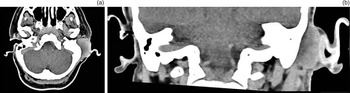
Blood tests demonstrated a normal white cell count with a C-reactive protein level of 63 mg/l. A computed tomography scan with contrast showed a 35 × 23 mm area reported as a fluid-filled collection with homogeneous enhancement overlying the external auditory canal, destruction of the posterior wall of the ear canal, and an opacified mastoid (Figure 1).
(a) Axial and (b) coronal computed tomography scans with contrast.

Initial aspiration of the mass did not yield any fluid. Intravenous antibiotics were commenced.
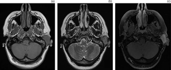
In light of the diagnostic uncertainty, the patient underwent an examination under anaesthetic with an open biopsy. The surgical findings revealed a solid mass. Histology findings were consistent with an extra-abdominal desmoid tumour. A magnetic resonance imaging (MRI) scan subsequently demonstrated a lesion with a low T1-weighted and intermediate T2-weighted signal, and gadolinium enhancement (Figure 2).
(a) T1-weighted, (b) T2-weighted and (c) T1-weighted plus gadolinium axial magnetic resonance imaging scans.

Following discussion in the sarcoma multi-disciplinary team meeting, the patient was initially managed expectantly. A repeat MRI scan conducted two months later showed tumour growth of 10 mm in the long axis. The patient was offered chemotherapy with vinblastine and methotrexate. This proved effective at reducing the tumour size.
• Desmoid tumours (aggressive fibromatosis) are rare, benign, locally invasive soft tissue tumours
• Of desmoid tumours, 7–15 per cent affect the head and neck; most are found in the neck
• Two cases of non-traumatic desmoid tumour of the ear have been described in paediatric patients (2 and 15 years old)
• Desmoid tumours have been associated with trauma, including surgical trauma
• This is the first reported case of a desmoid tumour of the ear following surgery
• Otolaryngologists should be aware of desmoid tumour as a possible diagnosis for a soft tissue mass at a surgical site
Discussion
This case represents an interesting diagnostic and therapeutic challenge. Head and neck desmoid tumours, though uncommon, are a known entity; only two cases affecting the ear have been described in case series, one on the earlobe skin in a 2 year oldReference Wang, Chang, Ko, Cheng, Yeh and Lou2 and one on the superior aspect of the pinna in a 15 year old.Reference Risoud, Mortuaire, Leroy, Leblond and Fayoux3
In our patient, the diagnosis was unlikely enough that it was not included in the differential, despite review by several otolaryngologists, and was identified only on histology. The patient's previous cholesteatoma and surgical interventions may have misguided the differential diagnosis; however, desmoid tumours have been reported to be related to trauma, and in particular surgical trauma, in 3–33 per cent of cases.Reference de Bree, Zoras, Hunt, Takes, Suarez and Mendenhall1 It can therefore be assumed that, in this case, the tumour may have been related to the previous operation. Abdominal wall desmoid tumours can be related to pregnancy or the use of a contraceptive pill; however, this association has not been described with other desmoid tumours.Reference de Bree, Zoras, Hunt, Takes, Suarez and Mendenhall1
Surgical management in this instance would have been locally destructive, particularly as resection with wide margins is necessary to avoid local recurrence. Medical management options include radiotherapy, non-steroidal anti-inflammatory drugs, cytotoxic or hormonal therapy. There is no clear consensus on the best therapeutic option; the different options must be considered in a multi-disciplinary setting, based on the individual characteristics of the patient and the tumour.Reference de Bree, Zoras, Hunt, Takes, Suarez and Mendenhall1,Reference Collins, Fischer and Tufaro4
Competing interests
None declared