Implications
Plantain (Plantago lanceolata L.) is a perennial herb having bioactive components. The present experiment evaluated the effect of plantain on the responses of plasma glucose metabolism to exogenous insulin in heat-exposed sheep. Plasma glucose turnover rate (TR) was affected neither by feeding plantain nor exposure to a hot environment (30°C, 70% relative humidity (RH)). However, the response of plasma glucose utilization to exogenous insulin infusion was reduced in sheep when 50% of the mixed hay of orchardgrass (Dactylis glomerata) and reed canarygrass (Phalaris arundinacea) diet was replaced by fresh plantain.
Introduction
Glucose metabolism is influenced by nutritional and physiological conditions (Evans and Buchanan-Smith, Reference Evans and Buchanan-Smith1975; Buckley et al., Reference Buckley, Herbein and Young1982; Sano et al., Reference Sano, Takahashi, Ambo and Tsuda1983). Sano et al. (Reference Sano, Takahashi, Ambo and Tsuda1983) reported that glucose metabolism declined during heat exposure (HE) in sheep. Sano et al. (Reference Sano, Takebayashi, Kodama, Nakamura, Ito, Arino, Fujita, Takahashi and Ambo1999) indicated that in fact blood glucose turnover was greater during cold exposure, and that the sensitivity of glucose metabolism to insulin was increased during cold exposure. Another report found that the tissue responsiveness to exogenous insulin was unchanged by HE (30°C) and there was a tendency for this to be reduced by chromium supplementation in sheep (Sano et al., Reference Sano, Konno and Shiga2000).
Plantain (P. lanceolata L.) is a perennial herb highly palatable for grazing animals, provides a mineral-rich forage and growing on a wide range of agricultural soils: it is tolerant to drought and of many common diseases and pests (Mook et al., Reference Mook, Haeck, van der Toorn and van Tienderen1989; Stewart, Reference Stewart1996; Rumball et al., Reference Rumball, Keogh, Lane, Miller and Claydon1997). Plantain has anti-bacterial properties (Ishiguro et al., Reference Ishiguro, Yamaki and Takagi1982), enzyme inhibitory activities (Nishibe and Murai, Reference Nishibe and Murai1995) and anti-inflammatory effects (Park and Chang, Reference Park and Chang2004; Beara et al., Reference Beara, Orčić, Lesjak, Mimica-Dukić, Peković and Popović2010). Plantain also has bioactive components, acteoside, aucubin and catalpol (Al-Mamun et al., Reference Al-Mamun, Abe, Kofujita, Tamura and Sano2008a) that express superoxide anion radical scavenging activities (Al-Mamun et al., Reference Al-Mamun, Hanai, Tanaka, Tamura and Sano2007a). It was reported that plantain had a positive influence on whole body protein synthesis (Al-Mamun et al., Reference Al-Mamun, Hanai, Tanaka, Tamura and Sano2008b) in sheep during HE (30°C, 70% RH). However, until recently, the effect of plantain on the intermediary metabolism of glucose during exogenous insulin infusion in sheep exposed to heat has not been reported.
The present experiment was conducted to determine the effect of plantain intake on glucose metabolism in sheep exposed to a controlled hot environment, using the stable isotope dilution method of [U-13C]glucose and exogenous insulin infusion.
Material and methods
Animals, diets and heat exposure
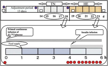
Six crossbred (Corriedale×Suffolk) shorn sheep (Ovis aries L.) of both sex (three wethers and three ewes), aged around 3 years and weighing 40±2 kg of BW were used. Two dietary treatments were used. One was mixed hay (MH-diet) of orchardgrass (D. glomerata L.) and reed canarygrass (P. arundinacea L.) ration mixed in the ratio of 60 : 40 (1.78 kcal metabolizable energy (ME)/g dry matter (DM), 115 g/kg CP and 650 g/kg NDF (aNDFom)). The second ration was the PL-diet in which 50% of MH-diet was replaced by fresh plantain (117 g/kg CP and 470 g/kg aNDFom). In both dietary treatments the ME and CP intakes were assumed to be offered at around maintenance level (National Research Council, 1985). The animals were fed once daily at 1400 h, and orts were collected daily. Sheep were offered water access ad libitum. The experiment was performed using a crossover design with two 23-day periods in which either PL- or MH-diets were fed (Figure 1). Fresh plantain grown in the experimental field of Iwate University was cut and delivered daily (for details about the location and cultivation please see Al-Mamun et al., Reference Al-Mamun, Abe, Kofujita, Tamura and Sano2008a). The mixed hay and fresh plantain was chopped and fed to the animals in order to avoid choice feeding, as the morphology, such as leaves size, stems physical properties, can stimulate foraging behavior of sheep (Provenza, Reference Provenza2003; Smit et al., Reference Smit, Tas, Taweel, Tamminga and Elgersma2005; Barre et al., Reference Barre, Emile, Betin, Surault, Ghesquière and Hazard2006). The sheep were housed in individual pens in a sheep barn during the first 13 days adjustment period. On day 14, the animals were moved to metabolic cages in a controlled environmental chamber (70% RH, thermoneutral temperature 20°C±1°C (TN) and with lighting from 0800 to 2200 h). On day 18, an isotope dilution method using [U-13C]glucose was conducted to determine the effect of exogenous insulin infusion on glucose metabolism during TN. Ambient temperature was then elevated and maintained at 28°C to 30°C (HE, 28°C from 2200 to 1000, and 30°C from 1000 to 2200 h), but the RH and lighting were unchanged (70% RH, and lighting from 0800 to 2200 h) for 5 days. The same isotope dilution method was performed on day 23 of the experimental period. Sheep were weighed on days 0 (start of experiment), 14, 18 and 23. Shearing was performed closely on days 0 (start of experiment), 13 and 18.
The experimental layout showing environmental condition, BW measurement, shearing, measuring physiological responses, blood sampling protocol and isotope dilution with date and time. TN=thermoneutral (20°C with 70% relative humidity); HE=heat exposure (30°C with 70% relative humidity).  , The sheep were weighed at starting of the experiment, on day 14 of the experiment and after finishing of each environmental treatment.
 , Shearing was performed closely on day 0 (start of experiment), 13 and 18.
 , Heart rate, respiration rate and rectal temperature were measured at 1400 h immediate before feeding for 3 successive days during both the TN (days 15 to 17) and HE (days 20 to 22) in both the dietary treatments.
 , On the day 18 during TN and day 23 during HE isotope dilution techniques were performed to determine the responses of plasma glucose metabolism to exogenous insulin infusion.
 , Blood samples (5 ml) were collected from the sampling catheter immediately before the isotope dilution technique, and 15 min and immediately before the insulin infusion, and during the last 120 min of isotope dilution techniques at 15-min intervals.

Blood sampling was performed without any noticeable stress to the sheep. Animal handling, cannulation and blood sampling were carried out according to the guidelines of the Animal Care Committee of Iwate University, Japan.
Daily physiological responses
Heart rate, respiration rate and rectal temperature were measured at 1400 h immediately before feeding for 3 successive days during both the TN (days 15 to 17) and HE (days 20 to 22) during both the dietary treatments.
Collection of feed samples
Mixed hay and fresh plantain were sampled twice a week. Samples were dried in a forced air oven at 60°C for 48 h, ground to 1-mm mesh using a micro grinder and kept at −30°C until further analyses.
Isotope dilution procedures
Two vinyl catheters, of which one was a multichannel catheter for infusion of [U-13C]glucose, and insulin and another for blood sampling, were inserted into the left and right jugular veins in the morning of each determination of isotope dilution method. Catheters were filled with a sterile solution of trisodium citrate (0.13 mol/l). Immediately after completion of exogenous insulin infusion, a 20% glucose solution was infused (100 ml/sheep) through the infusion catheter in order to avoid hypoglycemia. At 1300 h on the day of the isotope dilution method, 7.3 mg [U-13C]glucose (d-glucose-13C6, 99 atom% excess 13C; Cambridge Isotope Laboratories Inc., Highwood Drive, Tewksbury, MA, USA) dissolved in 10 ml of saline solution was infused as a priming injection (Wolfe, Reference Wolfe1984), and thereafter the same isotope was continuously infused at a rate of 8 µg/kg0.75 per min for 6 h.
Exogenous insulin was infused at a rate of 1 mU/kg0.75 per min during the last 2 h of the isotope dilution technique. Blood samples (5 ml) were collected from the sampling catheter immediately before the isotope dilution technique, and 15 min and immediately before the insulin infusion, and during the last 120 min of isotope dilution at 15-min intervals. Samples were transferred into heparinized centrifuge tubes and chilled until centrifugation. Plasma glucose concentration, [U-13C]glucose enrichment, plasma glucose TR and responses of plasma glucose utilization to exogenous insulin infusion were determined.
Analyses
The DM, ash, ether extract, and N contents were analyzed according to the procedure of Association of Official Analytical Chemists (1995). The NDF was analyzed according to the procedures of Van Soest et al. (Reference Van Soest, Robertson and Lewis1991).
Blood samples were centrifuged at 12 000×g for 10 min at 2°C (RS-18 IV, Tomy, Tokyo, Japan), and the plasma was stored at −30°C pending further analysis. Plasma (baseline samples from preinfusion period) concentration of retinol and α-tocopherol were determined using HPLC (Hitachi, U-2010, Tomy, EX-125, Taitec, DTU-1C Shizuoka, Japan) according the method described by Rupérez et al. (Reference Rupérez, Mach and Barbas2004) with slight difference in UV detection. The UV detection was performed at 325 nm excitation and 470 nm emissions for up to 6 min for retinol detection, and fluorescence was employed with excitation at 295 nm and emission at 330 nm for α- and γ-tocopherol for 6 to 13 min. Concentration of plasma nonesterified fatty acids (NEFA) was enzymatically determined with a kit (NEFA C test, Wako Pure Chemicals, Osaka, Japan).
Isolation and derivatization of plasma glucose was performed by the procedure of Tserng and Kalhan (Reference Tserng and Kalhan1983). The isotopic abundance of the glucose derivative was determined with a gas chromatography MS system (QP-2010; Shimadzu, Kyoto, Japan) with selected ion monitoring. The following ions were determined: [U-13C]glucose enrichment m/z 314 and m/z 319. Concentrations of plasma glucose were enzymatically determined by the method of Huggett and Nixon (Reference Huggett and Nixon1957).
Calculations
Mean values with standard errors of the mean (SEM) are presented. The blood glucose TR before starting of the insulin infusion was calculated using the equation described by Tserng and Kalhan (Reference Tserng and Kalhan1983):
 $$R\,{\equals}\,I{\times}\left( {1/E-1} \right)$$
where R is the TR of plasma glucose, I the infusion rate of [U-13C]glucose isotope and E the plasma glucose enrichments during steady states.
The rates of production (R p ) and utilization (R u ) of glucose during the last 120 min of [U-13C]glucose isotope dilution were determined using nonsteady-state equation (Cowan and Hetenyi, Reference Cowan and Hetenyi1971) as described by Sano et al. (Reference Sano, Fujita, Murakami and Shiga1996):
 $$\matrix{ {R_{p} \,{\equals}\,\left( {I{\minus}p \cdot V \cdot \left( {\left( {C_{1} {\plus}C_{2} } \right)} \right./2 \cdot \left( {\left( {E_{2} {\minus}E_{1} } \right)/\left( {T_{2} {\minus}T_{1} } \right)} \right)} \right) \cdot (1\!/\left( {E_{2} {\plus}E_{1} } \right) \cdot 2{\minus}1)} \hfill \cr {R_{u} \,{\equals}\,R_{p} {\minus}p \cdot V \cdot {\rm }\left( {\left( {C_{2} {\minus}C_{1} } \right)/\left( {T_{2} {\minus}T_{1} } \right)} \right)} \hfill \cr } $$
where I is the infusion rate of [U-13C]glucose, C 1 and C 2 are plasma glucose concentrations (mg/ml) and E 1 and E 2 are the enrichments of plasma [U-13C]glucose (mol % excess) at T 1 and T 2 (min), respectively. V is the distribution volume of glucose and p the pool fraction. The distribution volume was assumed to be 179 ml/kg BW (Weekes et al., Reference Weekes, Sasaki and Tsuda1983) and the pool fraction was assumed 0.65 (Brockman, Reference Brockman1979).
Statistical analysis
All data were analyzed with the MIXED procedure of SAS (1996) using the following statistical model:
 $$Y\,{\equals}\,\mu {\plus}\delta _{l} {\plus}\beta _{{{\rm i}(l)}} {\plus}\alpha _{j} {\plus}\gamma _{k} {\plus}\left( {\alpha \gamma } \right)_{{jk}} {\plus}{\epsilon}_{{ijkl}} $$
where Y ijkl is the response due to subject i (sheep), diet (j), period (l) and environment (k); µ the overall mean; δ l the fixed effect due to period l, β i(l) the random effect due to subject (sheep) i within periods; α j the fixed effect due to diet j; γ k the fixed effect due to environment k; (αγ) jk the fixed interaction effect due to diet j and environment k, ε the random error which follows normal distribution with mean 0 and variance σ 2.
The lsmeans statement was used to test the effects of period, diet, sex, environment and interaction of diet, environment and sex. The random effect was sheep. In most cases there were no significant effects of period, sex and diet×environment×sex interactions except heart rate, which differed between sexes (the results were not reported in the text rather supplementary data in Supplementary Table S1). Results were considered significant at the P<0.05 level, and a tendency was defined as 0.05⩽P<0.10. Repeated measures analyses were used for the time course of changes and interaction using the Tukey–Kramer adjustment test and the significance was considered as P<0.05.
Results
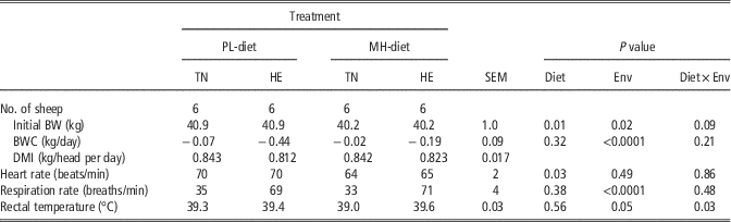
Body weight decreased (P=0.01) for the PL-diet than the MH-diet, and declined during the HE (P=0.02) compared with the TN treatment period in the present experiment (Table 1), and there was a tendency (P=0.09) of diet and environmental interaction. The dry matter intake (DMI) was comparable between diets and was lower (P<0.0001) during the HE compared with the TN. Heart rate was higher (P=0.03) when animals were offered the PL-diet relative to the MH-diet and remained similar between environments. Respiration rate and rectal temperature were higher during HE (P<0.0001 and P=0.05, respectively) and were comparable between diets. However, for rectal temperature there was a significant (P=0.03) diet and environmental interaction; that is the rectal temperature was lower when animals were offered the MH-diet during the TN period than the other treatments.
Effects of plantain (Plantago lanceolata L.) and heat exposure (30°C) on BW change (BWC), dry matter intake (DMI), heart rate, respiration rate and rectal temperature in sheep

PL-diet=MH-diet and fresh plantain (Plantago lanceolata L.) at a ratio of 1 : 1; MH-diet=mixed hay of orchardgrass (Dactylis glomerata) and reed canarygrass (Phalaris arundinacea) at a ratio of 60 : 40; TN=thermoneutral (20°C with 70% relative humidity); HE=heat exposure (30°C with 70% relative humidity); Env=environment; Diet×Env=diet and environmental interaction.
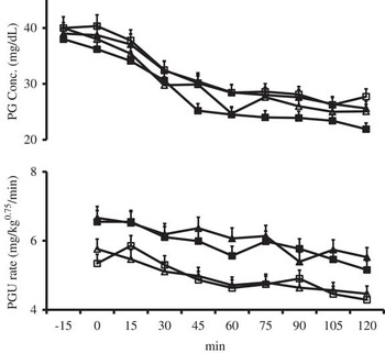
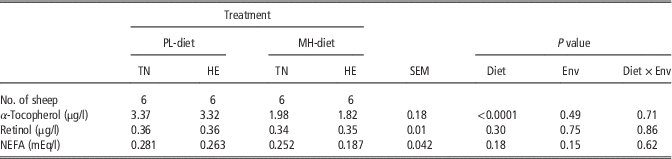
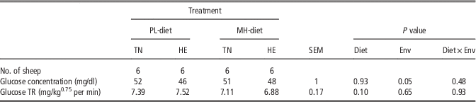
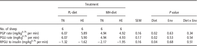
Plasma concentration of α-tocopherol was greater (P<0.0001) for the PL-diet than for the MH-diet and remained comparable between environments and no diet and environmental interactions were found (Table 2). Concentration of plasma retinol remained comparable between diets and between environments. Plasma concentration of NEFA did not differ between diets and between environments. Plasma glucose concentration before isotope dilution technique was reduced for sheep (P=0.05) during the HE compared with the TN and remained comparable between diets (Table 3). Plasma glucose TR during preinfusion period of insulin did not differ between dietary treatments (P=0.10) and between environments (P=0.65). Plasma glucose concentration and utilization during the last 120 min of [U-13C]glucose isotope dilution technique, while insulin was infused decreased gradually as shown in Figure 2. Plasma glucose production and utilization (during the last 120 min of [U-13C]glucose isotope dilution technique, while insulin was infused) were greater (P=0.02 in both cases) for the PL-diet than the MH-diet, and was not affected by environments (Table 4). Responses of plasma glucose utilization (difference between glucose utilization during insulin infusion and glucose TR before insulin infusion) to exogenous insulin infusion were lower (P=0.04) for the PL-diet compared with the MH-diet, and no significant environmental effects, nor diet and environmental interactions were found.
Time course changes of plasma glucose concentration, and plasma glucose utilization during the last 120 min of [U-13C]glucose isotope dilution, while exogenous insulin was infused at a rate of 1 mU/kg0.75 per min in sheep (n=6; three wethers and three ewes). The open triangle (∆) indicates mixed hay of orchardgrass (Dactylis glomerata) and reed canarygrass (Phalaris arundinacea) at a ratio of 60 : 40 (MH-diet) at thermoneutral 20°C with 70% relative humidity (TN); open square (□) indicates MH-diet at heat exposure 30°C with 70% relative humidity (HE); the solid black triangle (▲) indicates PL-diet, which was composed with MH-diet and fresh plantain (Plantago lanceolata L.) at a ratio of 1 : 1 at TN; the solid black square (■) indicates PL-diet at HE. PG=plasma glucose; Conc=concentration; PGU=plasma glucose utilization.

Effects of plantain (Plantago lanceolata L.) and heat exposure (30°C) on plasma concentrations of α-tocopherol, retinol and nonesterified fatty acids (NEFA) in sheep

PL-diet=MH-diet and fresh plantain (Plantago lanceolata L.) at a ratio of 1 : 1; MH-diet=mixed hay of orchardgrass (Dactylis glomerata) and reed canarygrass (Phalaris arundinacea) at a ratio of 60 : 40; TN=thermoneutral (20°C with 70% relative humidity); HE=heat exposure (30°C with 70% relative humidity); Env=Environment; Diet×Env=diet and environmental interaction.
Effects of plantain (Plantago lanceolata L.) and heat exposure (30°C) on plasma glucose concentration and turnover rate (TR) in sheep

PL-diet=MH-diet and fresh plantain (Plantago lanceolata L.) at a ratio of 1 : 1; MH-diet=mixed hay of orchardgrass (Dactylis glomerata) and reed canarygrass (Phalaris arundinacea) at a ratio of 60 : 40; TN=thermoneutral (20°C with 70% relative humidity); HE=heat exposure (30°C with 70% relative humidity); Env=environment; Diet×Env=diet and environmental interaction.
Effects of plantain (Plantago lanceolata L.) and heat exposure (30°C) on plasma glucose production (PGP) and utilization (PGU) rates and response of plasma glucose utilization (RPGU) to exogenous insulin infusion in sheep

PL-diet=MH-diet and fresh plantain (Plantago lanceolata L.) at a ratio of 1 : 1; MH-diet=mixed hay of orchardgrass (Dactylis glomerata) and reed canarygrass (Phalaris arundinacea) at a ratio of 60 : 40; TN=thermoneutral (20°C with 70% relative humidity); HE=heat exposure (30°C with 70% relative humidity); Env=environment; Diet×Env=diet and environmental interaction.
Discussion
In a previous experiment, Sano et al. (1983) stated that plasma glucose concentration was lower for sheep-fed roughage-based diets maintained at an ambient temperature of 30°C compared with 20°C, which agreed with our findings under the present experimental condition. Plasma glucose TR was not affected by diet that agrees with a previous finding evaluating diets with a 9 : 1 ratio of mixed hay and plantain (Al-Mamun et al., Reference Al-Mamun, Yamaki, Masumizu, Nakai, Saito, Sano and Tamura2007b). Plasma glucose TR was not significantly affected by HE. However, a previous study reported that plasma glucose metabolism decreased in sheep during HE (Sano et al., Reference Sano, Konno and Shiga2000). This inconsistency might be due to the fact that Sano et al. (Reference Sano, Konno and Shiga2000) determined the glucose TR on the 2nd day of HE, whereas in the present experiment it was evaluated on the 5th day. Physiological adjustment to prolonged heat may have contributed to a lack of response to heat stress in the present study (Christison and Johnson, Reference Christison and Johnson1972). The plasma glucose utilization rate decreased for both the diets and both environmental treatments during the last 120 min of the isotope dilution technique, while exogenous insulin was infused. However, the response of plasma glucose metabolism to exogenous insulin infusion was lower when animals were offered the PL-diet than MH-diet, indicating the PL-diet induced resistance to exogenous insulin. The resistance of PL-diet to exogenous insulin might be due to the presence of bioactive components present in plantain. Yoshida et al. (Reference Yoshida, Rikimaru, Sakai, Nishibe, Fujikawa and Tamura2012) added plantain to high-fat diets resulting in reduced epididymal white adipose tissue (WAT) weight and reduced blood serum glucose levels for C57BL/6J mice relative to diets without plantain without changing serum insulin levels. Therefore, plantain improved resistance to insulin in these C57BL/6J mice. Our results provide evidence for a similar effect in sheep. Plantain-fed mice exhibited marked increases in hormone-sensitive lipase, β-3 adrenergic receptor and carnitine palmitoyltransferase-2 and significant decreases in fatty acid synthesis transcripts in WAT (Yoshida et al., Reference Yoshida, Rikimaru, Sakai, Nishibe, Fujikawa and Tamura2012). The increase in insulin resistance that accompanies obesity is a consequence of several factors including impaired inhibition of glucose output by insulin, increased insulin and free fatty acids in plasma and decreased glucose clearance by the skeletal muscle (Purushotham et al., Reference Purushotham, Wendel, Liu and Belury2007).
Previously it was reported that the glucagon-like peptide-1 (GLP-1) receptor agonists added to basal insulin decreased postprandial glucose levels, decreased BW and reduced basal insulin requirements without increasing the risk of major hypoglycemic events (Cohen et al., Reference Cohen, Audehm, Pretorius, Kaye, Chapman and Colagiuri2013). It was also reported that hyperinsulinemia triggers the insulin resistance (Juan et al., Reference Juan, Fang, Kwok, Perng, Chou and Ho1999). Moreover, Cowan et al. (Reference Cowan, McKay and Fisher2012) found positive response of addition of GLP-1 receptor agonist with insulin in reducing insulin doses, further weight loss and risk of hypoglycemia in individuals having insulin resistance. Catalpol is one of the bioactive components of plantain (Al-Mamun et al., Reference Al-Mamun, Abe, Kofujita, Tamura and Sano2008a), which might be responsible for the insulin resistance. Because, the catalpol was found has the equal potentiality to work as like as GLP-1 receptor agonist (Jia et al., Reference Jia, Gong, Li, Zhu and Wang2015).
The contribution of higher plasma α-tocopherol in PL-diet group on insulin resistance is not clear, as there are controversial statements regarding its effect on the insulin resistance. For example, Rose et al. (Reference Rose, Obara, Fuse, Itoh, Ozawa, Takahashi, Hodate and Ohashi1996) demonstrated that elevated concentrations of growth hormone (GH) decreased the responsiveness of peripheral tissues to high concentrations of insulin during late lactation in the dairy cow. Further, it was reported that α-tocopherol increased the release of GH in the rat (Badamchian et al., Reference Badamchian, Spangelo, Hagiwara, Hagiwara, Veyama and Goldstein1994), insulin action is antagonized by GH in adipose tissue (Vernon, Reference Vernon1982; Etherton and Walton, Reference Etherton and Walton1986). Higher the plasma α-tocopherol most likely reduced the oxidation of lipid, which is commonly associated with insulin resistance (Leslie et al., Reference Leslie, Jill and Joseph2009). In another report, Costacou et al. (Reference Costacou, Ma, King and Mayer-Davis2008) stated that plasma concentration of α-tocopherol might improve insulin sensitivity and pancreatic compensation for insulin resistance, although it did not seemed to be related to the acute insulin response. Kim et al. (Reference Kim, Kim, Ham and Choue2013) found that oral administration of d-α-tocopherol with a high-fat diet exacerbated insulin resistance in male mice. In the present study, the infusion of exogenouse insulin might be partially responsible for the insulin resistance through raising the hyperinsulinemia. Because, in a previous study Gelardi et al. (Reference Gelardi, Rapoza, Renzulli and Cowett1999) stated that insulin resistance was related with hyperinsulinemic in lambs. Higher α-tocopherol is an indication of higher free radical scavenging activities (FRSA). It was reported that plantain was superior to other traditional pastures and herbs in Japan to FRSA (Al-Mamun et al., Reference Al-Mamun, Hanai, Tanaka, Tamura and Sano2007a). α-Tocopherol is the most frequently used lipid-soluble free radical scavenger, and it may scavenge the free radical on the surface of cell membrane efficiently and finally terminate the lipid oxidation (Buckley et al., Reference Buckley, Morrissey and Gray1995).
In the present experiment, respiration rate and rectal temperature increased, and BW decreased during HE compared with TN, which was in agreement with the previous findings (Sano et al., Reference Sano, Takahashi, Ambo and Tsuda1983; Achmadi et al., Reference Achmadi, Yanagisawa, Sano and Terashima1993; Itoh et al., Reference Itoh, Hodate, Koyama, Rose, Matsumoto, Ozawa and Obara2001). There was a diet and environmental interaction for rectal temperature, indicating that the response to heat is diet dependent. Rectal temperature was not affected by environment for sheep fed the PL-diet, but rectal temperature was higher during the HE than the TN period for sheep fed the MH-diet. It was found that roughage generated a higher heat increment than concentrate (Sudarman and Ito, Reference Sudarman and Ito2000). Sano et al. (Reference Sano, Tamura and Shiga2002) stated that plantain showed a more concentrate-like fermentation pattern than orchardgrass hay, which might be partially responsible for this higher rectal temperature. It is well known that, like the present experimental findings, HE increases rectal temperature in sheep. It was found that antioxidants ameliorated heat stress in sheep (Chauhan et al., Reference Chauhan, Celi, Fahri, Leury and Dunshea2014). Therefore, due to the antioxidant (Al-Mamun et al., Reference Al-Mamun, Hanai, Tanaka, Tamura and Sano2007a) and also the higher plasma α-tocopherol content of PL-diet, the rectal temperature was comparatively lower during the HE when animals were offered the PL-diet compared with the MH-diet. Environments did not affect heart rate, which was in accordance with the previous findings by Sano et al. (Reference Sano, Takahashi, Ambo and Tsuda1983) in sheep. In the present study, the reduction of BW during the HE and when animals were offered the PL-diet might at least partially relate to non-significant numerical variation in DMIs across treatments.
In a previous study, Sano et al. (Reference Sano, Tamura and Shiga2002) reported that diets containing plantain contained less gross energy than those with orchardgrass. Furthermore, the plasma concentration of NEFA is linearly and negatively correlated with feed intake of ewes (Landau et al., Reference Landau, Madar, Nitsan and Zoref1992). Slightly greater plasma NEFA concentration for PL-diet group might substantiate the theory that the DMI was numerically lower for PL-diet than that of the MH-diet.
Conclusion
Heat stress resulted in reduced plasma glucose concentration in sheep without altering the TR. Plasma concentration of α-tocopherol during preinfusion of insulin was higher in animals offered the PL-diet than the MH-diet. Plantain-induced resistance to exogenous insulin. However, additional research should be conducted to investigate the physiological consequences of the inclusion of plantain in diets for growing, pregnant or lactating ruminants.
Acknowledgments
The authors gratefully acknowledge the Grants-in-Aid for Scientific Research (P08442) from the Japan Society for the Promotion of Science for providing financial support. The authors are also grateful to Dr Andrea Hanson, USA and Prof. Dr Peter Wynn, Australia for their kind comments on this manuscript.
Supplementary material
To view supplementary material for this article, please visit https://doi.org/10.1017/S1751731116002846