INTRODUCTION
The project Latitudinal gradients of biodiversity in the deep sea of the Atlantic Ocean (DIVA) aims to study the biodiversity along an Atlantic latitudinal transect, from the Arctic to the Antarctic, in comparable abyssal and bathyal depths. During the expedition ‘Me48/1 DIVA 1’ on-board the ‘Meteor’ in July–August 2000, some Solenogastres belonging to three new species of the genus Kruppomenia Nierstrasz, Reference Nierstrasz1903 (Kruppomenia angolensis sp. nov., Kruppomenia glandulata sp. nov. and Kruppomenia macrodenticulata sp. nov.) were collected in the abyssal Angola Basin at 5125–5415 m depth. Solenogastres are a small class of vermiform molluscs that possess a mantle with chitinous cuticle and calcareous sclerites, a ventral pedal groove and a terminal or subterminal pallial cavity. The knowledge about the biodiversity of these molluscs is still meagre, therefore, the characteristics that define their taxa are incomplete in many cases. Although the abyssal plains represent around 40% of the oceanic bottoms, only 15 species of the 267 Solenogastres described so far are known from depths greater than 4000 m and of these, only 8 from deeper than 5000 m (García-Álvarez & Salvini-Plawen, Reference García-Álvarez and Salvini-Plawen2007; Gil-Mansilla et al., Reference Gil-Mansilla, García-Álvarez, Urgorri, Martínez Arbizu and Brix2008, Reference Gil-Mansilla, García-Álvarez and Urgorri2009, Reference Gil-Mansilla, García-Álvarez and Urgorri2011).
MATERIALS AND METHODS
The specimens were collected during the expedition ‘Me48/1 DIVA 1’ in the abyssal Angola Basin at depths between 5125 and 5415 m using an epibenthic sledge, on a bottom made up of white to light beige muddy sediment with a large amount of globularian foramiferans. The temperature on the bottom was 2.48°C and the salinity 34.8‰ (Kröncke & Türkay, Reference Kröncke and Türkay2003). The specimens were fixed and preserved in 70% ethanol. The specimens were photographed and measured and their external anatomy was described. The sclerites were studied by separating small pieces of the mantle. The specimens were decalcified with an EDTA solution and the anterior and posterior region of the specimens were embedded in paraffin and cut in serial transverse sections of 5 µm. The sections were dyed with Mallory's trichrome stain and the internal anatomy was manually reconstructed.
DIAGNOSIS
Common atriobuccal cavity. Radula with narrow biserial plates with numerous small denticles. Anteroventral radular sac (when present) paired. Ventrolateral foregut glandular organs epithelial in the shape of ampoules (type C according to Salvini-Plawen, Reference Salvini-Plawen1978; type Simrothiella according to Handl & Todt, Reference Handl and Todt2005). With seminal receptacles and/or seminal vesicles. With respiratory folds. With dorsoterminal sense organ.
Type species: Kruppomenia minima Nierstrasz, Reference Nierstrasz1903
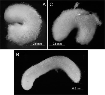
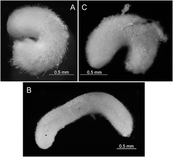
Habitus. (A) Kruppomenia angolensis sp. nov.; (B) Kruppomenia glandulata sp. nov.; (C) Kruppomenia macrodenticulata sp. nov.
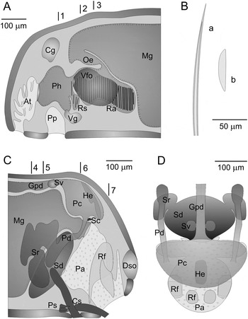
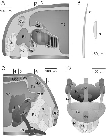
Kruppomenia angolensis sp. nov. (A) Anatomical reconstruction from histological sections of the anterior body; (B) sclerites: a. hollow acicular sclerite; b. blade scale-shaped sclerite; (C) anatomical reconstruction from histological sections of the posterior body; (D) anatomical reconstruction of reproductive system and pallial cavity (dorsal view). Lines 1–3 in (A) indicate the position of the transversal section in Figure 3A–C. Lines 4–7 in (C) indicate the position of the transversal section in Figure 4A–D. At, atrium; Cg, cerebral ganglion; Cs, copulatory stylets; Dso, dorsoterminal sense organ; Gpd, gonopericardioduct; He, heart; Mg, midgut; Oe, oesophagus; Pa, pallial cavity; Pc, pericardium; Pd, pericardioduct; Ph, pharynx; Pp, pedal pit; Ps, prepallial spicules; Ra, radula; Rf, respiratory fold; Rs, radula sack; Sc, supra-rectal commissure; Sd, spawning duct; Sr, seminal receptacle; Sv, seminal vesicle; Vfo, ventrolateral foregut glandular organs; Vg, ventral ganglion.
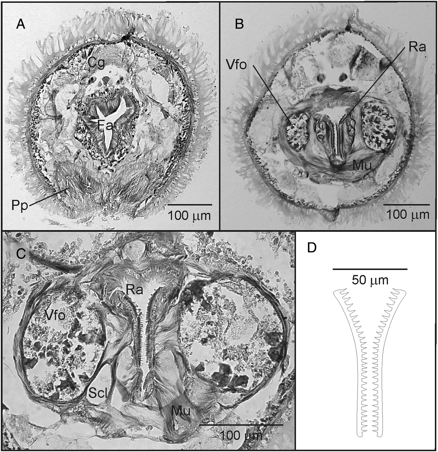
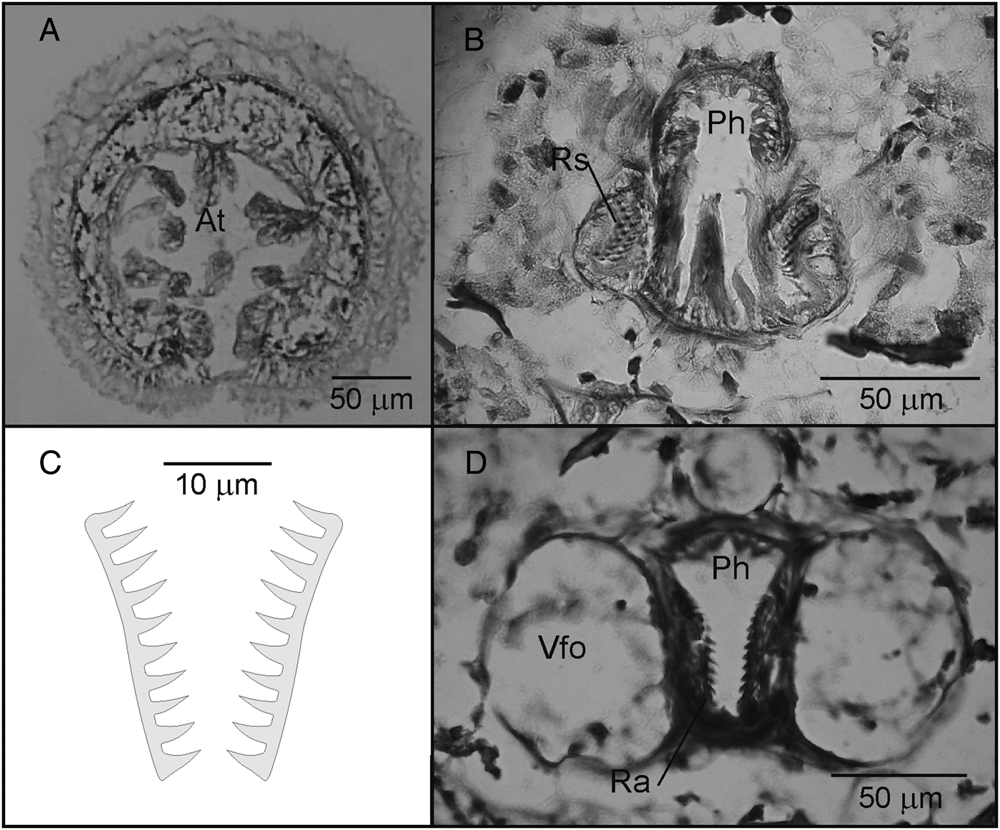
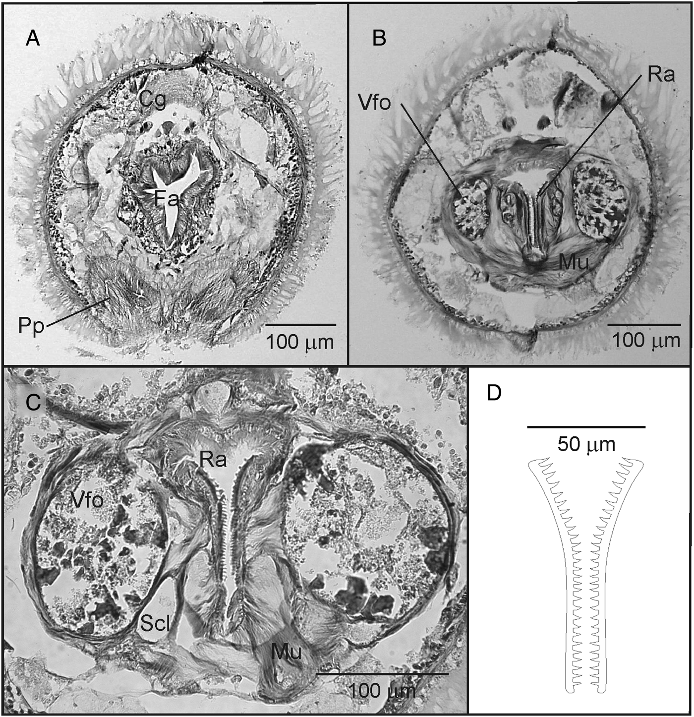
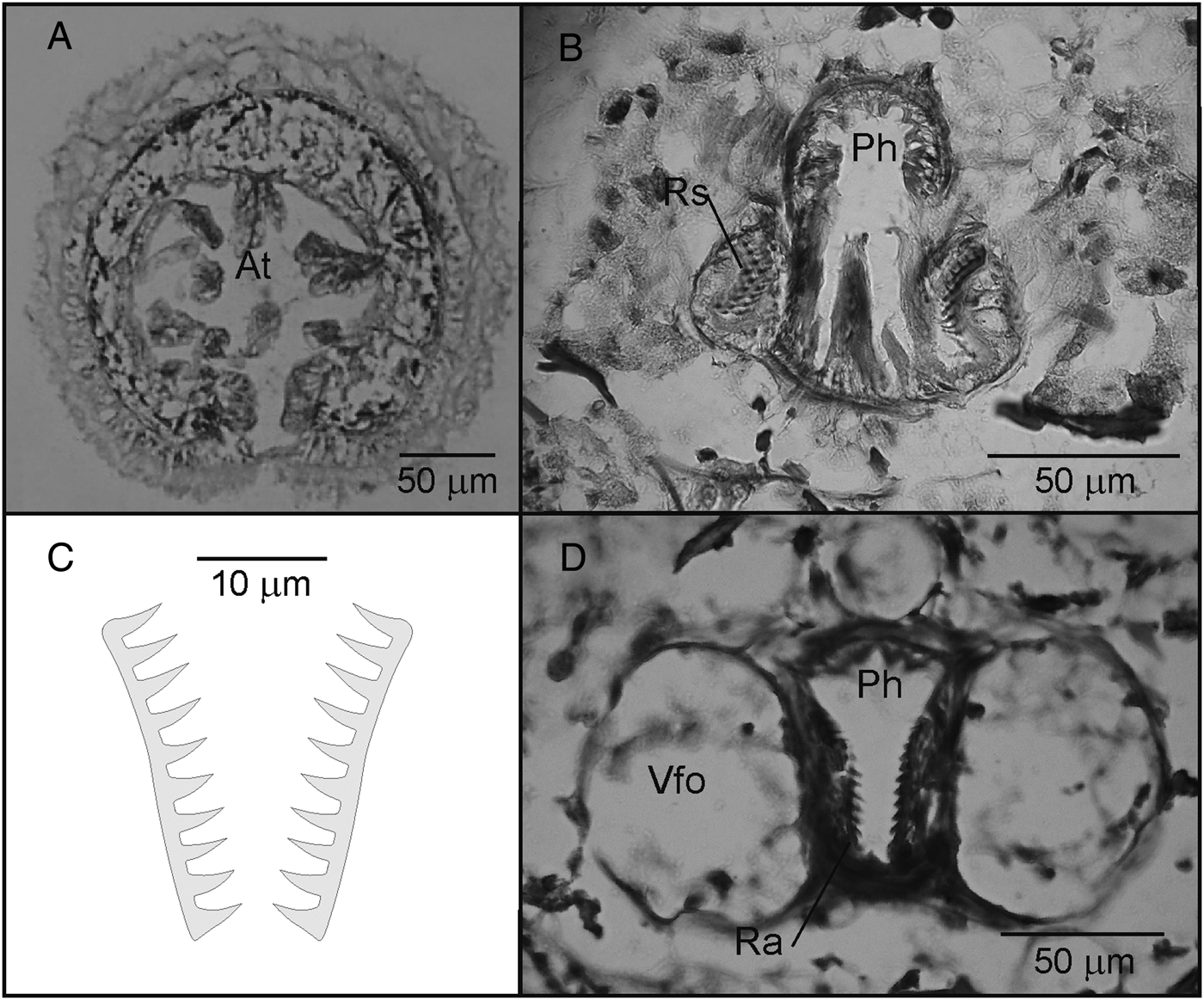
Kruppomenia angolensis sp. nov. (A–C) Sections through the anterior body corresponding to lines 1–3 in Figure 2A; (D) drawing of radula plates. Cg, cerebral ganglion; Scl, supporting cell; Mu, musculature; Ph, pharynx; Pp, pedal pit; Ra, radula; Vfo, ventrolateral foregut glandular organs.
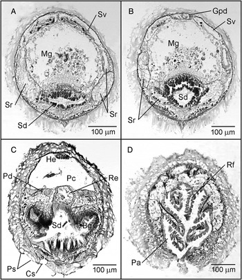
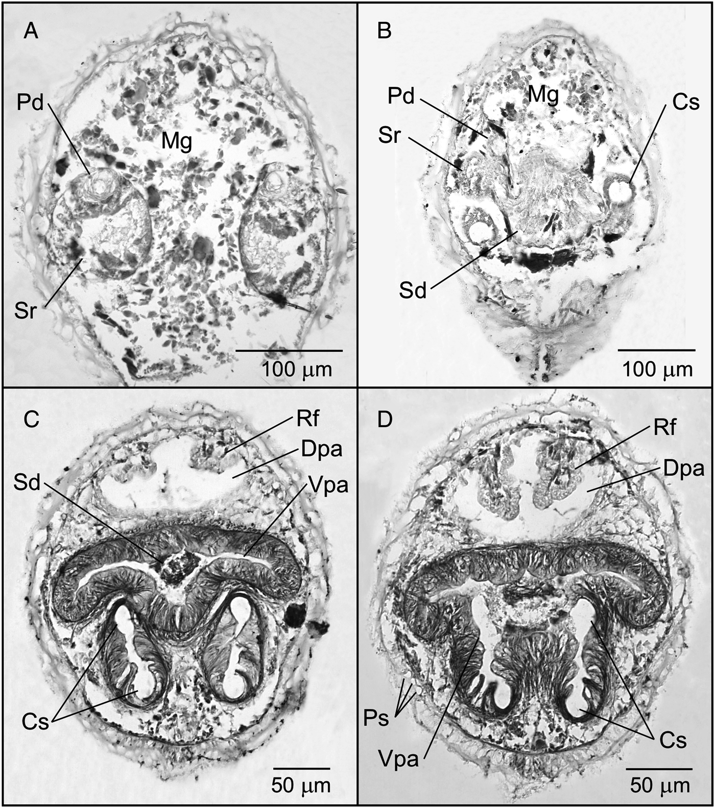
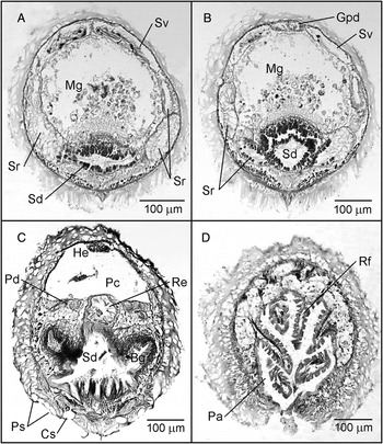
Kruppomenia angolensis sp. nov. (A–D) Sections through the posterior body corresponding to lines 4–7 in Figure 2C. Bg, anterolateral bag in pallial cavity; Cs, copulatory stylets; Gpd, gonopericardioduct; He, heart; Mg, midgut; Pa, pallial cavity; Pc, pericardium; Pd, pericardioduct; Ps, prepallial spicules; Re, rectum; Rf, respiratory fold; Sd, spawning duct; Sr, seminal receptacle; Sv, seminal vesicle.
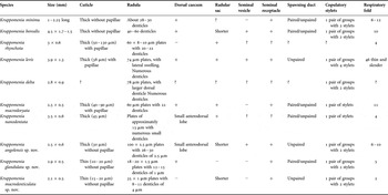
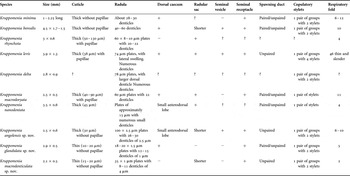
Differences between the species of the genus Kruppomenia. (+) present; (–) absent; (?) unknown.
TYPE MATERIAL
Two specimens
Holotype: sectioned and mounted on slides; sclerites on slides. Paratype (immature) sectioned and mounted on slides; sclerites on slides. Both from abyssal Angola Basin, Station 344 ‘Me48/1 DIVA 1’ (17°06′12″S 04°41′42″E–17°07′30″S 04°42′18″E; water depth: 5415 m).
Depository: holotype and paratype are deposited in the Zoologische Staatssammlung München (holotype: ZSM Mol 20080848; paratype: ZSM Mol 20080849).
ETYMOLOGY
Angolensis, from Angola. With regard to the basin where it was collected.
DIAGNOSIS
Body 2.5 mm long by 0.6 mm in diameter. Without lumps or keels. Cuticle moderately thick (30 µm) without epidermal papillae. With hollow acicular sclerites at least in two layers. Common atriobuccal cavity. Pedal groove with one fold that does not get into the pallial cavity. Biserial radula made up of homodenticulate plates with 26–30 triangular denticles. With a pair of short anterolateral radular sacs. Ventrolateral foregut glandular organs made up of a pair of ampoules with epithelial glandular cells encircled by musculature. Intestine with an anterodorsal lobe, which does not represent an authentic anterodorsal intestinal caecum; without lateral constrictions. With seminal vesicles in the gonopericardioducts. With seminal receptacles. Unpaired genital orifice. With a pair of groups of copulatory stylets with two stylets each. With 4–5 pairs of prepallial spicules. With 3–5 respiratory folds. Dorsoterminal sense organ in terminal position.
DESCRIPTION
Habitus: 2.5 mm long and 0.6 mm thick. Short and thick cylindrical body without lumps or keels. Sclerites with oblique insertion, protruding from the cuticle. In the posterior region of the body, there are longer sclerites that enclose the pallial cavity. White colour observed after fixation and preservation in 70% ethanol (Figure 1A).
Mantle: moderately thick cuticle (30 µm) without epidermal papillae. Sclerites with oblique insertion, in 1–2 layers. Hollow acicular sclerites, slightly sigmoid (fragmented 220 µm long) and sclerites type blade-shaped scale (65 µm long; 15 µm wide maximum), located on both sides of the pedal groove (Figure 2B).
Pedal groove and pallial cavity: the pedal groove starts in the pedal pit (90 µm long; 200 µm wide; 120 µm high) which has numerous long cilia. A pair of large pedal glands opens into the pedal pit. The pedal groove possesses a small fold that does not extend into the pallial cavity. The pallial cavity (180 µm long; 200 µm wide; 300 µm high) opens in subterminal position; it presents 3 pairs of respiratory folds in the holotype and 5 pairs in the paratype: one pair is joined to the dorsal wall and the other to the lateral walls (Figures 2C, D & 4D). The anterior region of the pallial cavity has a ciliated epithelium. The anus opens into the anterodorsal region. The medial part of the cavity becomes narrower laterally and widens anteroventrally forming a bag that extends laterally, into which the spawning duct opens (Figures 2C & 4C).
Digestive system: the mouth opens in the dorsoposterior region of the common atriobuccal cavity. It continues in the pharynx (275 µm long; 180 µm wide; 200 µm high) which is internally covered by a thin cuticle and its anterior region has a Y-shaped lumen (Figure 3A). The radular apparatus (270 µm long) is encircled by a strong muscular layer (Figure 3B) and possesses two short anterolateral radular sacs (35 µm long) that showed fragments of radula plates, more developed in paratype; a homodenticulate biserial radula, with 25–35 pairs of narrow and slightly curved plates in their distal region (100 µm long; 2.5 µm wide) with 26–30 triangular denticles (2.5 µm high), regularly arranged along the plate (Figure 3D); and a long radular sheath, where several developing plates can be observed. The radula plates are located upon musculature and several supporting cells (Figure 3B–D). The ventrolateral foregut glandular organs consist of a pair of ampoules with epithelial glandular cells and being encircled by musculature (type C, according to Salvini-Plawen, Reference Salvini-Plawen1978; type Simrothiella, according to Handl & Todt, Reference Handl and Todt2005). They open dorsolaterally into the pharynx, in the area of the radula beginning (Figures 2A & 3B, C). There is a narrow oesophagus that curves anteriorly until it joins the anteroventral region of the intestine through a narrow opening. The intestine lacks constrictions and shows a small anterodorsal lobe without forming an intestinal caecum (Figure 2A). Remains of cnidocytes were found in the intestine of the two specimens; besides, a fragment of a polychaete was observed in the paratype. The rectum is narrow and internally ciliated, the anus is located in the dorsoanterior region of the pallial cavity (Figures 2C & 3C).
Nervous system and sense organs: the cerebral ganglion (75 µm long; 150 µm wide; 100 µm high) has a quadrangular outline in section and is located dorsally to the anterior region of the pharynx. There are a couple of small ventral ganglia, ventrally to the pharynx behind the pedal pit (Figure 2A). The suprarectal commissure is located dorsally to the rectum (Figure 2C). The atrial sense organ is located in the anterior region of the common atriobuccal cavity and has numerous simple papillae (Figure 2A). The dorsoterminal sense organ is in terminal position (Figure 2C).
Reproductive system: the gonads presented ova in the anterior region and spermatozoids in the posterior. Narrow gonopericardioducts; each of them has a seminal vesicle with spermatozoids (Figures 2C, D & 4A, B). Large pericardium with a tubular heart that joins the dorsal wall of the pericardium through its anterior and posterior ends. Two narrow and ciliated pericardioducts come out from the ventral region of the pericardium, they get wider along their length and end in a pair of bilobed seminal receptacles, with a dorsal and a ventral lobe (Figure 2C, D). The seminal receptacles join the spawning duct laterally through the ventral lobe. The lobes present histological differences: the epithelium of the dorsal lobe is made up of smaller cells than those of the ventral lobe, being the epithelium of the latter histologically similar to that of the anterior region of the spawning duct (Figure 4A, B). Spawning duct unpaired along its whole length, it has a glandular and ciliated epithelium (Figure 4A, B). It shows a bag in the posteroventral region, where the genital orifice is located and it opens in the anteroventral region of the pallial cavity. A paired group of copulatory stylets, comprising two stylets each, are located in the ventroposterior region (Figures 2C & 4C). In each group, one stylet stretches anteriorly and has an oblique position in relation to the pedal groove (Figure 2C); this stylet is joined to developed musculature and extends ventrally to the exterior through the cuticle, anterior to the opening of the pallial cavity. The second stylet of each group also extends to the exterior through the cuticle, but posteriorly to the anterior and ventrally to the pallial cavity. Besides, there are 4–5 pairs of prepallial spicules located in the cuticle anterior to the opening of the pallial cavity (Figure 2C). In the paratype no copulatory stylets were observed, although some prepallial spicules are found; this may be related to the immature condition of the specimen.
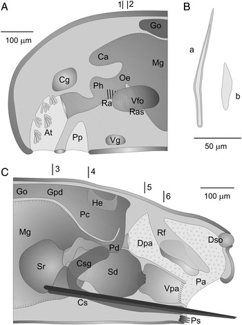
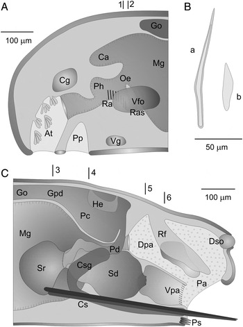
Kruppomenia glandulata sp. nov. (A) Anatomical reconstruction from histological sections of the anterior body; (B) sclerites: a. hollow acicular sclerite; b. blade scale-shaped sclerite; (C) anatomical reconstruction from histological sections of the posterior body. Lines 1 and 2 in (A) indicate the position of the transversal section in Figure 6A, C. Lines 3–6 in (C) indicate the position of the transversal section in Figure 7A–D. At, atrium; Ca, dorsal caecum; Cg, cerebral ganglion; Cs, copulatory stylets; Csg, copulatory stylets gland; Dpa, dorsal pallial cavity; Dso, dorsoterminal sense organ; Go, gonad; Gpd, gonopericardioduct; He, heart; Mg, midgut; Oe, oesophagus; Pa, pallial cavity; Pc, pericardium; Pd, pericardioduct; Ph, pharynx; Pp, pedal pit; Ps, prepallial spicules; Ra, radula; Ras, radula sheath; Rf, respiratory fold; Sd, spawning duct; Sr, seminal receptacle; Vfo, ventrolateral foregut glandular organs; Vg, ventral ganglion; Vpa, ventral pallial cavity.
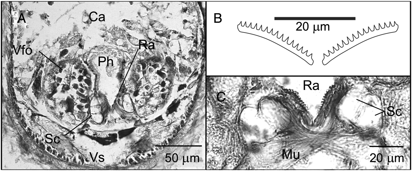
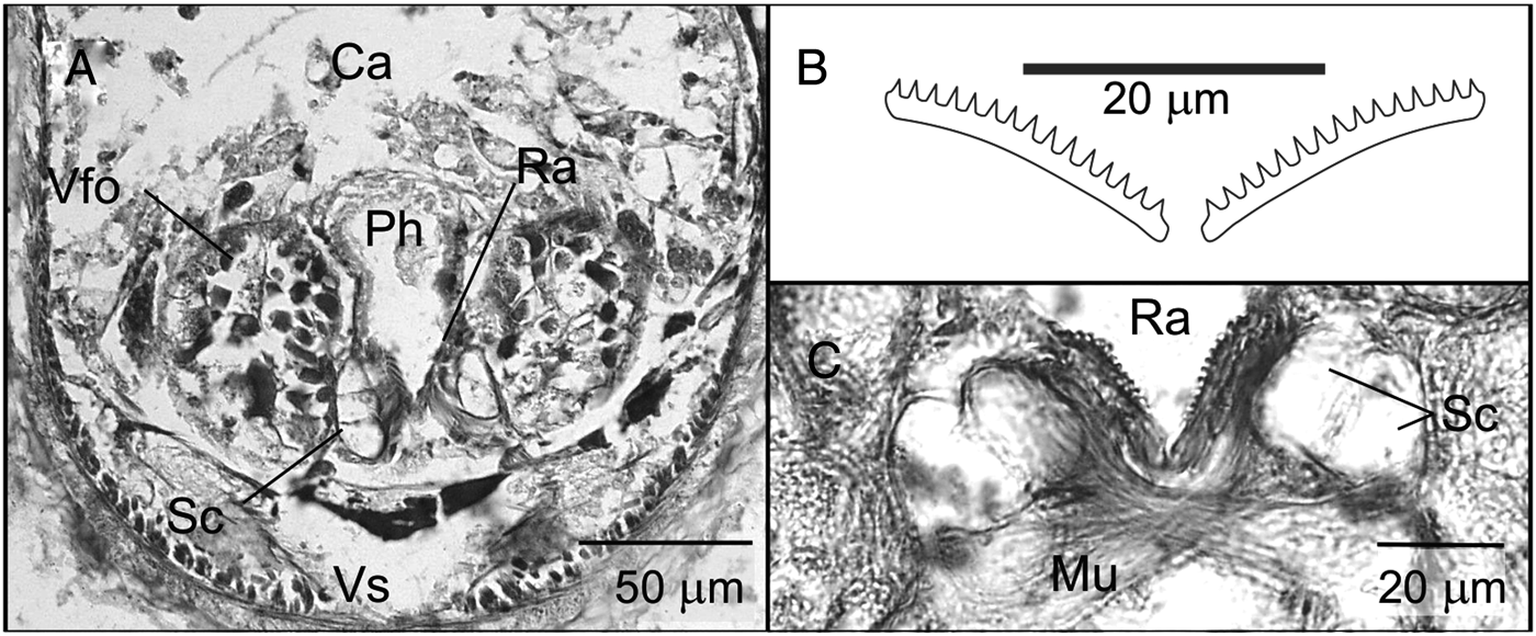
Kruppomenia glandulata sp. nov. (A & C) Sections through the anterior body corresponding to lines 1 and 2 in Figure 5A; (B) drawing of radula plates. Ca, dorsal caecum; Mu, musculature; Ph, pharynx; Ra, radula; Sc, supporting cell; Vfo, ventrolateral foregut glandular organs; Vs, ventral sinus.
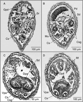
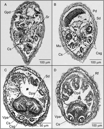
Kruppomenia glandulata sp. nov. (A–D) Sections through the posterior body corresponding to lines 3–6 in Figure 5C. Cs, copulatory stylets; Csg, copulatory stylet gland; Dpa, dorsal pallial cavity; Gpd, gonopericardioduct; Mg, midgut; Mu, musculature; Pc, pericardium; Pd, pericardioduct; Rf, respiratory fold; Sd, spawning duct; Sr, seminal receptacle; Vpa, ventral pallial cavity.
TYPE MATERIAL
Three specimens
Holotype sectioned and mounted on slides; sclerites on slides from abyssal Angola Basin Station 344 ‘Me48/1 DIVA 1’ (17°06′12″S 04°41′42″E–17°07′30″S 04°42′18″E; water depth: 5415 m). Paratype 1 sectioned and mounted on slides; sclerites on slides from abyssal Angola Basin Station 340 ‘Me48/1 DIVA 1’ (18°18′18″S 04°41′18″E–18°19′24″S 04°41′54″E; water deep: 5395 m). Paratype 2 sectioned and mounted on slides; sclerites on slides from abyssal Angola Basin Station 348 ‘Me48/1 DIVA 1’ (16°18′06″S; 05°27′12″E–16°19′18″S; 05°27′12″E; water deep: 5390 m).
Depository: holotype and paratypes 1 and 2 are deposited in the Zoologische Staatssammlung München (holotype: ZSM Mol 20080853; paratype 1: ZSM Mol 20080850; paratype 2: ZSM Mol 20080855)
ETYMOLOGY
Latin: glandula, organ that produces a secretion; Latin: -atus, provided with. With reference to the gland joined to the copulatory stylets.
DIAGNOSIS
Body up to 2.9 mm long by 0.5 mm wide. Without lumps or keels. Thin cuticle (10–20 µm) without epidermal papillae. Hollow acicular sclerites arranged in one layer. Pedal groove with one fold that does not extend to the pallial cavity. Common atriobuccal cavity. Biserial radula representing homodenticulate plates with 15–18 denticles. Without anterolateral radular sacs. Ventrolateral foregut glandular organs consisting of a pair of ampoules with glandular epithelial cells encircled by musculature. Intestine with anterodorsal paired caecum and without constrictions. With a pair of seminal receptacles, no seminal vesicles. Spawning duct paired in its anterior region. Unpaired genital orifice. With a paired group of copulatory stylets, each of them with two stylets, joined to a gland. With 7–8 pairs of prepallial spicules. Pallial cavity with a ventral and a dorsal chamber. With 5 respiratory folds. Dorsoterminal sense organ dorsally to the posterior pallial cavity.
DESCRIPTION
Habitus: 2.9 mm long and 0.5 mm thick. Without lumps or keels. Well-marked pedal groove. Sclerites with oblique insertion protruding from the cuticle. White colour observed after fixation and preservation in 70% ethanol (Figure 1B).
Mantle: thin cuticle (10–20 µm) without epidermal papillae. Sclerites with oblique insertion in one layer. Hollow acicular sclerites (130 µm long; 7.5 µm wide) slightly curved near their proximal end and blade-shaped scales (50 µm long; 12 µm wide maximum) located on both sides of the pedal groove (Figure 5B).
Pedal groove and pallial cavity: the pedal groove starts in a small pedal groove densely ciliated, into which a pair of pedal glands opens. It includes an only small-sized single fold that does not reach the pallial cavity. Wide pallial cavity that opens subterminally. In the anterior half, it divides in two chambers: dorsal and ventral (Figures 5C & 7C, D). The dorsal chamber has a thin epithelium; up to 5 respiratory folds and the anus are located in it. The ventral chamber has the shape of a bilobed bag, the epithelium is glandular, its posterior region with circular musculature, and the genital orifice are located in it. The exit of the copulatory stylets and prepallial spicules are located ventrally to the opening of the pallial cavity.
Digestive system: the mouth opens in the dorsoposterior region of the common atriobuccal cavity and continues in a narrow pharynx. The radular apparatus lacks anterolateral radular sacs and is made up of a homodenticulate biserial radula with 15–20 plates and a short radular sheath. Each radula plate (18–20 µm long; 1.5 µm wide) has 15–18 denticles approximately 1 µm high regularly arranged (Figure 6B). 3–6 large supporting cells are located ventrolaterally to the radula and ventrally a very developed musculature is present (Figure 6A, C). The ventrolateral foregut glandular organs consist of a pair of ampoules with epithelial glandular cells and being encircled by a thin layer of musculature (type C, according to Salvini-Plawen, Reference Salvini-Plawen1978; type Simrothiella, according to Handl & Todt, Reference Handl and Todt2005) (Figure 6A). The pharynx continues in a short oesophagus. The intestine presents a paired anterodorsal caecum and lacks constrictions. The anus opens in the anterior region of the dorsal chamber of the pallial cavity.
Nervous system and sense organs: the cerebral glanglion (75 µm long; 120 µm wide; 50 µm high) has a quadrangular transverse section and is located dorsally at the beginning of the pharynx. The ventral ganglia (70 µm long; 30 µm wide; 30 µm high) are located posteriorly to the pedal pit (Figure 5A). The atrial sense organ is located in the anterior region of the common atriobuccal cavity, it is ciliated and has numerous papillae in groups (Figure 5A). A dorsoterminal sense organ is located dorsally to the posterior region of the pallial cavity (Figure 5C).
Reproductive system: the gonads presented ova (15 µm diameter) joined to the medial wall and spermatozoids that took up the rest of the gonadal space. Large pericardium with a tubular heart that joins the dorsal wall of the pericardium by its anterior and posterior ends. Both pericardioducts come out from the posterior region of the pericardium, they lead anteriorly to join the spawning duct. There are no seminal vesicles. The pair of seminal receptacles (140 µm long; 30 µm wide; 140 µm high) full of spermatozoids opens into this same area (figures 5C; 7A). The spawning ducts possess a thin epithelium (Figure 7B), they remain separated in their anterior fourth, fuse posteriorly in a wide duct with glandular epithelium that opens onto the anterior wall of the ventral chamber of the pallial cavity through a narrow genital orifice (Figure 7C). There is a paired group of ventrolateral copulatory stylets (Figures 5C & 7). Each group is made up of two stylets and joined to a strong longitudinal musculature and a distally curved gland (Figure 7B, C). The gland opens separately anteriorly to the exit of the copulatory stylets. 7–8 pairs of prepallial spicules are located in the region anterior to the opening of the pallial cavity.
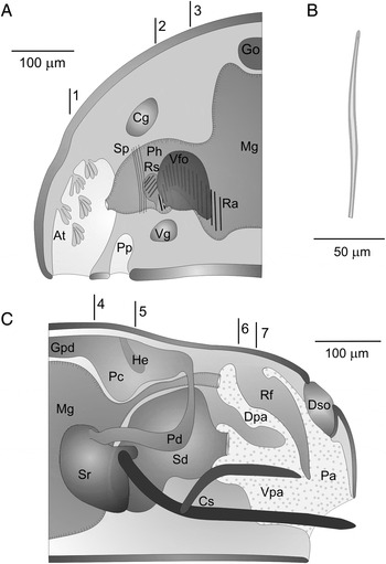
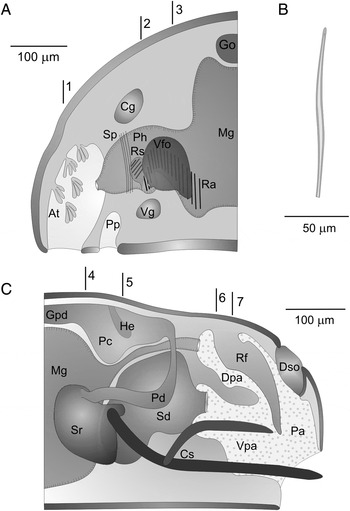
Kruppomenia macrodenticulata sp. nov. (A) Anatomical reconstruction from histological sections of the anterior body; (B) hollow acicular sclerite; (C) anatomical reconstruction from histological sections of the posterior body. Lines 1–3 in (A) indicate the position of the transversal section in Figure 9A, B & D. Lines 4–7 in (C) indicate the position of the transversal section in Figure 10A–D. At, atrium; Cg, cerebral ganglion; Cs, copulatory stylets; Dpa, dorsal pallial cavity; Dso, dorsoterminal sense organ; Go, gonad; Gpd, gonopericardioduct; He, heart; Mg, midgut; Pa, pallial cavity; Pc, pericardium; Pd, pericardioduct; Ph, pharynx; Pp, pedal pit; Ra, radula; Rf, respiratory fold; Rs, radula sac; Sd, spawning duct; Sp, sphincter; Sr, seminal receptacle; Vfo, ventrolateral foregut glandular organs; Vg, ventral ganglion; Vpa, ventral pallial cavity.
Kruppomenia macrodenticulata sp. nov. (A, B & D) Sections through the anterior body corresponding to lines 1–3 in Figure 8A; (C) drawing of radula plates. At, atrium; Ph, pharynx; Ra, radula; Rs, radular sac; Vfo, ventrolateral foregut glandular organs.
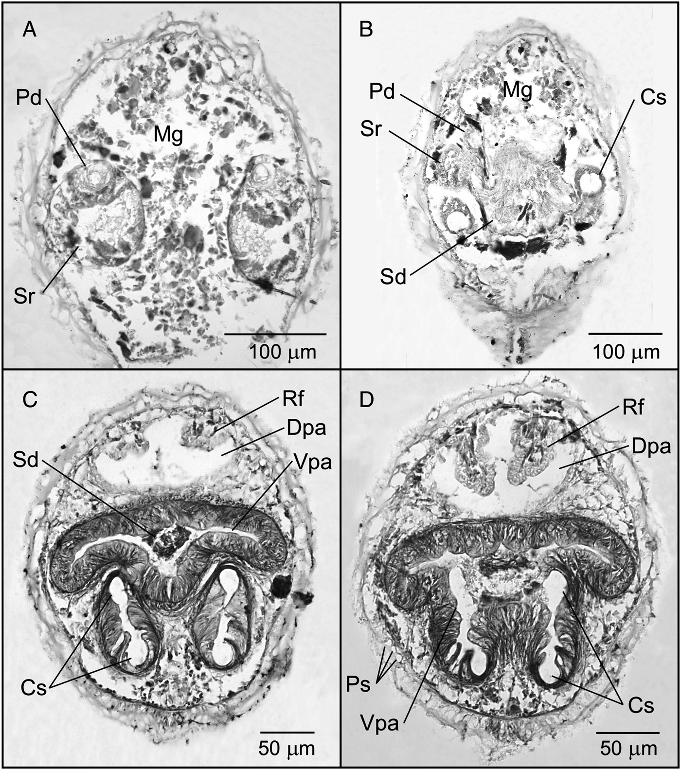
Kruppomenia macrodenticulata sp. nov. (A–D) Sections through the posterior body corresponding to lines 4–7 in Figure 8C. Cs, copulatory stylets; Dpa, dorsal pallial cavity; Mg, midgut; Pd, pericardioduct; Ps, prepallial spicules; Rf, respiratory fold; Sd, spawning duct; Sr, seminal receptacle; Vpa, ventral pallial cavity.
TYPE MATERIAL
Two specimens
Holotype sectioned and mounted on slides; sclerites on slides from abyssal Angola Basin Station 338 ‘Me48/1 DIVA 1’ (22°20′00″S 03°18′18″E–22°20′12″S 03°18′24″E; water deep: 5125–5144 m). Paratype sectioned and mounted on slides; sclerites on slides from abyssal Angola Basin Station 320 ‘Me48/1 DIVA 1’ (22°19′54″S 03°17′48″E–22°20′00″S 03°17′54″E; water depth: 5127 m).
Depository: holotype and paratype are deposited in the Zoologische Staatssammlung München (holotype: ZSM Mol 20080857; paratype: ZSM Mol 20080876).
ETYMOLOGY
Greek: macro-, large; Latin: denticulus, denticle; Latin: -atus, provided with. In connection with the size of the denticles of the radular plates.
DIAGNOSIS
Body 2.1 mm long by 0.55 mm thick. Without lumps or keels. Thin cuticle (15–20 µm) without epidermal papillae. Hollow acicular sclerites with oblique insertion in one layer. Common atriobuccal cavity. Pedal groove with one fold that does not extend into the pallial cavity. Pharynx with preradular sphincter. Biserial radula with homodenticulate radular plates with 8–11 denticles each. With a pair of anterolateral radular sacs. Ventrolateral foregut glandular organs consisting of a pair of ampoules with epithelial glandular cells and being encircled by musculature. Intestine without anterior caecum and without constrictions. With seminal receptacles. Unpaired spawning duct. Unpaired genital orifice. With a pair of groups of copulatory stylets with two stylets each. With prepallial spicules. Pallial cavity with a ventral and a dorsal chamber. With a pair of respiratory folds. Dorsoterminal sense organ above posterior pallial cavity.
DESCRIPTION
Habitus: 2.1 mm long by 0.55 mm thick. Without lumps or keels. With a clearly visible pedal groove. Sclerites with oblique insertion protruding from the cuticle. White colour observed in the fixed and preserved specimens in 70% ethanol (Figure 1C).
Mantle: thin cuticle (15–20 µm) without epidermal papillae, with sclerites of oblique insertion in one layer. Hollow acicular sclerites (fragmented, 140 µm long) slightly curved (Figure 8B).
Pedal groove and pallial cavity: the pedal groove starts in a ciliated pedal pit (25 µm long; 60 µm wide; 55 µm high) into which a pair of pedal glands opens. It shows one fold that does not extend into the pallial cavity. The pallial cavity opens subterminally and its anterior region is divided in a dorsal and ventral chamber. The dorsal chamber has a thin epithelium, a pair of dorsal respiratory folds and the anus opens in its anterior region. The ventral chamber has a glandular epithelium underlain by muscular fibres, the genital orifice opens in its anterior wall and the copulatory stylets are located ventrally. Both groups of stylets are separated by a medioventral longitudinal fold of the ventral pallial cavity (Figures 8C & 10C, D).
Digestive system: the mouth opens in the dorsoposterior region of the atriobuccal cavity. Short pharynx presenting a preradular sphincter in the medial region. The radular apparatus consists of a pair of short anterolateral radular sacs with remains of 5–6 radula plates and of a biserial radula with 20 pairs of homodenticulate plates (35 µm long and 1 µm wide) with 8–11 slightly hooked denticles 4 µm high (Figure 9B–D). 3 pairs of large supporting cells are located ventrally, below the end part of the radula. Together with the musculature surrounding the radular apparatus, they are a support for the radula. The ventrolateral foregut glandular organs (75 µm long; 60 µm wide; 75 µm high) are represented by a pair of ampoules with epithelial glandular cells and being encircled by a thin layer of musculature (type C, according to Salvini-Plawen, Reference Salvini-Plawen1978; type Simrothiella, according to Handl & Todt, Reference Handl and Todt2005) (Figure 9D); they open into the pharynx anterior of the radula. The intestine lacks an anterodorsal caecum and lateral constrictions and includes numerous remains of cnidocytes. The anus opens in the anterior wall of the dorsal chamber of the pallial cavity.
Nervous system and sense organs: the cerebral ganglion (65 µm long; 120 µm wide; 30 µm high) has a quadrangular outline in section and is located dorsally to the anterior region of the pharynx. The ventral ganglia (40 µm long; 35 µm wide; 35 µm high) are located behind the posterior region of the pedal pit (Figure 8A). The atrial sense organ is located in the anterior region of the common atriobuccal cavity and forms numerous papillae in groups of 4–5 (Figure 9A). The dorsoterminal sense organ is located in the posterior region of the body (Figure 8C).
Reproductive system: the pericardium includes a tubular heart (Figure 8C). Both pericardioducts come out from the posterior region of the pericardium, they lead anteriorly and open in the globular seminal receptacles, whose anterior region showed spermatozoids. There are no seminal vesicles. Both seminal receptacles join the spawning duct laterally, which is unpaired along its whole length (Figures 8C & 10A). The spawning duct shows a high and glandular epithelium and opens through a genital orifice in the anterior wall of the ventral chamber of the pallial cavity (Figures 8C & 10C). There is a paired group of copulatory stylets (Figures 8C & 10B–D). Each group consists of two stylets each encircled by a sheath. The ventral stylet is longer than the dorsal one and is joined to a strong musculature. The two final thirds of both stylets run parallel, separated by a space of 50 µm which is filled with glandular tissue. The stylets open into the ventral chamber of the pallial cavity, ventrally to the spawning duct, both groups being separated by a medioventral longitudinal fold. There are also 3 prepallial spicules on both sides of the pallial cavity (Figure 10D).
DISCUSSION
The three new species are classified within the order Cavibelonia Salvini-Plawen, Reference Salvini-Plawen1978, due to having hollow acicular sclerites and within the family Simrothiellidae, because they possess a biserial radula and ventrolateral foregut glandular organs type C, according to Salvini-Plawen, Reference Salvini-Plawen1978; type Simrothiella, according to Handl & Todt, Reference Handl and Todt2005. These new species are to be placed in the genus Kruppomenia, because they show a common atriobuccal cavity, a homodenticulate biserial radula, a spawning duct that opens through an unpaired genital orifice, copulatory stylets, respiratory folds and a dorsoterminal sense organ (Nierstrasz, Reference Nierstrasz1903; Todt & Salvini-Plawen, Reference Todt and Salvini-Plawen2003; García-Álvarez & Salvini-Plawen, Reference García-Álvarez and Salvini-Plawen2007).
So far, seven species of the genus Kruppomenia are known from different biogeographical areas in which the three new species originate (García-Álvarez & Salvini-Plawen, Reference García-Álvarez and Salvini-Plawen2007). Four of these species were collected on abyssal bottoms: Kruppomenia delta Scheltema & Schander, Reference Scheltema and Schander2000, Bay of Biscay (North Atlantic), 4307 m; Kruppomenia levis Scheltema & Schander, Reference Scheltema and Schander2000, Bay of Biscay (North Atlantic), 4327 m; Kruppomenia macrodoryata Todt & Salvini-Plawen, Reference Todt and Salvini-Plawen2003, Mozambique Channel (Indian Ocean), 3716 m; and Kruppomenia rhynchota Salvini-Plawen, Reference Salvini-Plawen1978, South Pacific, 3694 m (Salvini-Plawen, Reference Salvini-Plawen1978; Scheltema & Schander, Reference Scheltema and Schander2000; Todt & Salvini-Plawen, Reference Todt and Salvini-Plawen2003).
In Table 1 the main characteristics of the species of the genus Kruppomenia are summarized, including the three new species. The most significant differences of the three new species compared to those abyssal species of Kruppomenia relate to the radula structure and the copulatory stylets; they are commented on below.
The radula plates of K. delta exhibit one dorsal denticle larger than the other ones, whereas the radula plates of the three new species are homodenticulate. Regarding K. levis, the three new species lack the lateral swelling that characterizes the radula plates of K. levis; besides, the three new species have two copulatory stylets in each group instead of the four stylets per group in K. levis. On the other hand, the sizes of the radula plates of K. delta and K. levis are similar among them, but very different from the three new species: 1/3 longer in K. angolensis sp. nov., 1/4 shorter in K. glandulata sp. nov. and 1/2 shorter in K. macrodenticulata sp. nov. (Scheltema & Schander, Reference Scheltema and Schander2000).
As regards K. macrodoryata, the three new species have radula plates of different sizes: 1/3 longer in K. angolensis sp. nov., 1/3 shorter in K. glandulata sp. nov. and 1/2 shorter in K. macrodenticulata sp. nov; besides, the three new species have a larger number of copulatory stylets, a pair of groups with two stylets each, instead of the one pair of stylets in K. macrodoryata (Todt & Salvini-Plawen, Reference Todt and Salvini-Plawen2003).
The description of K. rhynchota was made from an immature specimen whose reproductive system was not developed yet (Salvini-Plawen, Reference Salvini-Plawen1978). Nevertheless, the structure of its radular apparatus allows a clear separation from the three new species. Kruppomenia angolensis sp. nov. possesses narrower radula plates, 1/3 longer than those of K. rhynchota, and in K. glandulata sp. nov. and K. macrodenticulata sp. nov. they are narrower and shorter (1/3 and1/2 of their length respectively).
These three new species collected in the abyssal Angola Basin present very significant differences among them in their radular apparatus, reproductive system and pallial cavity, which allow their identification as different taxa. In this way, K. angolensis sp. nov. has longer radula plates with more denticles, K. glandulata sp. nov. has smaller plates, and in K. macrodenticulata sp. nov. the denticles are larger. Both K. angolensis sp. nov. and K. macrodenticulata sp. nov. possess anterolateral radular sacs, which are absent in K. glandulata sp. nov. As regards the reproductive system of K. angolensis sp. nov. and K. macrodenticulata sp. nov., the spawning duct is unpaired along its whole length, whereas in K. glandulata sp. nov. the spawning duct exhibits a paired anterior region; in addition, K. glandulata possesses a paired copulatory stylet gland (see also K. minima). Finally, the pallial cavity of K. angolesis sp. nov. forms two lateral bags in the ventral region, whereas in K. glandulata sp. nov. and K. macrodenticulata sp. nov. the anterior region of the pallial cavity is divided in two chambers, a dorsal and a ventral one.
The absence of radular sacs in K. glandulata sp. nov. modifies the diagnosis of the genus so that the new diagnosis considers the possibility of the absence of radular sacs.
ACKNOWLEDGEMENTS
The cruise ‘M48/1 (DIVA–1)’ and ‘M63/2 (DIVA–2)’ was financed by the German Science Foundation (DFG). We are in debt to Dr M. Türkay, Professor Wägele and Professor Martínez Arbizu for inviting one of us (Professor V. Urgorri) to participate in this cruise. The authors are very grateful to Professor Luitfried Salvini-Plawen, for his help in improving this work. This publication is a contribution to the Census of Marine Life project Census of Abyssal Marine Life (CeDAMAr), under the FPU Programme (MEC—Spanish Government) and is also part of the research projects: DIVA–Artabria I (Xunta de Galicia Regional Government—PGIDT01PXI20008PR), DIVA–Artabria II (MEC—Spanish Government CTM–2004–00740) and DIVA–Artabria II–2009 (Xunta de Galicia Regional Government—PGIDIT07PXB000120PR).