INTRODUCTION
The term X-cell was first used by Brooks et al. (Reference Brooks, McArn and Wellings1969) to refer to large polygonal cells that were found in significant numbers in epidermal tumours occurring on pleuronectid fish (flatfish) from the north Pacific coast of the USA. Since then, numerous unidentified cells associated with significant fish pathologies and having a similar appearance to Brooks' X-cells have been reported from over 20 species of fish representing 10 families and spanning 5 teleost orders; the Pleuronectiformes (flatfishes), Perciformes (perch-like fish), Gadiformes (cods and hakes), Siluriformes (catfish) and Salmoniformes (salmonids). Numerous theories for the origin of these cells and the aetiology of the condition have been proposed over the years. These have included the possibility that they are derived from virally transformed fish cells (Wellings and Chuinard, Reference Wellings and Chuinard1964; Peters et al. Reference Peters, Peters, Stich, Acton and Bresching1978, Reference Peters, Stich and Kranz1981; Gorgollón et al. Reference Gorgollón, Alfaro and Kuznar1982) or that the abnormal growths arose due to exposure to coastal pollution (Stich et al. Reference Stich, Acton and Forrester1976; Wellings et al. Reference Wellings, McCain and Miller1976) or that the tumours or neoplasms were adenomas, adenocarcinomas or chemodectoma-like conditions (Alpers et al. Reference Alpers, McCain, Myers, Wellings, Poore, Bagshaw and Dawe1977; Lange and Johannessen, Reference Lange and Johannessen1977). However, X-cell disease is now widely accepted as being caused by an unknown protozoan parasite or group of parasites (Brooks et al. Reference Brooks, McArn and Wellings1969; Dawe, Reference Dawe, Dawe, Harshbanger, Kondo, Sugimura and Takayama1981; Myers, Reference Myers1981; Harshbarger, Reference Harshbarger1984; Shinkawa and Yamazaki, Reference Shinkawa and Yamazaki1987; Kent et al. Reference Kent, Myers, Wellings and Elston1988). Furthermore, recent small subunit ribosomal DNA (SSU rDNA) sequence data from epidermal tumours on fish from Japan suggest that they are related to members of the Haplosporidia (Miwa et al. Reference Miwa, Nakayasu, Kamaishi and Yoshiura2004; Miwa and Kamaishi, Reference Miwa and Kamaishi2009). The Haplosporidia are a group of spore-forming parasites infecting marine invertebrates, which, together with the Paramyxea, form the Ascetosporea (Sprague, Reference Sprague1979, Reference Sprague and Parker1982) now considered to be members of the phylum Cercozoa (Cavalier-Smith, Reference Cavalier-Smith2002).
Typical X-cell disease is known to develop in 3 main fish tissues. Large epidermal tumours can form in the skin epidermis, fins, head and inner opercular region of numerous pleuronectid fish species in the Pacific (Wellings et al. Reference Wellings, Chuinard, Gourley and Cooper1964; McArn et al. Reference McArn, Chuinard, Miller, Brooks and Wellings1968; Yamazaki et al. Reference Yamazaki, Hibino, Oishi, Harada, Stich and Acton1978), on gobiid fish in Japan and Europe (Ito et al. Reference Ito, Kimura and Miyake1976; Shinkawa and Yamazaki, Reference Shinkawa and Yamazaki1987), on marine catfish from the Gulf of Mexico (Diamant et al. Reference Diamant, Fournie and Courtney1994) and on notothenid rock cod from the Antarctic (Bucke and Everson, Reference Bucke and Everson1992). Pseudotumours form in the pseudobranchial tissue of gadoids (cod) of the Pacific and Atlantic Oceans (Alpers et al. Reference Alpers, McCain, Myers, Wellings, Poore, Bagshaw and Dawe1977; Morrison et al. Reference Morrison, Shum, Appy, Odense and Annand1982). Pale (yellowish) and swollen gill filament lesions develop in pleuronectid fish and Laval's eelpouts (Zoarcidae) from the North Atlantic (McVicar et al. Reference McVicar, Bucke, Watermann and Dethlefsen1987; Desser and Khan, Reference Desser and Khan1982; MacLean and Despres-Patanjo, Reference MacLean and Despres-Patanjo1993) and in various notothenid ice fishes and merluccid hake from the Southern Ocean (Gorgollón et al. Reference Gorgollón, Alfaro and Kuznar1982; Franklin et al. Reference Franklin, McKenzie, Davison and Carey1993; Montgomery and Wells, Reference Montgomery, Wells, Rankin and Jensen1993; Davison, Reference Davison1998). X-cells found at these three typical sites are usually surrounded by a matrix of host cell termed ‘envelope cells’ (Desser and Khan Reference Desser and Khan1982; Diamant et al. Reference Diamant, Fournie and Courtney1994) and have not been reported as intracellular. In addition to these three more common sites of infection, internal X-cell lesions have been reported in the spleen, kidney and ovary from the European dab (Limanda limanda L.) suffering with simultaneous X-cell gill lesions (Diamant and McVicar, Reference Diamant and McVicar1987). Internal lesions have also been reported from the visceral cavity of a black croaker (Cheilotrema saturnum) in the eastern Pacific (Kent et al. Reference Kent, Myers, Wellings and Elston1988) and from the liver parenchyma in cultured coho salmon (Oncorhynchus kisutch) in the Atlantic (Dyková et al. Reference Dyková, Figueras and Novoa1993), in both cases fish showed no signs of typical external X-cell lesions.
In the Atlantic, the European dab, L. limanda, from the North Sea was the first pleuronectid fish reported to have X-cell lesions in the gill lamellae (McVicar et al. Reference McVicar, Bucke, Watermann and Dethlefsen1987). A similar diseased condition in the gill lamellae also occurs in American plaice (Hippoglossoides platessoides) on the Atlantic coast of the USA (MacLean and Despres-Patanjo, Reference MacLean and Despres-Patanjo1993) and in Laval's eelpout (Lycodes lavalaei) from the Newfoundland coast of Canada (Desser and Khan, Reference Desser and Khan1982).
This study aimed to examine the histopathology and ultrastructure of the X-cell parasite from infected gill tissues from European dab and obtain a SSU rDNA sequence for comparison with the available sequence data from X-cells causing epidermal tumours on fish in Japan. Additional SSU rDNA sequence data from X-cells infecting Atlantic fishes will hopefully help to clarify their unresolved position within protist systematics.
MATERIALS AND METHODS
European dab exhibiting the typical clinical signs of X-cell disease, pale yellowish swollen gill filaments, were taken by trawl in the Moray Firth, Scotland. Gill arches were removed and fixed in 10% neutral buffered formalin for 48 h before being transferred to 70% ethanol and processed for routine histology. Additional infected gill arches were fixed directly in 95% ethanol for the molecular study. During dissection, any tissues from the opercular region appearing abnormal in colour or size were removed and fixed in 95% ethanol for DNA analyses.
Histological sections were cut at 5 μm thickness and stained using a standard haematoxylin and eosin methodology. In addition, 6 μm sections were cut and mounted on 3-(aminopropyl) triethoxysilane (APES)-coated slides and stored until required for in situ hybridization (ISH).
For transmission electron microscopy (TEM), infected gill lamellae and dissected cysts from gill arches were fixed in 2·5% glutaraldehyde in 0·1 M sodium cacodylate buffer (pH 7·2) at 4°C for 2 h. Specimens were then rinsed in 0·1 M sodium cacodylate buffer at 4°C overnight before being post-fixed in 1% osmium tetroxide for 1 h, dehydrated through an ethanol series, embedded in Spurr's resin and polymerized at 60°C for 48 h. Semi-thin sections of 0·5 μm thickness were first cut and stained with 1% Azur II followed by 1% methylene blue in 1% borax (50:50). Ultrathin sections (60–80 nm) were stained with uranyl acetate and lead citrate and examined using a Tecnai G2 Spirit BioTwin transmission electron microscope operating at 120 kV.
Infected primary gill filaments and cysts from the gill arches were dissected from the gill material, fixed in 95% ethanol, homogenized with a sterile Eppendorf pestle and digested overnight at 56°C in 0·4 ml of high concentration urea buffer containing 100 μg/ml proteinase K. DNA was extracted using a QIAamp DNA Mini Kit (QIAGEN Inc.) following the manufacturer's tissue protocol and used as the template for subsequent PCR reactions. SSU rDNA was amplified using the universal primers 18e (Hillis and Dixon, Reference Hillis and Dixon1991) and 18gM (Freeman et al. Reference Freeman, Yokoyama and Ogawa2008), which were used in combination with internal primers designed from unpublished sequence data from epidermal tumours from an X-cell infected pleuronectid fish (Pseudopleuronectes obscurus) from Hokkaido Japan: 280f 5′ ATCCATCAGCCATCGACGC 3′ and 1300r 5′ TCGTCCGATCCTCAGTCGG 3′ (Freeman and Ogawa, unpublished data). An additional specific primer 1320f 5′ AGGGAAGATAACTGGCGTGC 3′ was used with the downstream primer 18gM to help confirm the sequence in an indistinct region of the consensus sequence obtained using the other primers. All PCRs were performed in 20 μl volumes containing ~10 ng of genomic DNA, 15 pmol of each primer, 0·25 mM of dNTP, PCR buffer with a final MgCl2 concentration of 2 mM and 0·5 units of Taq DNA polymerase. After an initial denaturation at 95°C for 6 min, samples were subjected to 35 cycles of amplification (denaturation at 95°C for 30 sec, primer annealing at 55°C for 30 sec, and extension at 72°C for 1 min), followed by a 7-min terminal extension at 72°C. PCR amplicons were purified using a PCR purification kit (QIAGEN Inc) and used directly in sequencing reactions. The sequencing reactions were performed using BigDye™ Terminator Cycle Sequencing chemistry utilizing the same oligonucleotide primers that were used for the original PCRs. DNA sequencing was performed on infected tissue samples from 4 infected fish. Sense and anti-sense strands were sequenced for all PCR products and consensus sequences were obtained manually using CLUSTAL_X (Thompson et al. Reference Thompson, Gibson, Plewniak, Jeanmougin and Higgins1997) and BioEdit (Hall, Reference Hall1999). CLUSTAL_X was used for the initial sequence alignments with the settings for gap opening/extension penalties being adjusted manually to achieve optimum alignments. Regions of ambiguous sequence alignments were manually edited using the BioEdit sequence alignment editor. Alignments of specific taxa were generated using CLUSTAL_X and percentage divergence matrices constructed using the Neighbour-joining method (Saitou and Nei, Reference Saitou and Nei1987).
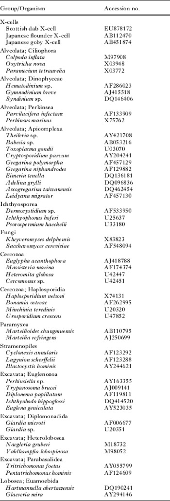
Appropriate taxa (Table 1) were chosen for the phylogenetic analyses by performing nucleotide BLAST searches with the dab X-cell sequence (Altschul et al. Reference Altschul, Gish, Miller, Myers and Lipman1990). BLAST settings were initially used at the default option, but subsequently set to be more stringent and hence more appropriate for divergent sequences by reducing the setting for the expect threshold from 10 to 2 and altering the ratio for match/mismatch scores from 0·5 (1/−2) (best for sequences that are well conserved (95%)) to a ratio of 1 (1/−1) which is more suitable for sequences that are less conserved (75%) (States et al. Reference States, Gish and Altschul1991). Additional taxa were chosen to further represent the groups highlighted during the BLAST searches and to represent other major protist phyla (Table 1).
List of taxa/small subunit ribosomal DNA sequences used in the phylogenetic analyses

Phylogenetic analyses were done using maximum likelihood (ML) methodologies in PAUP*4.0 beta10 (Swofford, Reference Swofford2002). For the ML analysis, the Akaike Information Criterion (AIC) implemented in Modeltest 3.7 (Posada and Crandall, Reference Posada and Crandall1998) was used to determine the most suitable model of evolution. Based on the AIC, the ML analysis was performed with the general time-reversible, GTR+I+G model of evolution with the settings as follows: Base=(0·2239 0·2301 0·2923) Nst=6 Rmat=(1·3071 3·5944 1·8363 0·8542 5·0656) Rates=gamma Shape=0·7091 Pinvar=0·0698. Clade support was assessed with bootstrapping of 100 replicates. Gaps were treated as missing data.
Bayesian inference (BI) analyses were conducted using MrBayes v. 3.0 (Ronquist and Huelsenbeck, Reference Ronquist and Huelsenbeck2003). Models of nucleotide substitution were evaluated for the data using MrModeltest v. 2.2 (Nylander et al. Reference Nylander, Ronquist, Huelsenbeck and Nieves-Aldrey2004). The most parameter-rich evolutionary model based on the AIC was the general time-reversible, GTR+I+G model of evolution. Therefore, the settings used for the analysis were nst=6, with the gamma-distributed rate variation across sites and a proportion of invariable sites (rates=invgamma). The priors on state frequency were left at the default setting (Prset statefreqpr=dirichlet (1,1,1,1)). Posterior probability distributions were generated using the Markov Chain Monte Carlo (MCMC) method with 4 chains being run simultaneously for 1 000 000 generations. Burn in was set at 2500 and trees were sampled every 100 generations making a total of 7500 trees used to compile the consensus trees.
The ISH methodology used in this study follows that of Morris et al. (Reference Morris, Adams and Richards1999) with the modification implemented by Holzer et al. (Reference Holzer, Sommerville and Wootten2003). In brief, mounted sections were hydrated and permeabilized with 100 μg ml−1 proteinase K in Tris-buffered saline (TBS) pH 8 for 25 min at 37°C. Sections were then washed in phosphate-buffered saline (PBS pH 7·4) and post-fixed for 15 min with 0·4% paraformaldehyde in PBS. After a distilled water wash, sections were exposed to 10% hydrogen peroxide (H202) in methanol for 10 min in order to prevent non-specific peroxidase binding. The H2O2 was washed off with distilled water and the sections dried at 45°C, in order to be able to omit the time-consuming pre-hybridization step. Sections were then encircled with a PAP-pen and equilibrated in hybridization buffer consisting of 4× saline-sodium citrate buffer (SSC) in TBS containing 0·5% Ficoll, 0·5% polyvinylopyrrolidone, 0·5% bovine serum albumin, 100 μg ml−1 calf thymus DNA, and 1·5 ng ml−1 of each oligonucleotide probe. Two 5′ biotin-labelled oligonucleotide probes were used in the hybridization; probe 1320f (as above) and the antisense probe R3M 5′ TAGGAATTCCTCGTTCAAGACG 3′. Formamide was not included in the buffer. Sections were sealed and denatured for 4 min at 95°C and left to hybridize for 1 h at 45°C. Hybridization was followed by non-stringent and stringent washes with 2× SSC and SSC 0·1% Tween 20 respectively. Signal detection was achieved using incubation with 1/100 horseradish peroxidase-labelled streptavidin (SAPU, Scottish Antibody Production Unit, Edinburgh, UK) for 20 min, followed by 3 washes with PBS and visualized with the substrate (VIP chromogen; Vector Laboratories, Peterborough, UK). Methyl green (0·5%) was applied for 5 min as a counter stain, after which sections were rapidly dehydrated in 95% and 100% ethanol, transferred to xylene and mounted in Pertex.
RESULTS
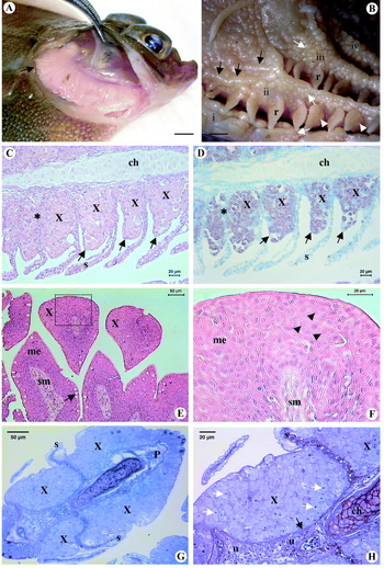
European dab ranging from 17 to 23 cm in total length were collected showing typical signs of X-cell disease, with pale yellow ‘fluffy-looking’ gill filaments (Fig. 1A). After complete removal of the whole gill arch complex, numerous raised round-shaped cysts could be seen on the internal surfaces of the gill arches and sometimes on the gill rakers (Fig. 1B). The cysts were present on all gill arches but seemed to be more established on the third and fourth arches, occasionally numerous adjacent cysts fused to form 1 long sausage-shaped cyst (Fig. 1B). A single fish was observed with a small swelling on the pseudobranch, which was removed and processed for DNA analysis.
Gross external appearance and histopathology of X-cell disease in European dab, Limanda limanda from the Moray Firth Scotland. (A) Fresh gills are pale and yellowish in colour with a ‘fluffy’ appearance; typically primary lamellae at the dorsal end of the gill arch (black arrow) are enlarged and lengthened due to infection and protrude from the operculum. Scale bar represents 1 cm. (B) Numerous raised round-shaped cysts (white arrows) are present on the internal surfaces of the 4 gill arches (i–iv) and occasionally seen on the gill rakers (white arrowhead). Cysts sometimes appear to fuse together forming an elongated sausage-shaped cyst (black arrows). Scale bar represents 4 mm. (C and D) Sagittal serial histological sections: H&E stained (C) and from the in situ hybridization (ISH) study (D), of an infected primary gill filament. Large clusters of X-cells (X) occupy the basal region between the secondary lamellae (s) adjacent to the primary lamellae, leading to total fusion of the secondary gill lamellae (black asterisk). (C) Shows the exact location of the X-cells, represented by the specific brown signal from the ISH study. (E and F) H&E-stained sections of cysts from the internal surfaces of the gill arches. (E) Section through numerous cysts showing lightly stained X-cells (X) proliferating in the mucosal epithelium (me) surrounding the uninfected central submucosa (sm). Numerous mucous cells are seen on the surface of the cysts (black arrow). (F) Enlargement of the boxed area in (E); revealing numerous cells with typical X-cell morphology, having large nuclei (black arrowheads). (G and H) Transverse semi-thin section of X-cell infected primary gill lamellae. (G) Reveals that the X-cells (X) extend around the entire circumference of the primary lamellae (P), sometimes proliferating along the entire length of the secondary lamellae (s). Higher magnification of the same section in (H) reveals X-cell nuclei containing large dark-staining nucleoli (white arrows) and a layer of uninfected undifferentiated cells (u) and chondrocytes (ch) lying beneath the basal lamina (black arrow).

Histological sections of the gill tissue showed considerable pathology in the primary gill lamellae, with masses of lightly-staining X-cells proliferating from the basal lamina between the secondary lamellae adjacent to the primary lamellae, in some cases leading to total fusion of the secondary gill lamellae (Fig. 1C, D). The dense proliferations of X-cells were contained by gill epithelium and did not fully penetrate the primary or secondary lamellae (Fig. 1C, D). However, functional gill tissues were replaced by X-cells, in some cases along the entire length of the primary lamellae. The specific oligonucleotide probes designed from the novel SSU rDNA sequence used in the ISH study successfully targeted the X-cell parasites. The X-cells are clearly visible, seen as brown signals and shown to be located along the primary gill filament clustering in the areas exhibiting abnormal gill histopathology (Fig. 1D). Histological sections of the gill arch cysts revealed lightly staining X-cells proliferating in the mucosal epithelium surrounding the uninfected central submucosal tissue layer (Fig. 1E). Numerous mucous cells were evident on the surface of the cysts. At higher magnification the X-cells looked similar to those seen in the gill lamellae, being lightly stained with large prominent nuclei (Fig. 1F).
Transverse semi-thin sections revealed that the X-cell proliferation also extended completely around the transverse circumference of the primary lamellae, extending to the ends of the secondary lamellae (Fig. 1G). At higher magnification the X-cells can be clearly seen as a mass of large polygonal cells with big nuclei and visible nucleolus surrounded by darker-staining host envelope cells (Fig. 1H).
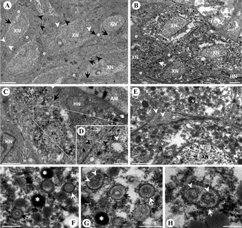
TEM showed 2 basic types of X-cells. The first type of X-cells has large nuclei occupying the majority of the cellular space, with little visible cytoplasm containing the occasional mitochondria-like organelle (Fig. 2A). These types of X-cells were predominately found occurring in the cysts on the inner surfaces of the gill arches. The distinction between X-cell and host envelope cells is difficult to assess, but the host envelope cell matrix is clearly supported by an extensive network of large desmosomes (Fig. 2A). The second type of X-cells was found in the gill lamellae, also containing large nuclei but occupying significantly less cell volume (Fig. 2B). The cytoplasm of these X-cells is often lacking material close to the nucleus but always contains numerous small vesicle-like structures that range from slightly to extremely electron dense and predominate towards the X-cell plasma membrane. The distinction between these X-cells and the host envelope cells is much more apparent and far fewer desmosomes are present between the surrounding envelope cells (Fig. 2B). Figure 2C shows an X-cell that is radically cleaved and contains 2 nuclei as if dividing. These cells were located deeper in the gill tissue and seen near gill filament abductor muscle (Fig. 2C). These cells contained cytoplasmic organelles that were bounded by a double membrane and resembled mitochondria, but had little or no discernable cristae (Fig. 2D). In the X-cells found in the gill lamellae the mitochondria-like organelles were not observed but the cytoplasm contained numerous other small vesicle-like structures, usually observed as circular in section, and ranging from 300 to 500 nm in diameter (Fig. 2E, F). Host envelope cells contained mitochondria that can be seen in the wider bands of cytoplasm at the junctions between X-cells (Fig. 2E). No clear Golgi dictyosome was observed in the X-cells, but rough endoplasmic reticulum in the form of a single membrane stack adjacent to nuclear pores of the nuclei were sometimes apparent, and vesicles of moderate electron density were associated with the trans-face of the Golgi network (Fig. 2E). At higher magnification, some of the small vesicle-like structures are seen to contain regular circular zones of varying electron density, in a concentric pattern, while others are much more intensely stained, appearing uniformly dark (Fig. 2F, G). The vesicles have a thickened membrane, which is clearly comprised of a double layer in places (Fig. 2F–H). Some vesicles are more oval in section and contain 2 electron-dense cores (Fig. 2G, H). Apart from the relative volume of the X-cell occupied by the nucleus, the main difference between the two types of X-cells are the numerous vesicles seen in the cytoplasm of the X-cells found in the gill lesions which are absent from the gill arch cyst X-cells.
Transmission electron micrographs (TEM) of infected gill material. (A) Cysts from a gill arch. X-cells have a very large nucleus (XN) and nucleolus (n) occupying nearly the entire cell. There is a narrow band of cytoplasm that contains infrequent mitochondria-like organelles (white arrowheads) before the X-cell plasma membrane (black arrows). Surrounding the X-cells are the more electron-dense host envelope cells (e) that are connected to each other via a network of large desmosomes (black arrowhead). Scale bar represents 2 μm. (B) Infected gill lamellae. X-cells have a more condensed nucleus (XN). The cytoplasm contains numerous small organelle-like structures that range from slightly to very electron dense and predominate towards the X-cell plasma membrane (white arrow). Host envelope cells (e) are more distinct and the host cell nucleus (HN) often visible. Scale bar represents 2 μm. (C and D) Dividing X-cell found deeper in the gill tissues, adjacent to gill filament abductor muscle (AM) and surrounded by host envelope cells with clearly visible nuclei (HN). The X-cell is radically cleaved in the central part of the cell (black arrow) and has a distinct nucleus (XN) in each lobe. Inset (D) is a magnification of the boxed section from (C) and shows the plasma membranes of the X-cell (black arrow-heads) in the cleaved part of the cell and a mitochondrion-like organelle. Scale bars represent 2 μm (C) and 500 nm (D). (E) Magnification of the central region of (B). Envelope cell (e) amid 4 adjoining X-cells. White arrowheads indicate host envelope cell mitochondria. The X-cell cytoplasm contains numerous vesicle-like structures (v), some of which are associated with the trans-Golgi network (G). Rough endoplasmic reticulum (rer) can be seen adjacent to the X-cell nucleus (XN) located in close proximity to nuclear pores (white arrow). Scale bar represent 1 μm. (F–H) Cytoplasmic vesicles in the X-cells. The vesicles range from being extremely electron dense (white asterisk) to containing regular circular zones of varying electron density in a concentric pattern (black arrows). The vesicles have a thickened membrane, which is clearly comprised of a double layer in places (white arrows). Some vesicles are more oval in section and contain 2 electron-dense cores (white arrowheads). Scale bars represent 500 nm (F and G) and 200 nm (H).

Analysis by specific PCR of all abnormal tissues observed from the gill/opercular region revealed that X-cell DNA was confirmed to be present in all cases: gill lamellae, gill arch cysts and abnormal pseudobranch. Positive PCRs from all abnormal tissues were used to obtain the SSU rDNA sequence. The consensus sequence obtained was 1763 base pairs in length and has been deposited in GenBank and assigned the Accession number EU878172. Searches conducted with the consensus sequence in nucleotide databases using BLAST with default settings, revealed only 2 sequences with an expectation value (E value) of zero, both from X-cells causing epidermal tumours in fishes from Japan. All other BLAST results using the default settings had low maximum identities given over extremely short query coverage and a high E value, indicating that any similarity present did not reflect a common descent. BLAST results with more stringent settings yielded far more sequences with low expectation values, mainly from the alveolate groups. Numerous initial phylogenetic trees were constructed using taxa taken from all of the BLAST results data, including some with high E values, in order to confirm their phylogenetic grouping with respect to the X-cell sequences. The non-X-cell taxa taken from the BLAST searches using the default settings were mainly basal fungal lineages which grouped within the fugal clade in the ‘eukaryotic crown’ and were distant to the X-cell grouping. Some of the unidentified environmental eukaryotic samples grouped within the main alveolate clade, but none grouped near the X-cell clade or significantly altered tree topology by their inclusion. Therefore, the environmental sequences were omitted from the final analyses as it was felt they could not contribute to X-cell identification as they themselves were mostly unidentified.
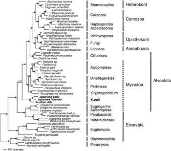
Phylogenetic trees created using Maximum Likelihood (ML) and Bayesian inference (BI) methodologies gave similar tree topologies with respect to the evolutionary positioning of the dab X-cells; the BI tree is shown in Fig. 3. The consensus tree from the BI analysis shows that the SSU rDNA sequence obtained from dab X-cells from Scotland forms a monophyletic clade with the sequences from X-cells infecting the Japanese goby and flounder. This grouping has a high external probability support value of 1·0. However, its relative positioning in the tree is less robust, but still well supported, with an external probability of 0·67. The X-cell clade is positioned within the myzozoan group, directly below the main group containing the dinoflagellates and the majority of the apicomplexans, but higher than the more basal eugregarine (apicomplexan) group.
Phylogenetic tree of protist groups, constructed using Bayesian Inference analyses using the general time-reversible (GTR+I+G) model of evolution. The numbers at the nodes represent the clade posterior probability, indicating the proportion of sampled trees that contained that branch. A solid circle at the node indicates a posterior probability of 0·9 and above. To the right of the tree are the currently accepted taxonomic rankings (order, class, phylum) far right are the higher scientific classifications (super-phylum). Scale bar represents 100 changes.

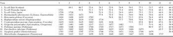
From tree topologies, representative sequences from various protist groups were taken to create a percentage identity matrix (Table 2). Dab X-cells were 89·3% and 86·7% similar to Japanese X-cell sequences from flounder and goby respectively. The two Japanese X-cell sequences were more closely related to each other being 91·8% similar. From other non-X-cell taxa used in the analysis, the dab X-cell sequence was most similar to Perkinsus marinus and Cryptosporidium parvum with 77·3% and 76·8% identities respectively.
Percentage identities (above diagonal) of the SSU rDNA of selected taxa with number of bases compared (below diagonal)

DISCUSSION
Typically, X-cell disease in the European dab, Limanda limanda, is characterized by lesions in the primary gill lamellae causing swollen and pale gill filaments (McVicar et al. Reference McVicar, Bucke, Watermann and Dethlefsen1987). However, X-cells have also been associated with pseudobranchial swellings and visceral lesions in L. limanda (Diamant and McVicar, Reference Diamant and McVicar1987) and may also be associated with an inflammatory condition of the subcutaneous connective tissue in skin (Watermann, Reference Watermann1982). Here we report X-cells from L. limanda in gill arch cysts as well as from abnormal pseudobranchial tissue and gill filaments (internal organs and skin epithelium were not examined during this study). X-cell infections among fish species generally appear to be tissue or site specific: gadoids with pseudobranchial tumours, Atlantic dab and plaice with gill lesions and Pacific flounder with epidermal tumours. General tissue specificity for X-cell disease among certain fish species remains unexplained, but may simply be due to a group of parasitic organisms, the X-cells, developing host, site and tissue specificities over time. Furthermore, most X-cell infected tissues are all of similar origins, being derived from the ectoderm (Alpers et al. Reference Alpers, McCain, Myers, Wellings, Poore, Bagshaw and Dawe1977). The fact that X-cell lesions may occur elsewhere in the host fish with simultaneous infection in the primary tissue location suggests that X-cells are relatively motile, or have motile stages, and that under certain conditions X-cell proliferation can result at a secondary infection site, normally atypical for that species. Cod have been reported to have gill lesions in addition to primary pseudobranchial tumours (McCain et al. Reference McCain, Gronlund, Myers and Wellings1979) and European dab have been reported to have pseudobranchial swellings in addition to primary gill lesions (Diamant and McVicar, Reference Diamant and McVicar1987; present study); there are no clear reports of infection at a secondary site without significant simultaneous pathology at the primary site of infection for these fish species. X-cells typically appear in histological sections in very close association with a complex matrix of host envelope cells that would not allow such motility within the host. However, Yamazaki et al. (Reference Yamazaki, Hibino, Oishi, Harada, Stich and Acton1978) described the presence of very small amoeboid-like ‘wandering’ X-cells, only 3–4 μm in diameter, at the boundaries of epidermal tumours in flatfish from northern Japan that appeared to be able to move freely within the intercellular spaces between basal cells, and the autofluorescence of X-cell cytoplasm revealed X-cells in cod tumours that were smaller than cod erythrocytes (Dawe, Reference Dawe, Dawe, Harshbanger, Kondo, Sugimura and Takayama1981). These small amoeboid-like X-cells may be responsible for autoinfection within the organ and similar tissues within the same host. Other reports from advanced cod pseudobranchial tumours describe free-floating X-cells displaying amoeboid shapes in the central part of the tumour (Watermann and Dethlefsen, Reference Watermann and Dethlefsen1982), and free X-cells in the interlamellar spaces of ruptured gill lesions in the American plaice H. platessoides from the Atlantic, suffering with similar X-cell gill lesions to L. limanda (MacLean and Despres-Patanjo, Reference MacLean and Despres-Patanjo1993). These descriptions of free-floating X-cells are from advanced tumours described as ‘close to rupture’, and may represent the infective stage for a different or alternate host. During the current study no such small or free-floating amoeboid X-cells were observed, which may have been due to the non-mature condition of the lesions examined.
The experimental transmission of X-cells between fish of the same species has been attempted but never successfully achieved. Ito et al. (Reference Ito, Kimura and Miyake1976) used a cell-free inoculum of neoplastic tumours from A. flavimanus to subcutaneously inoculate naïve A. flavimanus, but after 160 days no tumours had developed. Freeman et al. (unpublished data) have attempted the feeding of tumour tissues and inter-peritoneal injection of tumour homogenate to naïve fish in both Atlantic cod and Japanese flounder; neither trail resulted in the successful transmission of X-cell disease. The lack of direct transmission suggests that an obligate intermediate host or a free-living stage is required in the life cycle for X-cell organisms. This may also help to explain the unusual distribution patterns often associated with X-cell infections. Diamant and McVicar (Reference Diamant and McVicar1989) reported extreme variability in the prevalence of X-cell infected dab from the Moray Firth in Scotland, ranging from nil to 60% of dab collected from adjacent locations. As no spore-like forms or flagellated stages have ever been described from X-cell infected fish, despite numerous thorough histopathological studies, it can be assumed that transmission from the fish host is via the amoeboid stages observed and not via spores or flagellated forms. Fish species that develop X-cell disease are associated with the benthic substrate, and dab suffering with X-cell disease taken in trawls suffered very high mortality rates, >95% in the first week compared to <5% in non-affected dab (Diamant and McVicar, Reference Diamant and McVicar1987). High mortality of captured infected fish is thought to be due to respiratory restrictions caused by the substantial loss of functional gill tissue. Such respiratory limitation suggests that infected fish with advanced tumours would not be active and probably restricted to the benthic substrate where amoeboid X-cells could be released from the ruptured lesions directly into the substrate. Hence, the benthic substrate could be the location for the infection of an alternate host, such as a detritus feeder or a filter-feeding invertebrate.
Observations of dividing X-cells seem to correlate closely with host fish species. Dividing X-cells from epidermal tumours in Pacific flounder have never been reported; in fact researchers have even reported total cell inactivity (Peters et al. Reference Peters, Stich and Kranz1981, Reference Peters, Schmidt, Kranz and Stich1983), a phenomenon which added support, at the time, to the fact that X-cells were virally transformed cells that were recruited from pre-existing fish cells via infection with a lymphocystis-like virus. In this study, dividing cells with 2 nuclei were observed but were hard to find and could have easily been overlooked. Diamant and McVicar (Reference Diamant and McVicar1989) reported seeing various stages of apparent nuclear division in X-cells from dab, but only saw a single X-cell with 2 nuclei. Similar findings were also reported from ‘suspected’ X-cells causing an inflammatory epidermal condition in dab, where active division was only evident in one X-cell (Watermann, Reference Watermann1982). In contrast, X-cells in cod pseudobranchial tumours have been shown to form large plasmodia containing up to 60 nuclei, suggesting asynchronous division by plastotomy (Dawe, Reference Dawe, Dawe, Harshbanger, Kondo, Sugimura and Takayama1981; Watermann and Dethlefsen, Reference Watermann and Dethlefsen1982) or a failure in cytokinesis (Alpers et al. Reference Alpers, McCain, Myers, Wellings, Poore, Bagshaw and Dawe1977). However, the most convincing study confirming X-cell division has been demonstrated in the Japanese goby, Acanthogobius flavimanus, suffering from epidermal skin tumours. Amitotic division was shown to occur as the nucleus divides by constriction, without any sign of mitosis, to form bi- or polynuclear X-cells, which then divide by constriction of the cell membrane without any further sign of mitosis (Shinkawa and Yamazaki, Reference Shinkawa and Yamazaki1987). This apparent lack of mitosis and hence spindle and chromosome formation in the nucleus in some X-cells may explain the lack of reports of nuclear activity in X-cell prior to cell division, which must occur in order for such large X-cell masses to form. These differing reports of X-cell division support the theory that these organisms are not the same species, but a group of parasitic protozoans that infect fish and proliferate to form tumour-like masses of X-cells in certain tissues. TEM revealed two types of X-cells, those predominantly found in gill arch cysts and those in gill lesions. These X-cell types may represent different developmental stages and, although actively dividing stages were not frequently observed, such division may represent the transformation between the two X-cell types. Diamant and McVicar (Reference Diamant and McVicar1989) also described 2 types of X-cells from dab, but located within ‘nests’ and being considerably larger than the X-cells described here; such discrete ‘nests’ of large X-cells were not observed during this study. The more typical X-cells described by Diamant and McVicar (Reference Diamant and McVicar1989) correspond to the lamellar X-cells observed in this study. They also observed numerous electron-dense inclusions in the cytoplasm of these cells and suggested that they were formed from or were degenerate mitochondria, as numerous small round mitochondria of similar sizes and possible transitional stages between the two were seen alongside them. During the present study typical mitochondria were not observed in the X-cells. Double-membraned mitochondria-like organelles were seen in the dividing X-cells and in the immature lamellar X-cells prior to cytoplasmic vesicle formation. These mitochondria-like organelles were never seen in mature X-cells containing cytoplasmic vesicles. Host envelope cell mitochondria were often observed suggesting that tissue fixation was adequate for mitochondrial preservation. The fact that mitochondria-like organelles are present but disappear after the vesicles have formed does imply that they may have some involvement in the vesicle formation, as suggested by Diamant and McVicar (Reference Diamant and McVicar1989). Similar membrane-bound electron-dense cytoplasmic granules have been reported from X-cells infecting Pacific fish as discussed by Dawe (Reference Dawe, Dawe, Harshbanger, Kondo, Sugimura and Takayama1981), which led to the suggestion that due to their likeness to the haplosporosomes in the oyster parasite Minchinia (Perkins, Reference Perkins1969), X-cells were an amoeboid phase of a haplosporidian parasite.
Membrane-bound electron-dense cytoplasmic vesicles that migrate to the plasma membrane late in cell development or sporogenesis have been described from numerous parasitic groups: haplosporosomes from the Haplosporidia and Paramyxea and sporoplasmasomes from the Myxosporea (Lom et al. Reference Lom, Molnár and Dyková1986) and Malacosporea (Kent and Hedrick, Reference Kent and Hedrick1986). Genesis of these structures is initially associated with the trans-Golgi network and latterly with multivesicular bodies (MVBs) which contain numerous pleomorphic developing vesicles (Hine and Wesney, Reference Hine and Wesney1992; Morris et al. Reference Morris, Adams and Richards2000). Although Golgi dictyosomes were not routinely observed in this study, some association of the cytoplasmic vesicles in dab X-cells with the trans-Golgi face was noted, but no MVBs containing numerous developing vesicles, or vesicles with varying electron densities, were seen. The function of haplosporosomes remains unclear, but it has been suggested that they are associated with transport during spore wall formation (Ginsburger-Vogel and Desportes, Reference Ginsburger-Vogel and Desportes1979) or may act as an energy reserve for sporulation (Azevedo and Corral, Reference Azevedo and Corral1985). Sporoplasmasomes have been shown to be biochemically different from haplosporosomes, as they do not contain carbohydrates, but are also thought to be involved in the transport of materials to either the plasma membrane or spore wall (Morris et al. Reference Morris, Adams and Richards2000).
As gill lesion X-cells mature, the cytoplasmic vesicles are arranged along the plasma membrane, in a similar way to those in haplosporosomes and sporoplasmasomes, a phenomenon also noted by Diamant and McVicar (Reference Diamant and McVicar1989) in visceral X-cell lesions in dab. Such membrane association also suggests a possible transport or excretory role as hypothesized for both membrane-aligned haplosporosomes and sporoplasmasomes. However, as no spore formation is known to occur in fish X-cells, the vesicles may have a different role and could be a developmental precursor for a cellular structure that is required for motility or to aid infection of another host. X-cell vesicles may coalesce to form a rudimentary axoneme/basal body as seen in the amoeboflagellate group or a similar structure to the rhoptries or micronemes that develop in members of the alveolates.
The cytoplasmic vesicles observed in X-cells, haplosporosomes in the Haplosporidia and Paramyxea and sporoplasmasomes from the Myxozoa share some basic features, such as plasma membrane association and a degree of internal structure. However, these structures are seemingly unrelated as to their final function but may share a common transport role in the cell. Due to the distant phylogenetic positions between the Myxozoa (pluricelluar/related to Cnidaria), Paramyxea (basal eukaryotes) and Haplosporidia (Cercozoa), the use of these structures for wider taxonomic studies appears to be extremely limited.
X-cell parasites formed a monophyletic clade in all phylogenetic analyses. This grouping was consistently positioned directly below the main alveolate group containing all the dinoflagellates and the majority of the apicomplexans except for the eugregarines, which were located immediately basal to the X-cell clade. The alveolates are a major lineage of the protists comprised of 3 main groups: the Ciliophora (ciliates), the Apicomplexa and the Dinozoa (dinoflagellates). The Apicomplexa and the Dinozoa collectively form the Phylum Myzozoa (Cavalier-Smith and Chao, Reference Cavalier-Smith and Chao2004), and are more related to each other than either are to the ciliates. All alveolates have cortical alveoli which are flattened vesicles that form a layer supporting the plasma membrane, and all possess mitochondria with tubular cristae and have a distinct flagella structure. The X-cell clade is consistently located within the myzozoan group; however, our ultrastructure findings and those of other researchers do not support this alveolate grouping. No ultrastructural study on X-cells has been able to demonstrate cortical alveoli or the presence of either flagella structures or an apical complex. The lack of typical alveolate features at an ultrastructural level may be explained through the current lack of known developmental stages in the X-cell life cycle and no knowledge of the alternate and presumably definitive host. The sequencing of additional novel genes for the X-cell group, such as α and β-tubulin genes, is necessary to support this grouping within the Myzozoa.
From the percentage identity matrix, the dab X-cell sequence was most similar to the Japanese X-cell sequences, being more similar to the flatfish X-cell (89·3%) than to the goby X-cell (86·7%), suggesting that fish group (flatfishes) and hence parasite-host specificities may have played a role in the radiation of X-cell parasites. However, the two Japanese sequences are more similar (91·8%) than the flatfish X-cell sequences suggesting that geographical location is also an important factor and that host switching may have occurred. Such large percentage differences (<90%) in the rDNA would normally suggest that these organisms are only related at family level or higher. Apart from other X-cell sequences, the dab X-cell sequence was most similar to Perkinsus marinus (77·3%) and Cryptosporidium parvum (76·8%), with respect to percentage identity of the SSU rDNA, a trend that was also observed for both the Japanese X-cell sequences (Table 2) which supports the myzozoan placement in the phylogenetic analyses.
Perkinsus is known to occupy a basal position in the dinoflagellates (Saldarriaga et al. Reference Saldarriaga, McEwan, Fast, Taylor and Keeling2003) and Cryptosporidium is known to group with the gregarines which are thought to be the earliest lineage of the apicomplexans (Leander et al. Reference Leander, Clopton and Keeling2003), making them both important with respect to the divergence of the apicomplexan and dinoflagellate groups and hence the alveolates in general. The fact that all X-cell parasites sequenced to date show some similarity, albeit low, to both Perkinsus and Cryptosporidium compared to other protist taxa, and that the X-cell clade is immediately basal to the main Alveolate group in all our phylogenies, suggests that X-cell biology may well be important in understanding the early evolution of the Myzozoa and the alveolate group. However, it remains unusual that BLAST searches revealed no non-X-cell SSU rDNA sequences with an identity greater than 77%, suggesting that there is a current lack of data for an X-cell sister group, if one exists.
Dawe (Reference Dawe, Dawe, Harshbanger, Kondo, Sugimura and Takayama1981), Miwa et al. (Reference Miwa, Nakayasu, Kamaishi and Yoshiura2004) and Miwa and Kamaishi (Reference Miwa and Kamaishi2009) suggested that the X-cell group might be related to the haplosporidians. However, in this study the Haplosporidia consistently group with other members of the Cercozoa in phylogenetic analyses and the single nucleotide deletion, reportedly unique to all members of the Cercozoa at the end of helix 37 of the variable region V6 (Neefs et al. Reference Neefs and De Wachter1990; Cavalier-Smith and Chao, Reference Cavalier-Smith and Chao2003), is not a consistent feature in the X-cell SSU and, although present in both Japanese sequences, is absent from dab X-cell sequence. However, molecular phylogenetic analyses by Siddall et al. (Reference Siddall, Stokes and Burreson1995) and Flores et al. (Reference Flores, Siddall and Burreson1996) both placed the Haplosporidia within the Alveolata and it remains possible that the Haplosporidia had an alveolate ancestry and are hence related to basal alveolates, such as the X-cells.
From the phylogenetic analyses conducted during this study, it is clear that the Paramyxea do not group with other members of the Asceptospora/Cercozoa, and appear to occupy a basal position in all phylogenies, which is in agreement with previous reports (Berthe et al. Reference Berthe, Le Roux, Peyretaillade, Peyret, Rodriguez, Gouy and Vivarès2000).
X-cell parasites are a monophyletic group of protist parasites that form a clade within the Alveolates with respect to SSU rDNA analyses, and are probably basal myzozoans related to the dinoflagellates, apicomplexans, and potentially ancestral to both. However, their relative lack of ultrastructural features compared to other myzozoans, in particular the absence of cortical alveoli, makes it hard to confirm their actual systematic position. As experimental transmission of X-cell parasites from fish to fish has been unsuccessful, an alternate host is probably required in the life cycle. X-cell development in the alternate host may well expose the true identity of the group and reveal ultrastructural detail with which a satisfactory taxonomic placement for the X-cell parasites can be made.
This work was funded by The Royal Society of London. My sincere thanks go to David Bruno and Sarah Baker for supplying infected dab material and to Astrid Holzer for advice and assistance with the in situ hybridization part of the study. Sincere thanks also go to Linton Brown for his enthusiastic help with the electron microscopy.